Understanding the adaptive immune responses against newly emerged viruses, SARS coronavirus and avian h5n1 influenza a virus 3
Bạn đang xem bản rút gọn của tài liệu. Xem và tải ngay bản đầy đủ của tài liệu tại đây (2.26 MB, 179 trang )
1
CHAPTER 1: INTRODUCTION
1.1 Overview of emerging infectious diseases
Despite advances in medical research and treatments during the 20
th
century, infectious diseases remain the leading causes of death worldwide.
The elimination of smallpox in 1977 was a great achievement in the fight
against infectious diseases. However, infectious diseases continue to persist
and caused great losses both in the social and economic aspects. This is due to
the emergence of new infectious diseases, re-emergence of old infectious
diseases, and persistence of intractable infectious diseases. Emerging
infections can be defined as infections that have newly appeared in the
population, or have existed but are rapidly increasing in incidence or
geographic range (Morse & Schluederberg, 1990). Examples of recent
emerging diseases include Acquired Immunodeficiency Syndrome (AIDS)
cause by human immunodeficiency virus (HIV), hantavirus pulmonary
syndrome, Lyme disease and foodborne infection by O157:H7 Escherichia
coli.
There are several factors that contribute to the emergence of new
diseases. These include ecologic changes (e.g. deforestation), changes in
human demographics and behavior (e.g. urban migration and intravenous drug
use), and increased international air travel. Human populations were brought
closer and more frequently to the source of the pathogens. The outbreak of the
new infection, severe acute respiratory syndrome (SARS) may be the result of
increased use of exotic animals as food source. Close contact with palm civet
cats and raccoon dogs in China was identified as a potential route by which the
2
SARS coronavirus (SARS-CoV) was transmitted from animal to human (Guan
et al., 2003; Wang et al., 2005). In addition to the identification of new
human viruses, “old infectious pathogens” are also re-emerging. Microbial
adaptation and natural genetic recombination and mutation result in new
strains of known pathogens, which are not recognized by the human immune
system. One example is the emergence of H5N1 influenza A virus strain
which has caused human infection since 1997 (Subbarao et al., 1998).
The aim of the project was to understand the adaptive immune
response against two newly emerged viruses, SARS-CoV and the H5N1
influenza A virus. For SARS-CoV, the focus was mainly to investigate the T
cell response against the unique accessory 3a protein. The study was also
extended to T cell response against the structural nucleocapsid (N) protein in
SARS recovered individuals. The main objective for the H5N1 work was to
demonstrate the use of the recombinant baculovirus-expressed hemagglutinin
(HA) protein for use as vaccine and as a tool to generate neutralizing
antibodies. Further characterization of a potent monoclonal antibody (mAb)
against the HA protein was also performed.
1.2. Severe Acute Respiratory Syndrome (SARS)
1.2.1. Epidemiology of severe acute respiratory syndrome (SARS)
SARS first emerged in Guangdong, China in late 2002 (Zhong et al.,
2003). By March 2003, the disease spread to Hong Kong, and then to
Vietnam, Singapore and Canada (Lee et al., 2003; Poutanen et al., 2003;
Tsang et al., 2003). Those infected by SARS were mainly healthcare workers
or household members who had cared for patients with severe respiratory
3
illness. Contact tracing finally indicated the index case was a healthcare
worker from Guangdong province who visited Hong Kong and transmitted the
virus to several other guests who, further contributed to global dissemination
of the disease (Ksiazek et al., 2003). A novel coronavirus SARS coronavirus
(SARS-CoV) was identified as the causative agent (Drosten et al., 2003;
Ksiazek et al., 2003; Peiris et al., 2003b). The SARS-CoV went on to infect
more than 8000 people in 29 countries across 5 continents with 774 deaths
reported by World Health Organization (WHO) (WHO, 2003b). The SARS
epidemic was officially controlled by July 2003 with strict isolation of
patients.
The main route of transmission seems to be airborne droplets from
infected patients (Booth et al., 2005; Hui & Chan, 2010; Yu et al., 2004b).
Blood and fecal-oral transmission has also been suggested (Poon et al., 2003).
There are no known vectors for coronaviruses but epidemiological evidence
demonstrated that early cases of SARS were linked to exposure to wild game
animals in the live wet markets in Guangdong province (Guan et al., 2003). In
the nasal and fecal swabs from masked palm civets (Paguma larvata) and
raccoon dogs (Nyctereutes procyonoides) in the wet markets, SARS-like
viruses which are genetically and antigenically related to the human SARS-
CoV were detected using reverse transcriptase polymerase chain reaction (RT-
PCR) and electron microscopy of viral particles from infected cells. The
SARS-like viruses isolated from the animals were shown to have more than
99% homology with the human SARS-CoV. Interestingly, high
seroprevalence for the animal SARS-CoV antibodies were found in animal
traders working with these live animals in the wet markets, although they did
4
not have a history of SARS-like disease (MMWR, 2003). These observations
seem to suggest that the live animal market probably is the site for the animal
SARS-CoV to amplify and allow interspecies transfer of the animal virus to
the humans. However, it is not clear whether these animals are the natural
reservoirs of the SARS-CoV in the wild. In 2005, reports from several groups
identified a virus that was genetically closely related to human SARS-CoV in
the Chinese horseshoe bats, suggesting that they may be the natural source of
SARS-CoV although attempts to isolate the virus from bats have not been
successful (Lau et al., 2005; Li et al., 2005). Bats are known to be the
reservoir hosts of several zoonotic viruses, including the Hendra and Nipah
paramyxoviruses that have recently emerged in Australia and East Asia (Chua
et al., 2000; Murray et al., 1995). Finally, molecular epidemiology showed
that at least two strains of SARS-CoV have been found in patients in Hong
Kong (Guan et al., 2004a). This suggests that the virus had jumped from
animal source to human on two separate occasions. This indicates the
outbreak of SARS would have been inevitable and the potential of re-
emergence is also high.
1.2.2. Genome Organization
Coronaviruses are a diverse group of large, enveloped positive-
stranded RNA viruses. They belong to the order Nilovirales, family
Coronaviridae, genus Coronavirus and cause respiratory and enteric diseases
in humans and other animals. Three serologically distinct groups of
coronaviruses have been identified. Group I and II contain mammalian
viruses, whereas group III contains only avian viruses. After its discovery, the
5
SARS-CoV was initially placed in a new group (IV) of coronavirus as the
sequence is distinct from those previously reported in animals and humans
(Marra et al., 2003; Rota et al., 2003). However, examining sequences within
regions of ORF 1a of SARS-CoV showed domains that are unique to the
group II coronaviruses, suggesting that SARS-CoV may be more directly
related to group II viruses (Snijder et al., 2003). Group II coronaviruses
include the bovine coronavirus, human OC43 virus and murine hepatitis
coronavirus (MHV). More evidence demonstrated that the 3’UTR of SARS-
CoV could substitute functionally for that of MHV, but not with 3’ UTR from
group I coronavirus (Goebel et al., 2004). Thus some research groups
suggested that SARS-CoV belongs within group II coronaviruses, in a
subgroup IIb.
The genome of SARS-CoV is approximately 30 kb, with
polyadenylated positive-stranded RNA (Marra et al., 2003; Rota et al., 2003).
The genomic organization is typically of a coronavirus, with the characteristic
gene order, with the first two open reading frames (1a and 1b) encoding the
viral replicase and the downstream mRNAs encoding structural proteins spike
(S), envelope (E), membrane (M) and nucleocaspid (N). However, the gene
encoding hemagglutinin-esterase found in group II and some group III
coronaviruses was not found in SARS-CoV (Rota et al., 2003). The RNA is
packaged by the N protein into a helical nucleocapsid, with the S protein
forming morphologically characteristic projections on the virion surface.
6
1.2.2.1. Replicase genes
The viral replicase genes (ORF 1a and 1b) translate into two
polyproteins, pp1a (486 kDa) and pp1ab (790 kDa) (Thiel et al., 2003).
Expression of the pp1ab is predicted to involve a ribosomal frameshifting into
the -1 frame just upstream of the ORF1a translation termination codon. The
pp1a and pp1ab polyproteins undergo proteolytic processing by viral cysteine
proteinases to yield the functional components of the membrane-bound
replicase complex and a group of 16 non-structural proteins (nsp) (Ziebuhr et
al., 2000). SARS-CoV uses only two proteinases, PL2
pro
, a papain-like
cysteine proteinase (nsp3) and 3CL
pro
, a 3C-like proteinase (nsp5) , in contrast
to most coronaviruses that use three proteinases (Gao et al., 2003; Rota et al.,
2003; Snijder et al., 2003). The replicase complex, which includes an RNA-
dependent RNA polymerase and RNA helicase mediates both genome
replication and transcription of a “nested” set of subgenomic mRNAs. The
putative functions of the non-structural proteins are summarized in Figure 1.1.
The functions have been shown by biochemical assays or predicted based on
their functional domains or structural similarities to other proteins while others
remain to be further characterized [reviewed by (Cheng et al., 2007)].
7
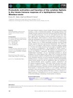
Fig. 1.1. Summary of the SARS-CoV genome organization and viral protein
expression. Replicase (ORF 1a and 1b), constituting the first 2/3 of the genome,
which translates into two polyproteins, pp1a and pp1ab. The putative functions of
each of the nsps are shown in the text boxes. Open reading frames (ORFs) in the
remaining 1/3 of the genome are translated from eight subgenomic mRNAs. Four of
the ORFs encode the structural proteins, spike (S), membrane (M), and envelope (E)
and nucleocapsid (N). Another eight unique ORFs encode accessory proteins (3a, 3b,
6, 7a, 7b, 8a, 8b and 9b), which have no significant sequence homology to viral
proteins of other coronaviruses. This figure was adapted and modified from Cheng,
V. C. C. et al. 2007. Clin. Microbiol. Rev. 20(4): 660-694.
P
Expression promoted degradation
of host endogenous mRNAs,
which may inhibit host protein
synthesis and prevented
endogenous IFNβ mRNA
accumulation
Deletion attenuates viral
growth and RNA synthesis
Papain-like protease 2;
ADP-ribose 1-phosphatase.
Not known
3C-like protease
Three-dimensional structure
found potential sites for protein-
protein interaction
Not known
Putative RNA-dependent RNA polymerase; crystal structure of
the hexadecameric nsp7-nsp8 possesses a central channel with
dimensions and positive electrostatic properties favorable for
nucleic acid binding; it is probably another unique RNA-
dependent RNA polymerase for its large genome
Three-dimensional crystal structure of a
dimer which binds viral RNA and interacts
with nsp8
Crystal structure suggests a nucleic acid
binding function within a larger RNA
binding protein complex for viral gene
transcription and replication
RNA-dependent RNA polymerase
Helicase (dNTPase and RNA
5’-triphosphatase activities)
3’→5’-exoribonuclease; supplements
the endoribonuclease activity in the
replication of the giant RNA genome
Uridylate-specific
endoribonuclease; Involved in
the coronavirus replication cycle
Putative 2’-O-ribose
methyltransferase
8
1.2.2.2. Structural and accessory proteins
The subgenomic mRNAs encode the structural proteins, S, E, M and
N, and the set of accessory proteins (namely ORF 3a, 3b, 6, 7a, 7b, 8a, 8b and
9b). The surface S protein is involved in the attachment and entry of the host
cell. The N together with M and E are involved in the assembly of the virion.
The accessory proteins have no significant homology to viral proteins of other
coronaviruses. These proteins are dispensable for virus replication in cell
culture while some appear to contribute to viral pathogenesis (Narayanan et
al., 2008; Tan et al., 2006). The focus of our study is the largest SARS-CoV
accessory protein, 3a. It is also known as U274 or X1 and this protein will be
discussed in detail in section 1.2.2.3. The characteristic, functions, and/or
putative roles of the four structural and seven other accessory proteins of the
SARS-CoV are outlined in Table 1.1 and Table 1.2 respectively.
9
Table 1.1 Summary of the SARS-CoV structural proteins
Protein (No. of
amino acid
residues in
protein)
Protein characteristic [ref(s)] Protein’s function(s) or putative function(s) [ref(s)] Effect on cellular response of host [ref(s)]
Spike protein
(1,255)
A type I integral membrane glycoprotein which
is N-glycosylated, trimerized in endoplasmic
reticulum (ER) (Bosch et al., 2003; de Groot et
al., 1987; Rota et al., 2003).
It is divided into 2 subdomains of similar size,
S1 and S2 with distinct functions. S1 domain
forms the globular portion of the spike,
mediating binding to host cell receptor,
angiotensin-converting enzyme 2 (ACE2) (Li
et al., 2003). The receptor binding domain
(RBD) is localized to amino acids 318 to 510.
The S2 ectodomain contains two regions with a
4, 3 hydrophobic (heptad) repeat, HR1 and
HR2 and a putative, internal fusion peptide
(Bosch et al., 2004; Sainz et al., 2005).
Biochemical studies have shown that peptides
corresponding to the HR1 and HR2 of the SARS-
CoV S protein can associate into an anti-parallel six-
helix bundle with structural features typical of class I
fusion proteins (Ingallinella et al., 2004; Liu et al.,
2004; Tripet et al., 2004). This HR1-HR2 structure
brings the fusion peptide in close proximity to the
transmembrane domain (Bosch et al., 2004), leading
to the fusion of the viral and cellular membrane, and
consequently the viral entry.
The S protein was known to be responsible
for inducing host immune responses and is
the primary target for viral neutralizing
antibodies (Keng et al., 2005; Zhou et al.,
2004).
The functional region of S protein from
amino acids 324-688 can induce the release
of IL-8 in lung cells (Stevens et al., 2006).
It induced unfolded protein response in
cultured cells as SARS-CoV with
accumulation of S protein in the ER, may
modulate viral replication (Yamada et al.,
2006).
10
Table 1.1 Continued
Protein (No. of
amino acid
residues in
protein)
Protein characteristic [ref(s)] Protein’s function(s) or putative function(s) [ref(s)] Effect on cellular response of host [ref(s)]
Envelope protein
(76)
The E protein of SARS-CoV (9-12 kDa)
has a short 7-9 amino acid hydrophilic
region and a 21-29 amino acid hydrophobic
region, followed by a hydrophilic C-
terminal region (Shen et al., 2003).
It was demonstrated that the E protein does form
ion channels, which are are more selective for
monovalent cations than monovalent anions
(Wilson et al., 2004).
It was shown to be important for viral assembly as
demonstrated by the formation of VLPs (Ho et al.,
2004; Nal et al., 2005; Vennema et al., 1996)
.
It induced apoptosis in transfected Jurkat T
cells in the absence of growth factors. A novel
BH3-like region located in the C-terminal
cytosolic domain of SARS-CoV E protein can
bind Bcl-xL, whose overexpression can
antagonize apoptosis (Durrer et al., 1996).
Membrane
protein (221)
The M protein contains a long cytoplasmic
tail, 3 hydrophobic transmembrane
domains, and a short glycosylated N-
terminal ectodomain. It has been shown to
be N-glycosylated at asparagines residue at
position 4 (
Nal et al., 2005; Voss et al.,
2006)
.
Functional analysis showed that the N-terminal
region of the M protein, comprising of the 3
transmembrane domains, is sufficient to mediate
accumulation of M in the Golgi complex and
recruit the S protein to the sites of viral assembly
and budding in the ER Golgi-intermediate
compartment (
Voss et al., 2009).
M protein induced apoptosis in HEK293T
cells, which could be suppressed by caspase
inhibitor (
Tsurudome et al., 1992).
11
Table 1.1 Continued
Protein (No. of
amino acid
residues in
protein)
Protein characteristic [ref(s)] Protein’s function(s) or putative function(s) [ref(s)] Effect on cellular response of host [ref(s)]
Nucleocapsid
protein (422)
The N protein is highly charged basic protein
of 422 amino acids (~48 kDa) with seven
successive hydrophobic residues near the
middle of the protein. At the N terminal, it
contains a highly conserved motif
[FYYLGTGP] which is observed in all
coronavirus N proteins (Rota et al., 2003). It
is reported to be abundantly found in the
cytoplasm and nucleus of SARS-CoV
infected cells.
Properties of the N protein include self-dimerization
(He et al., 2004), RNA-binding capabilities (Huang
et al., 2004), cleavage by caspase 3 (Ying et al.,
2004) and activation of signal transduction
pathways.
The N protein was shown to induce
apoptosis in COS cells in the absence of
growth factors (Surjit et al., 2004). It
antagonized interferon (IFN) by inhibiting
synthesis of IFNβ (Weber et al., 1994).
Nuclear factor kappa B (NF-κB) activation
was observed in Vero E6 cells expressing the
N protein in dose dependent manner (Zhou
et al., 2004). It may cause inflammation of
the lungs by activating COX-2 gene resulting
in inflammation through multiple COX-2
signaling cascades (Keng et al., 2005).
Most of the sera obtained from convalescent
SARS patients have antibodies against N
(Leung et al., 2004; Shi et al., 2003; Tan et
al., 2004a). In addition, it was reported that
the N protein can induce specific T cell
responses (Kim et al., 2004).
12
Table 1.2 Summary of SARS-CoV accessory proteins
Protein (No. of
amino acid
residues in
protein)
Protein characteristic [ref(s)] Protein’s function or putative function(s) [ref(s)] Effect on cellular response of host [ref(s)]
ORF 3b (154) 3b was expressed from an ORF which
overlaps the ORF3a and E by using an
internal ribosomal entry site (Rota et al.,
2003).
One study revealed 3b initially accumulates in the
nucleus and subsequently translocates to the
mitochondria (Freundt et al., 2009). However, the
exact mechanism in which 3b contributes to
SARS-CoV pathogenesis is still not known.
Overexpression of 3b has been shown to induce
cell cycle arrest at the G0/G1 phase and
apoptosis (Yuan et al., 2006a). It is also
suggested to be a type I IFN antagonist (Freundt
et al., 2009; Kopecky-Bromberg et al., 2007).
ORF 6 (63)
ORF 6 protein is a 63 amino acid,
membrane associated protein (Geng et al.,
2005; Pewe et al., 2005). It is expressed in
virus-infected Vero E6 cells as well as in the
lungs and intestine specimen of SARS
patients. It is mainly localized in the ER and
Golgi compartments. It is shown to
incorporated into SARS-CoV virus particles
although the protein is also secreted from
infected cells or cells transiently expressing
ORF6 (Huang et al., 2007a).
Several studies have indicated that ORF 6 might
be involved in viral replication and play a role in
SARS pathogenesis. It interacts with SARS-CoV
nsp 8, which was proposed to be a low-fidelity
primase producing short RNA primers utilized by
the primer-dependent nsp 12 for initiation of viral
RNA replication (
Kumar et al., 2007). It also
partially colocalized with nsp3, a marker for virus
replication complexes and induced membranous
structures similar to vesicles involved in virus
replication (Zhou et al., 2010).
In the mouse study done with attenuated MHV
expressing SARS-CoV ORF6 showed the virus
replicating to higher titers and exhibited higher
virulence in mice (
Zhao et al., 2009a). This
could be explained by studies revealed that ORF
6 is a type I IFN antagonist, preventing the
nuclear translocation of signal transducer and
activator of transcription (STAT) 1 (Frieman et
al., 2008; Kopecky-Bromberg et al., 2007). A
conformation-dependent motif involving both
the C and N terminal of the ORF6 was shown to
be important for impeding nuclear import
(Hussain & Gallagher, 2010; Zhou et al., 2010
).
13
Table 1.2 Continued
Protein (No. of
amino acid
residues in
protein)
Protein characteristic [ref(s)] Protein’s function or putative function(s) [ref(s)] Effect on cellular response of host [ref(s)]
ORF 7a (154)
and ORF 7b (44)
The 7a protein (also known as X4, U122
protein) is a type I transmembrane protein
consisting of a 15 amino-acid signal peptide
sequence at its N terminus, and 81 amino-
acid luminal domain, a 21 amino-acid
transmembrane domain, and a short C-
terminal tail (Nelson et al., 2005). It has an
ER retrieval motif (KRKTE), located at the
C-terminus, important for transport of
proteins back to the ER and mediates the
recycling of 7a between the ER and Golgi
apparatus (Fielding et al., 2004).
The 7b protein is a 44 amino-acid integral
membrane protein expressed in SARS-CoV
infected cells and its transmembrane domain
is essential for its Golgi compartment
localization (Schaecher et al., 2008).
7a also interacts with 3a, which interacts with M,
E, S suggesting that they form complexes (Tan et
al., 2004b). Huang et. al. showed that in addition
to 3a, 7a protein was also identified as a SAR-
CoV structural protein when co-expressed with
the other structural proteins (Huang et al., 2006).
However interactions between the proteins seem
to be non-essential for the incorporation of 7a
protein into VLPs.
Both 7a and 7b proteins seems to be dispensable
for virus replication when SARS-CoV mutant
lacking the 7a and 7b genes can be replicated
efficiently in cell culture and mice (Yount et al.,
2005).
Some biological effects of 7a include induction of
apoptosis in various cell lines through caspase-
dependent pathway and interaction with Bcl-X
L
protein (Schaecher et al., 2007; Tan et al., 2007);
inhibition of cellular protein synthesis; activation
of p38 mitogen-activated protein kinase (MAPK)
(Kopecky-Bromberg et al., 2006); and suppression
of cell cycle progression at the G
0
/G
1
phase (Yuan
et al., 2006b). It is also a potent suppressor of
host RNA silencing mechanism during infection
(Karjee et al., 2010).
Other studies have shown that expressed 7a
protein interacts with small glutamine-rich
tetratricopeptide repeat-containing protein
(Fielding et al., 2006) and human Ap4A-hyrolase
(Vasilenko et al., 2010) with biological
significance needed to be elucidated.
It has been suggested that 7b is a potential
attenuating factor in the SARS-CoV genome, with
enhanced viral replication in hamsters (Pfefferle et
al., 2009).
14
Table 1.2 Continued
Protein (No. of
amino acid
residues in
protein)
Protein characteristic [ref(s)] Protein’s function or putative function(s) [ref(s)] Effect on cellular response of host [ref(s)]
ORF 8a (39)
and ORF 8b
(84)
Accessory proteins 8a and 8b are distinct from
ORF 8 (122 amino-acids) in conformation (Keng
et al., 2006). Epidemiology studies showed that
most human isolates of SARS-CoV had a 29
nucleotide deletion in ORF 8, resulting in two
discrete ORFs 8a and 8b. The early human and
animal isolates contained only one ORF 8 (Guan
et al., 2003). This loss of 29-nt might be an
evolutionary adaptation of the virus to infect
humans. Insertion of this 29-nt sequence using
reverse genetics did not have much impact on
virus growth and RNA replication in cell culture
(Yount et al., 2005). Thus it probably does not
play any role in pathogenesis.
8a protein can interact with S protein, while 8b
interacts with M, E, 3a and 7a. On the other hand,
ORF 8 interacts with S, 3a and 7a.
It was shown that 8a not only enhances
viral replication but also induces
apoptosis through a mitochondrion-
dependent pathway (Chen et al., 2007).
The expression of 8b significantly down-
regulated the E protein level, but not its
mRNA level (Keng et al., 2006).
However, the biological significance of
these proteins is not known.
ORF 9b (98)
The 9b protein is encoded by an internal ORF
within the N gene and is translated via a leaky
ribosomal scanning mechanism into a 98 amino
acid protein (
Xu et al., 2009). The expression of
this protein is demonstrated in SARS-CoV
infected cells and in clinical specimens (Chan et
al., 2005b).
Crystal structure of 9b revealed a novel dimeric tent-
like structure with amphipathic surface and a central
hydrophobic cavity, which binds lipid molecules that
probably allow it to associate with intracellular
vesicles (
Meier et al., 2006). Thus, 9b has been
suggested to contribute to virus assembly but the
precise function still need to be further investigated.
There has been evidence showing the presence of 9b
in VLPs and purified virions, indicating that it is also
a structural protein (Xu et al., 2009).
Antibodies against 9b have been detected
in serum of SARS patients (Qiu et al.,
2005).
15
1.2.2.3. Accessory protein, ORF 3a
3a, also known as ORF3, X1 or U274, is the largest of these
accessory proteins with 274 amino acids. It is an O-glycosylated protein. It is
expressed from subgenomic RNA3, which contains the 3a and 3b ORFs
(Marra et al., 2003; Rota et al., 2003). The topology of 3a was suggested to
be its N terminus facing the extracellular matrix, followed by three
transmembrane domains and its C terminus facing the cytoplasm (Tan et al.,
2004b; Yu et al., 2004a; Zeng et al., 2004). 3a can be detected in alveolar
lining pneumocytes and some intra-alveolar cells of lung specimens of SARS
patients as well as in SARS-infected cells. It has been reported to localize to
the Golgi apparatus, the plasma membrane and intracellular vesicles of
unknown origin (Yu et al., 2004a; Yuan et al., 2005). Transportation of 3a to
the cell surface depends on the juxtaposition of two sorting motifs, YxxΦ
(where x is any amino acid and Φ is an amino acid with a hydrophobic side
chain) and ExD (diacidic) motif, and it can subsequently undergoes
endocytosis (Tan et al., 2004b). Zeng et. al. reported disulfide-linked
complexes of S and 3a in the medium of SARS-CoV infected cells, suggesting
that 3a was secreted together with S through the formation of virus particles
(Zeng et al., 2004). It was later confirmed that 3a indeed was a novel
structural component of the SARS-CoV virion (Ito et al., 2005; Shen et al.,
2005). However, 3a protein is dispensible for VLP and SARS-CoV assembly
(Hsieh et al., 2005; Mortola & Roy, 2004). Studies also revealed that 3a can
interact specifically with structural proteins, M and E and accessory protein 7a
(Tan et al., 2004b; Yuan et al., 2005).
16
The cysteine-rich domain of 3a was known to be responsible for
homo- and hetero-dimerization, which is crucial for its ion channel activity
(Lu et al., 2006b). Although the functional significance of the 3a’s ion
channel activity is still not well defined, it has been shown to be linked to its
pro-apoptotic function (Oostra et al., 2006). Two early studies have reported
3a protein to be pro-apoptotic (Law et al., 2005b; Wong et al., 2005). It was
shown that 3a triggers apoptosis via capase-8 dependent pathway (Law et al.,
2005b). More recently, Padhan et al. reported that 3a activates caspase-9 and
other elements of the intrinsic pathway including cytochrome c release, Bax
oligomerization, p53 upregulation and p38 MAP kinase (MAPK) activation
(Voss et al., 2009). This group later showed that the 3a protein elicits
apoptotic conditions by specific activation of ER stress and PERK signaling
pathway (Voss et al., 2006). They further proposed a potential role of 3a in
attenuating IFN responses and innate immunity through the induction of
degradation of IFN alpha-receptor subunit 1 (IFNAR1). However, further
studies are needed in animal model to confirm the extent of these effects in
SARS-CoV pathogenesis.
3a seems to play an important immunological role in SARS-CoV
infection. A strong and potentially protective humoral response is directed
against the amino terminus of 3a protein in SARS patients. Sera of SARS
convalescent patients showed immunoreactivity to 3a (Zhong et al., 2006).
48.8% of the recovered SARS patient had antibodies against 3a as compared
to only 7.4% of those who died from the disease. Amino acids 15-28 in the
ectodomain of 3a protein was able to induce neutralizing antibodies and
17
inhibit SARS-CoV propagation in Vero E6 cells (Akerstrom et al., 2006). 3a
was also reported to up-regulate the expression and secretion of fibrinogen
(Tan et al., 2005) and augment interleukin 8 (IL-8) and NF-κB promoter
activities (Kanzawa et al., 2006), possibly through its RNA-binding activity of
its C-terminal domain (Nal et al., 2005). These observations seem to suggest
that 3a might contribute to SARS pathogenesis by enhancing cytokine
production.
Some SARS patients were also reported to have bone problems
during the convalesecent phase. One study demonstrated that 3a does promote
osteoclastogenesis by direct and indirect mechanisms (Obitsu et al., 2009).
Other cellular host responses induced by 3a include perturbing Arf1-mediated
vesicle trafficking, leading to vesicle formation and Golgi fragmentation
(Freundt et al., 2010); and inhibiting cell proliferation caused by a block in the
G1 phase of cell cycle (Yuan et al., 2007).
18
1.3. H5N1 Influenza A
1.3.1. Epidemiology of H5N1 influenza A virus
Influenza A viruses can be divided into high and low pathogenicity
(Swayne & Suarez, 2000). The low pathogenic strain of virus cause mild
respiratory disease, a decrease in egg production, and/or depression in
chickens. In contrast, the highly pathogenic avian influenza (HPAI) viruses
cause significant mortality in chickens and more recently in ducks, geese and
wild waterfowl. HPAI H5N1 influenza viruses were first isolated from sick
geese in Guangdong, China in 1996 (Xu et al., 1999). In 1997, these viruses
caused outbreaks in chickens in Hong Kong and were transmitted to humans
(Claas et al., 1998; Subbarao et al., 1998). It resulted in 18 infected cases with
6 deaths. Infections were acquired by humans directly from the chickens.
Although the HPAI H5N1 virus was successfully eradicated by slaughtering
all the poultry in Hong Kong, the donor of the hemagglutinin (HA) gene of the
1997 H5N1 strain, A/goose/Guangdong/1/96 continued to circulate in geese in
southeastern China. From 1997 through 2001, the HA on the various
genotypes remained antigenically homogenous, but in 2002 it underwent
marked antigenic drift, which rendered the virus highly pathogenic in the
ducks and other aquatic birds (Guan et al., 2004b; Sturm-Ramirez et al.,
2004). In 2003, the H5N1 virus re-emerged in a family in Hong Kong,
causing 2 deaths (Peiris et al., 2004). The strain was found to be antigenically
and molecularly similar to the strain that was pathogenic for ducks and
chickens. The HPAI H5N1 influenza virus then continued to cause a bird flu
epidemic in Southeast Asia and then later spread to more than 60 countries
19
(Neumann et al., 2010). Although the outbreaks of the HPAI H5N1 virus in
these countries were mainly confined to poultry, the virus was transmitted to
humans. Since 2003, 504 individuals in 15 countries have been infected, with
299 fatal cases, a mortality rate of more than 60% (WHO, 12 August 2010).
Multiple genotypes were isolated from the infected birds, although one
particular genotype Z dominated (Li et al., 2004; Lipatov et al., 2004). It
drifted antigenically and the current circulating strain appears to be
antigenically and genetically similar to A/Vietnam/1203/04 (VN04), which
infected some humans in Vietnam and Thailand (Lipatov et al., 2004). All the
HPAI H5N1 HAs possess a series of basic amino acids at the cleavage site (-
RRKKR-), a characteristic of HPAI virus and most of their HA gene belong to
the A/goose/Guangdong/1/96 lineage (Hoffmann et al., 2000b; Li et al.,
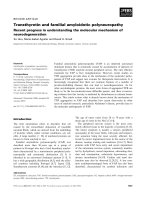
2004). Based on the HA sequences, HPAI H5N1 viruses are now divided into
10 different clades as shown in Figure 1.2 (WHO, March 2009).
Human infection of the H5N1 virus was most likely acquired directly
from the poultry and does not seem to spread efficiently among humans
(Wang et al., 2008a). However, cases of possible human-to-human
transmission have been reported (Kandun et al., 2006; Wang et al., 2008a;
WHO, 2010). The continued evolution of the virus or a single reassortment
with human influenza strains could render the virus successful in transmission
between humans. Thus there is a fear of possible H5N1 pandemic.
20
Fig. 1.2. Diagram showing the phylogenetic tree for the hemagglutinin gene of
highly pathogenic avian influenza A (H5N1) viruses. The geographic distributions
refer to avian isolates, and the tree is based on publicly available sequences. This
figure was adapted from WHO, Continuing progress towards a unified nomenclature
system for the highly pathogenic H5N1 avian influenza viruses (WHO, March 2009).
21
1.3.2. Genome organization
Influenza A is the prototype of the family, Orthomyxoviridae, which
comprises enveloped viruses with segmented negative-sense RNA genome.
Based on the antigenicity of the HA and neuraminidase (NA) surface
glycoproteins, influenza A viruses currently form 16 HA (H1-H16) and 9 NA
(N1-N9) subtypes. Over the past century, the only viruses that had infected
and circulated in humans are the H1N1, H1N2, H2N2, and H3N2 subtypes.
However, recently humans infection are now associated with the H5, H7 and
H9 subtypes.
The genome of influenza A virus is composed of eight single-stranded
negative-sense RNA segments which encodes for the 11 genes: HA, NA,
matrix 1 (M1), matrix 2 (M2), nucleoprotein (NP), nonstructural protein 1
(NS1), nonstructural protein 2 (NS2, also known as nuclear export protein,
NEP), polymerase acidic protein (PA), polymerase basic protein 1 (PB1),
polymerase basic protein 2 (PB2) and polymerase basic protein 1-F2 (PB1-
F2). The most striking feature of the influenza A virion is the layer of
projections made of 2 distinct glycoproteins, HA and NA. Embedded also in
the viral membrane is the M2 ion channel protein. The M1 protein, sitting just
underneath the lipid membrane, forms a matrix holding the viral
ribonucleoproteins (vRNPs). The vRNPs are made up of viral RNA wrapped
up around NP and very small amounts of NEP. At one end of the vRNPs are
the three polymerase proteins, PA, PB1, PB2, which make up the RNA
polymerase complex (Nayak et al., 2009; Nayak et al., 2004).
22
The properties and functions of the hemagglutinin of the H5N1 virus
will be discussed in details in section 1.3.2.2. The characteristics, functions
and/or putative roles of the other structural and non-structural proteins are
outlined in Table 1.3.
23
1.3.2.1 Structural and non-structural proteins
Table 1.3 Summary of structural and non-structural proteins of the H5N1 virus
Protein (No. of
amino acid
residues in
protein)
Protein characteristic [ref(s)] Protein’s function or putative function(s) [ref(s)]
Effect on cellular response of host
[ref(s)]
Nucleocapsid
protein (498)
The NP protein (~ 56 kDa) is rich in arginine
residues and has a net positive charge at pH
6.5 (Winter & Fields, 1981). This protein is
phosphorylated but it is unclear what percent
of the protein is phosphorylated and whether
phosphorylation is essential for its function
(Davey et al., 1985).
The primary function of NP is to encapsidate the virus
genome for the purposes of RNA transcription,
replication and packaging. It is suggested to interact with
cellular polypeptides, including actin, components of the
nuclear import and export apparatus and a nuclear RNA
helicase, indicating multiple functions of the protein
(Baudin et al., 1994; Portela & Digard, 2002). Recent
evidence showed that the NP can directly interact with
the viral polymerase and mediates the switch from
capped-primed viral mRNA synthesis to unprimed viral
RNA replication (Newcomb et al., 2009
).
Lysine at position 184 of the H5N1 NP
protein seems to induce earlier mortality
in chickens, with increased virus titers
and nitric oxide levels and upregualated
host immune genes, IFNα, IFNγ, Mx1
and inducible nitric oxide synthetase
(Wasilenko et al., 2009).
Neuraminidase
protein (453)
The NA protein is homotetramer (~ 220 kDa)
with each polypeptide made up of 453 amino
acid residues. The protein is a prototype class
II integral membrane protein, with its N-
terminus in the cytoplasm, a transmembrane
domain, the stalk, the head with the catalytic
active site (
Varghese et al., 1983).
The role of NA in the influenza virus life cycle is still
unclear. NA can catalyze the cleavage of the α–ketosidic
linkage between a terminal sialic acid and an adjacent D-
galactose or D-galactosamine (
Palese et al., 1974). It is
suggested that NA functions to remove sialic acid from
HA, NA and the cell surface, thus the release of virus
from the host cell. It may also allow the transport of the
virus through the mucin layer in the respiratory tract to
the target epithelial cells.
Immunization with H5N1 NA was
shown to induce high titers of HPAI-
neutralizing serum, although with lower
response as compared to the HA protein
(
Nayak et al., 2010). Anti-N1 cross-
protecting antibodies against H5N1 can
be detected in some H1N1 infected
individuals (Frobert et al., 2010).
24
Table 1.3 Continued
Protein (No. of
amino acid
residues in
protein)
Protein characteristic [ref(s)] Protein’s function or putative function(s) [ref(s)] Effect on cellular response of host [ref(s)]
Matrix protein,
M1 (252) and
M2
(97)
Three mRNA transcripts have been identified
from the RNA segment 7, a transcript encoding
M1 protein; a spliced mRNA encoding the M2
protein; and an alternatively spliced mRNA,
which encode 9 amino acid peptide that has not
been recognized (Lamb et al., 1981).
M1 is a 28 kDa protein and constitutes the
most abundant protein in the virion. The M2
protein is type III integral membrane protein,
with N-terminal extracellular domain,
transmembrane domain, and a cytoplasmic tail.
Post-translation modifications such as
phosphorylation on serine and palmitylation on
cysteine can be found in the cytoplasmic
domain (Holsinger et al., 1994). The native
form of the M
2
protein is homotetramer, either
existing as a pair of disulfide-linked dimers or
disulfide-linked tetramers (Lamb et al., 1985).
The M1 protein underlies the viral lipid envelope
and provides rigidity to the membrane of the virion
(Martin & Helenius, 1991). It has been
demonstrated that the M1 protein interacts with
RNA, although a zinc-binding motif within the M1
protein does not seems to influence binding to
RNA. The transport the M1 protein into the
nucleus is required for the exit of the newly
assembled RNPs from the nucleus An interaction
between M1 and NS2 protein in purified virions
has also been proposed (Yasuda et al., 1993).
The M2 protein functions as an ion channel that
permits ions to enter the virion during the
uncoating as well as modulates the pH of
intracellular compartments (Pinto & Lamb, 2006
).
There has been evidence that the transmembrane
domain constitutes the pore of the channel as
specific changes in this domain alter the kinetics
and ion selectivity of the channel.
Amino acids Asp at position 30 and Ala at
position 215 in the M1 protein are necessary
for the H5N1 lethality in mice (Fan et al.,
2009a).
Capase motif found in the N terminal of M2
protein has been suggested to play a
significant role in virus pathogenicity shown
in chickens (Zhirnov & Syrtzev, 2009).
25
Table 1.3 Continued
Protein (No. of
amino acid
residues in
protein)
Protein characteristic [ref(s)] Protein’s function or putative function(s) [ref(s)] Effect on cellular response of host [ref(s)]
Polymerase
proteins, PA
(716), PB1 (757),
PB2 (759), and
PB1-F2 (87)
The three largest RNA segments encode the
PB1 (96 kDa), PB2 (87 kDa) and PA (85
kDa) proteins. PB1 and PB2 are found to
be basic and PA is acidic based on their
behavior on isoelectric focusing gels
(Horisberger, 1980). The polymerase
complex is a heterotrimer of subunits of
PA, PB1 and PB2 (~250kDa).
The polymerase complex is involved in both mRNA
transcription and viral replication. PA subunit has
endonuclease and protease activities; involved in
vRNA/cRNA promoter binding; interacts with the PB1
subunit. The PB1 subunit contains the RNA-dependent
RNA polymerase active site and interacts with PA and
PB2. The PB2 subunit is responsible for cap binding
and contains a bipartite nuclear localization signal at C
terminus for nuclear import from the cytoplasm
[Reviewed by (Das et al., 2010)].
Lys at PB2-627 plays a role in the
pathogenicity of the influenza virus.
(Shinya et al., 2004). It supports efficient
virus replication in mammalian but not
avian species. It has been found in a
substantial number of H5N1 viruses
isolated from infected humans.
PB1-F2 induces apoptosis, thus it has been
proposed to kill host immune cells
responding to influenza virus infection
(Chen et al., 2001). The presence of Ser in
place of Asn at position 66 of PB1-F2
conferred high pathogenicity to H5N1
virus in mice (Conenello et al., 2007).