Dynamics of liver fatty acid binding protein
Bạn đang xem bản rút gọn của tài liệu. Xem và tải ngay bản đầy đủ của tài liệu tại đây (4.08 MB, 178 trang )
DYNAMICS OF LIVER FATTY ACID BINDING
PROTEIN
LONG DONG (B.Sc.)
A THESIS SUBMITTED FOR THE DEGREE OF
DOCTOR OF PHILOSOPHY
DEPARTMENT OF BIOLOGICAL SCIENCES
NATIONAL UNIVERSITY OF SINGAPORE
2010
I
ACKNOWLEDGEMENTS
I would like to express my deepest gratitude to my supervisor, Associate Professor
Yang Daiwen, for his persistent support, inspiration and guidance during the course
of this research project. Without his encouragement and effort, completion of this
thesis would not have been possible.
Special thanks to Assistant Professor Mu Yuguang (Nanyang Technological
University) for his kind guidance and support in my computational studies, as well as
for allowing me to access the computational facility in NTU. The helpful discussions
with Dr. Dai Liang (Indiana University, USA) on various simulation techniques are
also acknowledged.
I would like to thank Associate Professor Martin J. Scanlon (Monash University,
Australia) for sharing his research experience and results on fatty acid binding
proteins, and thank Mr. Song Wei (Xiamen University, China) for sharing his
experimental results on the interaction between liver fatty acid binding protein and
various ligands.
The critical research comments from Associate Professor Henry Mok in the group
meeting discussion and technical assistance from Dr. Fan Jingsong are also
acknowledged.
II
Thanks to my colleagues, labmates and friends in Singapore, with whom I enjoyed a
pleasant learning experience. In particular, I would like to thank B. C. Karthik, Chen
Shuting, Dai Xuhui, Iman Fahim Hameed, Jiang Ping, Li Weifeng, Li Yanfu, Lim
Jack Wee, Dr. Lin Zhi, Meng Dan, Wang Shujing, Dr. Xu Weixin, Dr. Xu Yingqi,
Yong Yee Heng, Dr. Zhang Jingfeng, and Zheng Yu.
Many thanks to my parents who have been always encouraging and supporting me for
the choices I made in my life.
Finally, the NUS research scholarship, which supported my graduate research work,
is gratefully acknowledged.
III
TABLE OF CONTENTS
ACKNOWLEDGEMENTS I
TABLE OF CONTENTS III
SUMMARY VI
LIST OF FIGURES IX
LIST OF TABLES XII
LIST OF ABBREVIATIONS XIII
CHAPTER 1 INTRODUCTION 1
1.1 Overview of the fatty acid binding protein family 2
1.2 Overview of liver fatty acid binding protein (LFABP) 5
1.3 Mechanisms of the FABP-ligands interaction 6
1.4 Aim of the current studies 7
CHAPTER 2 LITERATURE REVIEW 9
2.1 Structural studies on fatty acid binding proteins: implication for ligand
entry and exit mechanisms 10
2.1.1 Structural studies on intestinal fatty acid binding protein 10
2.1.1.1 Three dimensional structure of holo-intestinal fatty acid binding
protein 11
2.1.1.2 Structural studies on apo-intestinal fatty acid binding protein 12
2.1.2 Structural studies on liver fatty acid binding protein 13
2.1.2.1 Crystallographic study on holo-liver fatty acid binding protein 14
2.1.2.2 Structural studies on apo-liver fatty acid binding protein 15
2.1.3 A brief summary 15
2.2 Characterization of the dynamics of fatty acid binding proteins 16
2.2.1 Fast dynamics of fatty acid binding proteins on the picosecond to
nanosecond timescales 16
2.2.2 Slow dynamics of fatty acid binding proteins on the microsecond to
millisecond timescale 18
2.2.3 Molecular dynamics simulation of fatty acid binding proteins 19
2.2.4 Summary of the previous dynamics studies and the aim of current
studies 21
2.3 NMR relaxation in liquids 22
2.3.1 Theory of spin relaxation in liquids 22
2.3.1.1 The master equation 22
2.3.1.2 Relaxation mechanisms: DD, CSA, and relaxation interference 24
2.3.2 NMR relaxation parameters and model free formalism 25
2.3.3 Conformational/chemical exchange effects in NMR spectroscopy 27
2.3.4 Transverse relaxation dispersion experiment 32
2.4 Molecular dynamics simulation 34
2.4.1 Basic principles 34
2.4.2 Force field for biomolecular simulations 35
IV
2.4.3 Limitations of MD simulation 37
CHAPTER 3 NMR SAMPLE PREPARATION, RESONANCE ASSIGNMENT
AND STRUCTURE CALCULATION OF LFABP 39
3.1 Materials and methods 40
3.1.1 Media 40
3.1.2 SDS-polyacrylamide gel (SDS-PAGE) electrophoresis 40
3.1.3 Expression and purification of LFABP 41
3.1.4 NMR experiments for structure determination of LFABP 43
3.1.4.1 4D time-shared
13
C/
15
N,
13
C/
15
N-edited NOESY 44
3.1.4.2 MQ-CCH-TOCSY 46
3.1.4.3 HNCA experiment 46
3.1.5 Resonance assignment 47
3.1.5.1 Sequential assignment 47
3.1.5.2 Sidechain assignment 48
3.1.5.3 NOE assignments and structure calculation 48
3.2 Results and Discussion 49
3.2.1 Sample preparation of LFABP 49
3.2.2 NMR assignment of LFABP 54
3.2.2.1 Sequential assignment 54
3.2.2.2 Sidechain and NOE assignment 54
3.2.3 Structure calculation 58
3.3 Conclusion 63
CHAPTER 4 PROBING SLOW MOTIONS OF LFABP ON MILLISECOND
TIMESCALES 64
4.1 A general overview 65
4.2 Accurately probing millisecond timescale dynamics: The theory and
method 65
4.2.1 Introduction: the theoretical background of the existing problem 66
4.2.2 Results and Discussion 67
4.2.2.1 Numerical optimization of the phase cycling scheme for relaxation
dispersion experiment 67
4.2.2.2 NMR experimental evaluation 78
4.2.2.2.1 Methods and materials 78
4.2.2.2.2 Results and discussion 79
4.3 Probing slow dynamics of LFABP: A test of the assumptive model 83
4.3.1 Methods and Materials 83
4.3.2 Results and Discussion 85
4.3.2.1 An assumptive model 85
4.3.2.2 Intrinsic millisecond timescale dynamics of apo-LFABP 86
4.3.2.3 ANS binding studied by NMR titration 90
4.3.2.4 Kinetic rates of ANS binding at the high affinity site 92
4.3.2.5 Kinetic rates of ANS binding at the low affinity site 94
4.3.2.6 Chemical shift perturbation pattern an implication for the
nature of the minor state 97
V
4.3.3 Conclusion 100
CHAPTER 5 BUFFER INTERFERENCE WITH PROTEIN DYNAMICS: A
CASE STUDY ON LFABP 101
5.1 Introduction 102
5.2 Methods and materials 104
5.3 Results and discussion 106
5.3.1 MES binding inducing chemical shift perturbation 106
5.3.2 Effects of MES binding on protein dynamics on the picosecond to
nanosecond timescale 110
5.3.3 Effects of MES binding on protein dynamics on the microsecond to
millisecond timescale 112
5.3.4 Commonality of buffer interference with protein dynamics 114
5.4 Conclusion 117
CHAPTER 6 MOLECULAR DYNAMICS SIMULATION OF LIGAND
DISSOCIATION FROM LFABP 118
6.1 Introduction 119
6.2 Methods 119
6.2.1 Molecular dynamics simulation 119
6.2.2 Parameters for random expulsion simulation 121
6.2.3 Identification of residues constituting the portals 123
6.3 Results and Discussion 123
6.3.1 Dissociation of OLA128 from holo-LFABP 123
6.3.2 Dissociation of OLA129 from holo-LFABP 126
6.3.3 Residues constituting individual portals 130
6.3.4 Root mean squared fluctuations (RMSF) during the dissociations 133
6.3.5 Which portal does OLA129 dissociate from when OLA128 still binds
the protein? 135
6.3.5 Dissociation of 1,8-ANS from LFABP 137
6.3.6 Comparative study between intestinal FABP and liver FABP 139
6.4 Conclusion 141
CHAPTER 7 GENERAL CONCLUSIONS 142
REFERENCES 147
VI
SUMMARY
Over a decade, scientists have been attempting to know more about the
conformational dynamics of fatty acid binding proteins (FABPs), in order to answer
the puzzling question – how ligands could access the internalized binding site(s) of
FABPs. Despite numerous efforts made in this field, the appreciation of this question
is still relatively poor nowadays. In the current study, we continued the effort to
explore the dynamical properties of liver fatty acid binding protein (LFABP) using
NMR spectroscopy and MD simulation techniques, aiming at advancing our
knowledge on this interesting topic.
The microsecond to millisecond timescale dynamics of FABPs was historically
hypothesized to represent a dynamical equilibrium between the “open” and “closed”
states, regulating the ligand entry/exit processes. Despite the potential significance,
the validity of this hypothesis has not yet been demonstrated. In the current study, the
slow dynamics of LFABP was quantitatively characterized using relaxation
dispersion NMR spectroscopy, which shows that LFABP is indeed highly flexible on
the millisecond timescales. In order to further examine the hypothetical role of the
millisecond dynamics of LFABP, the potential correlation between slow dynamics
and ligand entry/exit processes was modeled and evaluated by analyzing the kinetic
rates of LFABP-ANS interaction. The experimental result demonstrates that the
intrinsic millisecond dynamics of LFABP, somewhat disappointedly, does not
represent a critical conformational reorganization required for ligand entry due to the
VII
contradiction of timescales, but implies that it may represent a dynamical equilibrium
between the apo-state and a state resembling the singly-bound conformation. Analysis
of the kinetic rates of the ligand association shows that the ligand-entry related
dynamics could occur on the microsecond or sub-microsecond timescales, which is
much faster than previously assumed.
Despite fast advancement of experimental techniques for exploring protein dynamics,
direct visualization of ligand entry/exit processes which potentially involves multiple
transient steps is still formidable nowadays. In silico simulation, thus, provides a
good alternative way to investigate such dynamical details. However, the ligand
exit/entry is a slow event which could hardly be accessed by standard MD
simulations. In order to overcome this problem, random expulsion simulation, which
accelerates ligand motions with a randomly oriented external force, was applied to
investigate the ligand dissociation processes (in Chapter 6). Different ligand egress
routes were identified for LFABP in this work, which furthered our understanding on
the protein-ligand interplay. Future mechanistic studies on the ligand release and
uptake would benefit from the experimental and computational studies shown in this
thesis.
As a fortuitous discovery during our experimental studies, the millisecond timescale
dynamics of LFABP was found to be perturbed by the presence of buffer agents.
Although not being our initial aim, we characterized the amplitude of such buffer
perturbation to the slow motions of LFABP (in Chapter 5). This case study offers an
VIII
example of how the biophysical properties of proteins could be influenced by buffer
molecules, which would deserve the attention of scientists in the in vitro manipulation
of protein molecules.
IX
LIST OF FIGURES
Figure 1.1.1 Three-dimensional structures of FABPs. 4
Figure 2.3.1 Lineshapes of two-state chemcial exchange. 31
Figure 3.1.1 The pET-32a derived plasmid (pET-M). 42
Figure 3.1.2 Pulse sequence for recording 4D time-shared
13
C/
15
N,
13
C/
15
N-edited
NOESY. 45
Figure 3.2.1 Expression and purification of LFABP. 50
Figure 3.2.2
15
N-
1
H HSQC spectra of LFABP. 51
Figure 3.2.3 Overlay of HSQC spectra of LFABP with and without His-tag. 53
Figure 3.2.4 The chemical shift correlation of the HNCA, 4D NOESY, and MQ-
CCH-TOCSY spectra. 55
Figure 3.2.5 Sequential connectivity for a stretch of residues (T96-K98). 56
Figure 3.2.6 Backbone assignment of LFABP. 57
Figure 3.2.7 Superimposition of ten NMR conformers. 60
Figure 3.2.8 Ramachandran plot of LFABP. 61
Figure 3.2.9 Alignment of the NMR and X-ray structures of LFABP. 62
Figure 4.2.1 Dependence of average rotation on resonance offsets and pulse
imperfection. 69
Figure 4.2.2 Pulse scheme for the measurement of relaxation dispersion. 70
Figure 4.2.3 Dependence of
15
N
eff
R
2
on ν
CP
for different ∆ω
N
and pulse
imperfection for two different pulse schemes. 73
X
Figure 4.2.4 Contour plot showing the dependence of
eff
R
2
values on resonance
offsets. 74
Figure 4.2.5 Contour plot showing the dependence of
eff
R
2
values on pulse
imperfection. 75
Figure 4.2.6 Effects of the CSA-dipole cross-correlated relaxation and
1
H-
1
H
cross-relaxation on the dependences of R
2
eff
values on
ν
CP
. 77
Figure 4.2.7 Representative residues without R
ex
. 80
Figure 4.2.8 Relaxation dispersion profiles for representative residues. 82
Figure 4.3.1. LFABP residues undergoing conformational exchange. 88
Figure 4.3.2. Relaxation dispersion profiles of the residues with intrinsic
conformational exchange. 89
Figure 4.3.3. Representative residues in NMR titration experiments. 91
Figure 4.3.4. Kinetics of LFABP-ANS interaction. 93
Figure 4.3.5. The relaxation dispersion profiles of representative residues. 95
Figure 4.3.6. Histogram of CCSP of LFABP upon addition of ANS (at ~1:1 ratio).
98
Figure 4.3.7. Comparison of protein regions showing resonance shift and those
showing conformational exchange in the absence of ligands. 99
Figure 5.3.1. MES titration of LFABP. 107
Figure 5.3.2 Histogram of CCSP of LFABP upon titration with MES. 108
Figure 5.3.3. Residues that were significantly perturbed by MES. 109
Figure 5.3.4. Fast dynamics parameters of LFABP in the presence and absence
of MES. 111
XI
Figure 5.3.5. Relaxation dispersion curves of LFABP measured in the absence
and presence of MES. 113
Figure 5.3.6. Chemical shift perturbation by Bis-tris. 115
Figure 5.3.7. Relaxation dispersion curves of LFABP measured in the absence
and presence of Bis-tris. 116
Figure 6.2.1 The flow scheme of random expulsion simulation. 122
Figure 6.3.1 Three-dimensional structure of LFABP-oleate complex. 124
Figure 6.3.2. Dissociation of OLA128. 125
Figure 6.3.3. Dissociation of OLA129. 127
Figure 6.3.4. Front views of three portals. 132
Figure 6.3.5. Backbone residue-wise root mean square fluctuations (RMSF) of
LFABP. 134
Figure 6.3.6. Dissociation of OLA129 from portal II without displacing OLA128.
136
Figure 6.3.7. Snapshots of 1,8-ANS exiting the cavity. 138
Figure 6.3.8. Dissociation of palmitate (purple) from the cavity of IFABP (dark
green). 140
XII
LIST OF TABLES
Table 3.2.1 Summary of assignments and structure of LFABP 59
Table 6.3.1 REMD simulation of ligand exiting from the cavity of IFABP and
LFABP. 128
Table 6.3.2 Residues constituting the portal regions 131
XIII
LIST OF ABBREVIATIONS
AMBER Assisted model building with energy refinement
ANS 1-anilinonaphthalene-8-sulfonic acid
Bis-tris Bis(2-hydroxyethyl)-amino-tris(hydroxymethyl)-methane
CCSP Combined chemical shift perturbation
CHARMM Chemistry at Harvard molecular mechanics
CPMG Carr-Purcell-Meiboom-Gill
CSA Chemical shielding anisotropy
DD Dipole-dipole
DdCAD-1 Dictyostelium discoideum Ca
2+
dependent cell adhesion molecule-1
E.coli Escherichia coli
EDTA Ethylenediaminetetraacetic acid
FPLC Fast performance liquid chromatography
GROMACS Groningen machine for chemical simulations
HMQC Heteronuclear multiple quantum coherence
HSQC Heteronuclear single quantum coherence
INEPT Insensitive nuclei enhanced by polarization transfer
IPTG Isopropyl-beta-D-1-thiogalactopyranoside
kD kiloDalton
LB Luria Bertani
(L/I/A/M)FABP (Liver/Intestinal/Adipocyte/Muscle) fatty acid binding protein
XIV
MD Molecular Dynamics
MES 2-(N-morpholino)ethanesulfonic acid
MQ Multiple-quantum
NMR Nuclear Magnetic Resonance
NOE Nuclear Overhauser enhancement
OLA Oleate
OPLS Optimized potential for liquid simulations
PAGE Polyacrylamide gel electrophoresis
PLM Palmitate
ppm parts per million
ps/ns/µs/ms picosecond/nanosecond/microsecond/millisecond
REMD Random expulsion molecular dynamics
RF Radio frequency
RMSD Root mean square deviation
RMSF Root mean square fluctuation
SDS Sodium dodecyl sulphate
SQ Single-quantum
SW Spectral width
TOCSY Total correlation spectroscopy
TPPI Time proportional phase increment
TROSY Transverse relaxation optimized spectroscopy
XV
PUBLICATIONS
Long, D., Yang, D. (2010) Millisecond timescale dynamics of human liver fatty acid
binding protein: testing of its relevance to the ligand entry process. Biophys. J.
98:3054-3061
Long, D., Mu, Y., Yang, D. (2009) Molecular dynamics simulation of ligand
dissociation from liver fatty acid binding protein. PLoS ONE 4:e6081
Long, D., Yang, D. (2009) Buffer interference with protein dynamics: a case study on
human liver fatty acid binding protein. Biophys. J. 96:1482-1488
Long, D., Liu, M., Yang, D. (2008) Accurately probing slow motions on millisecond
timescales with a robust NMR relaxation experiment. J. Am. Chem. Soc.
130:2432-2433
Xu, Y., Long, D., Yang, D. (2007) Rapid data collection for protein structure
determination by NMR spectroscopy. J. Am. Chem. Soc. 129:7722-7723
1
CHAPTER 1
INTRODUCTION
2
Chapter 1: Introduction
Fatty acids, as one of the most abundant lipid molecules in vivo, play a critical role in
the living processes of cells. Not only are they key metabolites involved in energy
generation and storage, fatty acids, being crucial components of the lipids in cellular
membranes, directly affect the structure and function of membranes (Pohl et al.,
2004). However, the solubility of these molecules, due to their long hydrophobic
alkyl chains, is poor in the aqueous physiological environment. Thus, specific protein
molecules, which have high affinity with fatty acids, have evolved to assist the
transportation of fatty acids in aqueous environments. Serum albumin is one of such
proteins, which binds a variety of fatty acids and is responsible for extracelluar
transportation. Intracellularly, this work is done by fatty acid binding proteins
(FABPs) (Hamilton, 2004). A thorough understanding of the biophysical basis for
FABP-fatty acid interaction requires the knowledge of the protein three-dimensional
structure as well as structural dynamics, which has been puzzling scientists for a few
decades. The studies on the function, structure, and dynamics of fatty acid binding
proteins will be overviewed in the following sections.
1.1 Overview of the fatty acid binding protein family
Fatty acid binding proteins form an intracellular protein family with a low molecular
weight (about 15 kD). As a common feature of this family, all FABPs have high
affinity but low binding capacity (stoichiometry: one or two) of fatty acids. Tissues
(e.g. intestine, liver, adipose, and muscle) with active lipid metabolism or high rate of
fatty acid uptake generally have a high level of expression of one or more FABPs
3
(Hamilton, 2004; Storch & McDermott, 2009). While several FABPs could co-exist
in the same tissue, individual FABPs are named after the tissues where they were first
found and isolated.
Although the physiological functions of FABPs have not been completely elucidated
yet, their roles in facilitating the cellular transportation of poorly soluble ligands seem
irrefutable (Thompson et al., 1999a; Storch & Corsico, 2008). Two different
mechanisms of fatty acids transportation by FABPs have been revealed by in vitro
experiments (Storch & Thumser, 2000; Storch & Corsico, 2008). The first class of
FABPs is known to transfer/deliver their ligands through direct contact/collision with
biomembranes. Most FABPs (including the intestinal-type, adipose-type, heart-type,
keratinocyte-type, and myelin-type) belong to this class. By contrast, Liver-type
FABP (LFABP) represents the second class of FABP which transfers the ligands to
and from membranes through aqueous-phase diffusion. Diffusional protein (LFABP)
probably functions as a cytosolic reservoirs for the poorly soluble ligands (Storch &
Corsico, 2008).
Structural biology studies (X-ray crystallography and NMR spectroscopy) on the
FABP family have shown a conserved folding pattern for all family members (Storch
& Corsico, 2008) (Figure 1.1.1). Ten anti-parallel -strands form a cage-like -barrel
structure, composed of two roughly orthogonal -sheets. Two short -helices, located
between the first and second -strands, form a helix-turn-helix motif, which cap one
end of the binding cavity. Shown from the ligand-bound structure, the -barrel cavity
4
is where the binding site(s) are located. Crystallographic studies on the structure of
the apo- FABPs showed very little difference from that of the holo-proteins.
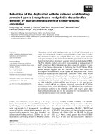
Figure 1.1.1 Three-dimensional structures of FABPs. Ribbon structures of
Intestinal FABP, Liver FABP, and Adipocyte FABP are displayed, with PDB entries
(apo-/holo-) of 1IFB/2IFB, 2F73/1LFO, and 1LIB/1LID, respectively. The -helix II
and the turns between C/D and E/F of holo-IFABP are shown in magenta.
In the family of FABPs, LFABP has some unique features (e.g. binding stoichiometry,
volume of the -cavity, lengths of -strands, etc.), besides its transportation
mechanism. A general overview of liver-type FABP is given in the following section.
5
1.2 Overview of liver fatty acid binding protein (LFABP)
LFABP mainly exists in the tissues of liver and intestine, and to a lesser extent, in
kidney and colon (Thompson, et al., 1999a). Along with other FABPs, LFABP
interacts with fatty acids and facilitates their transportation.
Crystallographic studies on oleate-bound LFABP have showed that the overall
conformation of LFABP is very similar to other FABPs (Thompson et al., 1997).
Two molecules of oleic acid are accommodated in the -barrel cavity of LFABP,
which agrees with the fluorescence study on LFABP-fatty acid interactions (Richieri
et al., 1994). One fatty acid molecule adopts a bent conformation in the inner position
of the cavity, with its carboxylate group interacting with arginine 122 of LFABP. The
other fatty acid adopts a more extended conformation, located near the helix-turn-
helix motif. The unique stoichiometric binding between LFABP and different ligands
could contribute to the specific functions of LFABP.
In addition to the unique binding stoichiometry, the ligand spectrum of LFABP is the
broadest in the FABP family. The ligands of LFABP include saturated and
unsaturated long chain fatty acids, bile salts, lysophosphatidic acid, heme, 1,8-ANS,
lipophilic drugs, etc. (Thompson et al., 1999a; Thompson et al., 1999b; Chuang et al.,
2008). The interaction with lipophilic drugs suggests an important role of LFABP in
the cellular transportation of these drugs. In addition, the localization studies using
laser scanning microscopy showed the colocalization of LFABP and peroxisome
proliferator-activated receptor (PPAR) in the nucleus and demonstrated the direct
6
interaction between these two proteins, indicating that LFABP is involved in
transportation of nucleus-targeted signaling molecules and affects the expression of
PPAR-sensitive genes (Wolfrum et al., 2001). Other functional studies on LFABP
also suggested tentative roles in cell proliferation (Bassuk et al., 1987), production of
hepatic very-low-density lipoprotein (VLDL) (Spann et al., 2006), and budding of
prechylomicron transport vesicles (PCTVs) from the endoplasmic reticulum (Neeli et
al., 2007)
1.3 Mechanisms of the FABP-ligands interaction
Despite the rich information obtained from functional and structural studies on fatty
acid binding proteins, the fundamental processes of FABP-ligand interaction have not
been fully understood yet. In particular, the mechanisms of how the ligands access the
internalized binding sites have been a puzzling question over two decades. Twenty
years ago, the crystallographic study on the palmitate-bound IFABP revealed that the
ligand, instead of binding at the cleft(s) on the surface of the protein, is situated
internally in the -barrel cavity (Sacchettini et al., 1989). In addition, a number of
three-dimensional structures of different FABPs determined later (Xu et al., 1993;
Young et al., 1994; Lassen et al., 1995; Thompson et al., 1997; Hohoff et al., 1999;
Balendiran et al., 2000), using either X-ray crystallography or NMR spectroscopy,
also confirmed the internalized location of the ligand binding site(s) for this protein
family. However, based on these static structures, the pathways for ligand entry and
exit could not be intuitively identified, since the dominant FABP conformation does
7
not show any obvious openings for ligand entry. Thus, conformational dynamics
appears to play a critical role for the function of FABPs.
Experimental and computational studies on the dynamics of fatty acid binding
proteins were hence conducted aiming at addressing the question of ligand entry and
exit (e.g. Hodsdon & Cistola, 1997a; Hodsdon & Cistola, 1997b; Zhang et al., 2006;
Friedman et al., 2005; Friedman et al., 2006). However, these dynamics studies,
which mainly focused on the protein motions on the picosecond to nanosecond
timescales, have not satisfactorily unraveled the dynamical mechanisms of ligand
entry and exit, which might be due to the very fast motional timescales they were
restricted to. Therefore, probing protein motions on much slower timescales would be
necessary in the current study.
1.4 Aim of the current studies
In this work, the primary purpose was to investigate the dynamical properties of liver
fatty acid binding protein both experimentally and computationally. This work was
expected to advance our understanding of how ligand molecules access the
internalized binding sites of LFABP. Since the internalized location of ligand binding
sites is found in many protein systems, the result of the current study may provide
general insights into the fundamental mechanisms of this type of protein-ligand
interactions. Considering the wide existence of fatty acid binding proteins in living
organisms, the knowledge on the structure-function-dynamics relationship should be
helpful for better appreciating their biological roles. Comprehensive literature review
8
on the previous structural and dynamical studies on FABPs, as well as the major
methodologies employed in the current study, will be presented in the second chapter.
9
CHAPTER 2
LITERATURE REVIEW