Development of high contrast coherent anti stokes raman scattering (CARS) and multiphoton microscopy for label free biomolecular imaging
Bạn đang xem bản rút gọn của tài liệu. Xem và tải ngay bản đầy đủ của tài liệu tại đây (7.81 MB, 145 trang )
DEVELOPMENT OF HIGH CONTRAST COHERENT
ANTI-STOKES RAMAN SCATTERING (CARS) AND
MULTIPHOTON MICROSCOPY FOR LABEL-FREE
BIOMOLECULAR IMAGING
LU FAKE
NATIONAL UNIVERSITY OF SINGAPORE
2010
DEVELOPMENT OF HIGH CONTRAST COHERENT ANTI-STOKES RAMAN SCATTERING
(CARS) AND MULTIPHOTON MICROSCOPY FOR LABEL-FREE BIOMOLECULAR IMAGING LU FAKE 2010
DEVELOPMENT OF HIGH CONTRAST COHERENT
ANTI-STOKES RAMAN SCATTERING (CARS) AND
MULTIPHOTON MICROSCOPY FOR LABEL-FREE
BIOMOLECULAR IMAGING
LU FAKE
A THESIS SUBMITTED
FOR THE DEGREE OF DOCTOR OF PHILOSOPHY
DIVISION OF BIOENGINEERING
NATIONAL UNIVERSITY OF SINGAPORE
2010
Acknowledgements
The work presented in this thesis was primarily conducted in Optical Bioimaging
Laboratory in the Division of Bioengineering at the National University of Singapore
during the period from January 2006 to January 2010. In the past four years, I met
many nice people who gave me big encouragement and kindly help. Here I would like
to thank them sincerely:
First and foremost, I would like to express my sincere appreciation to my advisor
Assistant Professor Huang Zhiwei, who offered me the opportunity in the very
beginning to pursue the PhD degree in his group. I am indebted to Dr Huang for his
professional advice, guidance, and patience throughout my studies. His fully financial
support on my experiments boosted the overall progress greatly. I believe and
appreciate that Prof Huang has an extraordinary impact on my future research career.
I greatly appreciate the generous support and guidance from Professor Colin Sheppard,
who is a very nice person as a great scientist in Optics. His equations and scientific
discussions gave me deep impression and positive affection. I would like to thank
Assistant Professor Chen Nanguang, who helped me a lot throughout my studentship.
Great appreciation and respect to Professor Dietmar W. Hutmacher and Professor
Hanry Yu and their group members, who taught me useful knowledge on biology and
biomedicine research and offered me cellular and tissue samples for my study.
I would also like to acknowledge my coworkers and team members in Optical
Bioimaging Laboratory: Dr Zheng Wei, Dr Liu Cheng, Dr Yuen Clement, Dr Yew Yan
Seng Elijah, Mo Jianhua, Teh Seng Knoon, Shao Xiaozhuo, Lin Kan, Lin Jian for their
kind discussions, suggestions and guidance on my research work.
I wish to thank my dear parents, darling wife, close brother and all my lovely
classmates and friends, with whom I kept walking through these hardworking days.
Last but not least, I would like to acknowledge the financial support from the Ministry
of Education of Singapore, the President Graduate Fellowship (PGF) of National
University of Singapore (NUS) for my research at NUS.
I
Table of Contents
Acknowledgements I
Table of Contents II
Abstract IV
List of Figures V
List of Abbreviations VII
Chapter 1 Introduction 1
1.1 Background 1
1.2 Motivations 4
1.3 Research Objectives 6
1.4 Thesis Organization 7
Chapter 2 Literature Review 9
2.1 Basic Theory 9
2.1.1 Rationale of Raman spectroscopy 9
2.1.2 Fundamental theory of CARS 11
2.2 Experimental Instrumentations of CARS Microscopy 14
2.2.1 Laser sources for CARS microscopy 14
2.2.2 Laser scanning CARS microscope 16
2.2.3 Multiplex CARS microspectroscopy 17
2.3 Suppression of Nonresonant Background in CARS Microscopy 19
2.3.1 Backward (Epi-) detection CARS 20
2.3.2 Counter-propagating CARS 21
2.3.3 Polarization-sensitive CARS 21
2.3.4 Time-resolved CARS 23
2.3.5 Pulse shaping in femtosecond excitation CARS 24
2.3.6 Interferometric CARS 24
2.4 CARS Applications in Life Sciences 25
2.4.1 Cellular imaging 26
2.4.2 Tissue imaging 29
2.5 Integrated CARS and Multiphoton Multimodal Nonlinear Optical Microscopy
3
2
2.6 Liver Steatosis and Liver Fibrosis 36
2.6.1 Liver steatosis 36
2.6.2 Liver fibrosis 37
2.6.3 Relationship between liver steatosis and liver fibrosis 38
2.6.4 Diagnosis of liver diseases 38
Chapter 3 Polarization-Encoded CARS for High Contrast Vibrational
Imaging 40
3.1 Linearly Polarized CARS with Heterodyne-Detection for Low Concentration
Biomolecular Imaging 40
3.1.1 Interferometric polarization (IP-) CARS 40
3.1.2 Phase-controlled P-CARS 47
II
III
3.1.3 Heterodyne polarization (HP-) CARS 60
3.2 Elliptically Polarized CARS for Intrinsic Nonresonant Background
Suppression 68
Chapter 4 CARS Imaging using Tightly Focused Radially Polarized Light
77
4.1 Radial Polarization (RP-) CARS with Annular Detection for High Contrast
Imaging 77
4.1.1 Introduction 77
4.1.2 Theory 78
4.1.3 Results and discussions 80
4.1.4 Summary 84
4.2 RP-CARS for Sensing Molecular Orientation with High Sensitivity 84
4.2.1 Principle 86
4.2.2 Experiment 90
4.2.3 Results and discussions 91
Chapter 5 Integrated CARS and Multiphoton Microscopy for Assessment
of Fibrotic Liver Tissues 95
5.1 Integrated CARS and Multiphoton Microscopy using Dual Paired-Gratings
Spectral Filtering of a Femtosecond Laser Source 95
5.2 Multimodal Nonlinear Optical (NLO) Imaging of Fibrotic Live Tissues 101
5.2.1 Sample preparation: the BDL rat model 101
5.2.2 Results and discussions 102
5.2.3 Summary 108
Chapter 6 Conclusions and Future Directions 110
6.1 Conclusions 110
6.2 Future Directions 114
List of Publications 118
References 121
Abstract
Coherent anti-Stokes Raman scattering (CARS) microscopy has received much interest
for imaging cells and tissues due to its outstanding capabilities of biochemical
selectivity using molecular vibrations, high sensitivity, as well as intrinsic
three-dimensional optical sectioning ability. In this thesis, the polarization effects in
CARS microscopy have been comprehensively studied and thereby several novel
CARS microscopic techniques for high contrast vibrational imaging and high sensitive
molecular orientation detection have been reported. An advanced interferometric
polarization CARS imaging technique was developed to effectively suppress the
nonresonant background, while greatly enhance the weak resonant signals of low
concentration biochemicals for high contrast and high sensitive biomolecular imaging.
To further reduce the excitation power for minimizing the photodamage to the
specimens, a unique heterodyne-detected polarization CARS technique by utilizing
interference of the relatively intense local oscillator CARS signal and the weak
resonant CARS signal generated simultaneously within the focal volume of the sample
was also developed for high sensitive CARS imaging. In addition, employing an
elliptically polarized pump field combined with a linearly polarized Stokes field,
intrinsic background-free CARS imaging was realized with much higher resonant
signal intensities to be detected as compared to conventional polarization CARS. To
facilitate the three dimensional molecular orientation sensing, a radial polarization
CARS microscope was demonstrated for improving the detection of longitudinally
oriented molecules in the samples. Further, an integrated CARS and multiphoton
microscopy technique by implementing a dual 4-f configured paired-gratings spectral
filtering module on a dual-color femtosecond laser source has also been successfully
developed for biomolecular imaging. It was demonstrated that high contrast CARS and
high quality multiphoton microscopy imaging could be acquired in tandem on the
same platform for quantitative assessment of biomolecular changes associated with
liver disease transformations (e.g., fatty/fibrotic liver). This research indicated the
great applicable potential of the integrated CARS microscopy and multiphoton
microscopy for label-free biomolecular imaging in biological and biomedical systems.
IV
List of Figures
Fig. 2.1
Energy diagram of light scattering.……………………………… …….10
Fig. 2.2
Energy diagram and phase matching condition of CARS radiation…… 11
Fig. 2.3
Schematic of laser scanning CARS microscope……………….… 17
Fig. 2.4
Illustration of electric vectors in polarization CARS………….… 23
Fig. 2.5
Raman spectrum and CARS image of lipid droplets in water… ………27
Fig. 2.6
CARS image of normal and mutant yeast cells…………… ………… 28
Fig. 2.7
CARS and SHG images of mouse skin in both hypodermis and dermis
layers………………………………………………………………… 35
Fig. 3.1
Polarization vectors of the pump and Stokes fields in interferometric
polarization (IP-) CARS……………………………………….… 40
Fig. 3.2
Schematic of IP-CARS microscope………………………… …………42
Fig. 3.3
Comparison of CARS images of 4.69 μm polystyrene beads in water by
conventional CARS, P-CARS and IP-CARS………………… 44
Fig. 3.4
CARS images of unstained human epithelial cell in aqueous environment
with conventional CARS, P-CARS and IP-CARS… 45
Fig. 3.5
Schematic of the phase-controlled polarization CARS microscope 52
Fig. 3.6
Comparison of spontaneous Raman spectrum, conventional and phase-
controlled P-CARS spectra of a polystyrene bead in water 54
Fig. 3.7
Phase-controlled P-CARS signals of a 1 μm polystyrene bead in water as
a function of voltages applied to the PZT……………………………….55
Fig. 3.8
CARS images of a 1 μm polystyrene bead in water for constructive
interference, destructive interference, phase-controlled P-CARS and the
conventional P-CARS 57
Fig. 3.9
CARS images of unstained epithelial cells in water for constructive
interference, destructive interference, phase-controlled P-CARS and the
conventional P-CARS………… ……………………………………….58
Fig. 3.10
The conventional CARS image of unstained epithelial cells in water due
to the induced polarization P
2
, and the correspondingly retrieved P-CARS
image through calculation………….….……….……… 59
Fig. 3.11
Principle of heterodyne polarization (HP-) CARS…………… 61
Fig. 3.12
Experimental schematic of the HP-CARS microscope………………….63
Fig. 3.13
Comparison of CARS images of polystyrene beads for local oscillator
CARS, P-CARS and HP-CARS……………………… ………… 65
Fig. 3.14
Comparison of CARS images of epithelial cells for local oscillator CARS,
P-CARS and HP-CARS………………………… ……… 67
Fig. 3.15
Principle of elliptically polarized (EP-) CARS………………………….69
Fig. 3.16
CARS images of 1.5μm polystyrene beads in water for normal CARS,
V
EP-CARS and P-CARS…….…………………………….…………… 73
Fig. 3.17
CARS images of lipid droplets in an unstained fibroblast cell in water for
EP-CARS and P-CARS………….………………… ……………….…75
Fig. 4.1
Illustration of the annular-detected RP-CARS microscopy………….….79
Fig. 4.2
Far-field RP-CARS radiation pattern………………………………… 82
Fig. 4.3
Calculated forward-detected RP-CARS intensities of different scatters 85
Fig. 4.4
Calculated epi-detected RP-CARS intensities of different scatters…… 86
Fig. 4.5
Calculated intensity distribution of the longitudinal and transverse
components on the focal plane of RP-CARS 88
Fig. 4.6
Schematic of RP-CARS microscope………………………………….…89
Fig. 4.7
RP-CARS and LP-CARS images of cottonwood leaf vascular bundles 91
Fig. 4.8
Changes of RP-CARS and LP-CARS signal intensities against the
polarization analyzer angle.………………………………………… 93
Fig. 5.1
Schematic of the integrated CARS and multiphoton microscopic platform
for bioimaging…………………….….……………………………… 96
Fig. 5.2
The measured pulse spectral FWHM and temporal duration as a function
of the slit width………………….…………………… …………… 99
Fig. 5.3
Comparison of fs- and ps-CARS spectra and images of 465 nm
polystyrene beads in water…………….………………… ………… 100
Fig. 5.4
Illustration of bile duct ligation (BDL) surgery on rats…………… ….102
Fig. 5.5
Comparison of normal and fibrotic liver tissue sample imaged by CARS
and SHG….………….………………………… …………………….103
Fig. 5.6
Multimodal imaging of fibrotic liver tissue using CARS, SHG and
TPEF 103
Fig. 5.7
Resonant and nonresonant CARS image of lipid droplets in diseased liver
tissue………………………………………………………………… 105
Fig. 5.8
CARS and TPEF images of ORO-stained fat droplets in liver…….… 106
Fig. 5.9
Digital mask processing for quantitative assessment of lipid droplets in
diseased liver tissue……….………………………………… ……….107
Fig. 5.10
Quantitative analysis of hepatic fat by CARS and collagen by SHG in
liver…………….……………………………………… …………… 108
VI
VII
List of Abbreviations
BDL = Bile duct ligation
CARS = Coherent anti-Stokes Raman scattering
C-CARS = Counter propagation CARS
DIC = Differential inference contrast
E-CARS = Epi-detected CARS
EP-CARS = Elliptically polarized CARS
F-CARS = Forward-detected CARS
FDTD = Finite-difference time-domain
FWHM = Full width at half maximum
fs = Femtosecond
HP-CARS = Heterodyne polarization CARS
IP-CARS = Interferometric polarization CARS
LP-CARS = Linearly polarized CARS
M-CARS = Multiplex CARS
NLO = Nonlinear optics
NA = Numerical aperture
NAFLD = Nonalcoholic fatty liver disease
NIR = Near infrared
OCT = Optical coherent tomography
OPO = Optical parametric oscillator
PCF = Photonics crystal fiber
ps = Picosecond
P-CARS = Polarization CARS
PMT = Photomultiplier tube
RP-CARS = Radially polarized CARS
SERS = Surface enhanced Raman scattering
SHG = Second harmonic generation
SFG = Sum frequency generation
THG = Third harmonic generation
TPEF = Two photon excited fluorescence
Chapter 1 Introduction
1.1 Background
Laser-scanning confocal fluorescence microscopy has been widely used in material
and life sciences for submicron level investigations through a fast imaging approach,
allowing the specific visualization of microscopic structures of the stained molecular
composition with both chemical specificity and three-dimensional sectioning
capability [1]. However, for biomolecular species and cellular components that cannot
tolerate fluorescence staining, other complementary contrast mechanisms with
noninvasive characterization are needed. Phase contrast and differential interference
contrast (DIC) microscopy [2, 3] rely on the minor differences of the refractive index
across the label-free sample to highlight the small particles and interfaces with index
mismatch. From this view, both of them are index-sensitive, not chemical-selective.
Vibrational microscopies, such as infrared spectroscopy and Raman spectroscopy [4-6],
have been used for chemically-selective imaging. Unfortunately, infrared absorption
microscopy suffers from low spatial resolution due to the long excitation wavelength
(diffraction limitation), while the sensitivity of Raman spectroscopy is limited by the
inherently very weak Raman scattering mixed with the strong fluorescence background.
Surface enhanced Raman scattering (SERS) detection schemes can be sensitive enough
for single molecule detection, due to the enhancement of Raman scattering by
molecules attached on rough metal surfaces, but the additional requirement of a
tedious preparation of substrates with nano-level metal structures makes it hard to be
used for most biological applications in vivo [7].
1
The technical achievements on femtosecond or picosecond pulsed laser sources
triggered the rapid development of nonlinear optical (NLO) microscopy for life
science applications [8]. The most commonly used and well developed nonlinear
modalities include two-photon excitation fluorescence (TPEF) [9-11], second
harmonic generation (SHG) [12-14], and third harmonic generation (THG) [15, 16].
The label-free biological application of TPEF imaging is hindered by the limited
endogenous fluorophores, while exogenous labeling also suffers from the drawback
that staining may alter the physiological environment of the biological/biomedical
systems. SHG imaging requires the local break of inversion symmetry in the molecules
and is only sensitive to few biochemicals, such as collagens. THG can work based on
the differences of third-order nonlinear susceptibility or refractive index, both of which
are nonresonant processes.
Recently, coherent anti-Stokes Raman scattering (CARS) imaging has been
developed as a useful complementary technique for video-rate vibrational imaging
based on the coherently enhanced Raman-active vibrations [17-19]. CARS as a typical
third-order nonlinear process, was first reported in 1965 by Maker and T
erhune at the
Ford Motor Company [20]. Thereafter CARS spectroscopy has been widely used as a
viable m
eans for chemical analysis in both gas and liquids [21-23]. In 1982, Ducan et
al. reported the first CARS microscope using a non-collinear configuration of pump
and Stokes beams to image onion cells with chemical specificity [24]. In that
experiment, the visible light excitation resulted in relatively larger nonresonant
background due to two-photon electronic resonance. On the other hand, the
2
non-collinear excitation geometry lowered down the spatial resolution and also made
the system unsuitable for microscopy applications. Until 1999, Zumbusch et al.
demonstrated the first CARS microscopy with collinear beam geometry for unstained
live bacteria and cell imaging [18]. Soon after, it was proved that in CARS microscopy
the interaction length is only several micrometers or less under tightly focusing
condition using large NA microscope objectives, thus the phase-mismatching condition
can be relaxed within the large cone angle with collinear beam geometry. Collinear
beam geometry is considered to be the key simplification strategy on CARS
implementation for its successful revival in the last decade [25].
The advantages of CARS microscopy has been concluded as follows [26-28]: (i)
Natural or artificial fluorescence probes are usually unnecessary in CARS imaging,
since its contrast mechanism is based on molecular vibrations that are intrinsic to the
samples. (ii) CARS signal is orders of magnitude more sensitive than Raman signal,
which yields much higher sensitivity with relatively lower average excitation power.
(iii) The third-order nonlinear signal generation dependence leads to inherent 3D
sectioning capability. (iv) CARS signal is blue-shifted from both pump and Stokes
frequencies, and can thus be easily detected avoiding the fluorescence background. (v)
The use of near-infrared (NIR) wavelength excitation minimizes the photodamage
(mainly water absorption) to the sample and also provides a large penetration depth for
thick samples or tissues. However, despite all its advantages, one major drawback of
CARS microscopy is the existence of the nonresonant background due to the electronic
contributions to the third-order nonlinear susceptibility from both the sample and the
3
solvent environment, which is independent of the resonant Raman scattering [23, 29].
The nonresonant background seriously destroys the vibrational contrast and sometimes
even overwhelms the weak resonant signals. Various methods have been developed for
suppression of the nonresonant background to improve the detection sensitivity and
spectral specificity in CARS imaging. These works will be comprehensively reviewed
in Chapter 2.
1.2 Motivations
The motivations of the study in this thesis are summarized as follows:
1) Although many techniques have been developed to suppress the nonresonant
background for high contrast CARS imaging, these methods either make the
system too complex or attenuate the resonant CARS signals seriously, limiting the
wide applications of CARS microscopy for imaging of low-concentration
biocompounds. It is highly desirable to develop robust and easy-to-operate CARS
microscopic techniques with high vibrational contrast for biological and
biomedical applications.
2) CARS radiation shows strong polarization sensitivity depending on both the
polarization direction of excitation (pump and Stokes) beams and the orientation
of the molecules under investigation. Polarization-sensitive CARS imaging has
been demonstrated. However, the comprehensive mechanism and its applicable
potential of polarization-encoded techniques for high sensitive CARS imaging,
such as elliptical polarization and radial polarization, has not been fully
understood.
4
3) Femtosecond (fs) pulse lasers have been widely used for multiphoton microscopy.
In contrast, picosecond (ps) pulse lasers are ideal for CARS imaging. In a
multimodal nonlinear optical (NLO) microscopy integrating CARS, TPEF, SHG,
THG, or SFG, both fs and ps laser sources are involved to make the technique very
costly and inconvenient for operation, especially in biological laboratories. To
facilitate the applications of multimodal NLO microscopy in biological and
biomedical systems, it is very necessary to simplify the technique by only
employing one fs laser source, while still being accessible to different nonlinear
optical microscopy imaging modalities for tissue imaging.
4) For liver disease diagnosis, the current available noninvasive tests lack sensitivity
and specificity and have limited utility in general. They are far not enough for
acute disease staging or grading for the establishment of a stable scoring system.
Thus, liver biopsy remains the only reliable way for screening and diagnosing of
liver diseases. There is an urgent need to develop and validate simple,
reproducible, noninvasive tools that accurately distinguish NASH from NAFLD
and determine the stage or grade of the diseases. Multimodal nonlinear optical
microscopy modality provides label-free imaging and quantitative assessment of
different biochemical compounds in tissue samples. It could be a very powerful
tool for liver disease (fibrosis and steatosis) diagnosis, especially for early stage
detection. Moreover, recent study has shown that liver fibrosis or even cirrhosis is
reversible, indicating that early disease diagnosis would be very important from
the clinical view.
5
1.3 Research Objectives
The main aims of this research are (1) to study the polarization effects in CARS and
investigate their applications for effective suppression of the nonresonant background
and facilitation of molecular orientation sensing, and (2) to establish a fs/ps swappable
multimodal nonlinear optical microscopy platform for high sensitive label-free liver
disease diagnosis at tissue level.
The specific objectives of this research are as follows:
1) To develop a novel interferometric polarization CARS (IP-CARS) imaging
technique to effectively suppress the nonresonant background, while greatly
enhance the weak resonant signals from low concentration biochemicals for high
contrast and high sensitive CARS imaging.
2) To propose a phase-controlled polarization CARS approach to avoid the use of fast
phase modulation for heterodyne detection in IP-CRAS by direct subtraction
between in-phase and out-of-phase images, providing a simple method to realize
background-free CARS imaging.
3) To propose a simplified heterodyne polarization (HP-) CARS scheme only using
single pump-Stokes beam to further reduce the excitation power for minimal
photodamage to the specimens, which utilizes interference of the relatively intense
idle CARS signal and the weak resonant CARS signal generated simultaneously
within the focal volume of the sample of conventional P-CARS for heterodyne
detection.
6
4) To explore the unique polarization effects in CARS with elliptically polarized light
and develop its potential application for intrinsic background-free CARS imaging
for the first time.
5) To investigate CARS microscopy with radial polarization illumination, a novel
annular aperture detection scheme was proposed in radially polarized (RP-) CARS
to significantly remove the nonresonant background for high contrast vibrational
imaging through finite-difference time-domain (FDTD) simulations. On the other
hand, since tightly focusing of radially polarized light generates strong
longitudinal electric fields within the focal volume, it would be interesting to
investigate experimentally RP-CARS imaging for facilitating longitudinally
oriented molecule detections and sensing.
6) To apply a unique implementation of a dual 4-f configured paired-gratings spectral
filtering of a femtosecond (fs) laser source to realize high contrast CARS and high
quality multiphoton microscopy on the same platform for label-free biomolecular
imaging through in tandem swapping the 4-f grating filtering between the ps mode
and fs mode.
7) To apply the integrated CARS and multiphoton imaging system for qualitative and
quantitative assessment of hepatic fats, aggregated collagens and hepatocyte
morphology in diseased liver tissues induced by bile duct ligation (BDL) in a rat
model.
1.4 Thesis Organization
The thesis is organized as follows: Chapter 1 introduces the background, motivations
7
and research objectives of this thesis. Chapter 2 firstly generalizes the fundamental
theory and instrumentation for CARS microscopy, and then reviews the major
technical aspects for suppression of the nonresonant background in CARS, followed
by reviewing the biological and biomedical applications of CARS imaging. Finally, a
brief review about liver steatosis and liver fibrosis diseases and their diagnosis
approaches is presented. Chapter 3 reports on the development of polarization-encoded
techniques in CARS for high contrast and high sensitive cellular imaging. In Chapter 4,
CARS microscopy using radially polarized (RP-) light illumination is reported, and the
potential using RP-CARS microscopy for high sensitive molecular orientations sensing
is discussed and demonstrated. Chapter 5 presents the development of an integrated
CARS and multiphoton microscopy platform and its application for quantitative
assessment of fibrotic liver tissue samples for the purpose of liver disease diagnosis.
Final conclusions and future directions are summarized in Chapter 6.
8
Chapter 2 Literature Review
2.1 Basic Theory
2.1.1 Rationale of Raman spectroscopy
Raman scattering is an inelastic scattering process of incident light photons interacting
with materials. It was first discovered by C. V. Raman in 1928 [30]. The classical
theory of light scattering from molecules describes the electric field of the scattered
radiation, E
sc
, as the result of an oscillating dipole induced on the molecule by the
presence of an incident field, E
in
. The dipole moment, μ, can be generally described by
the following equation [31],
),()()( tEtt
in
(2.1)
where α(t) is the polarizability tensor with time-dependence. Because there are beat
patterns between E
in
and α, μ will contain a number of frequency components. The
component of μ at ν
sc
=ν
in
, which is responsible for Rayleigh scattering, corresponding
to the linear component (elastic) of the polarizability tensor. On the other hand, Raman
scattering is due to the nonlinear harmonic terms (inelastic) in the molecule’s
polarizability.
Another theoretical explanation for light scattering is semi-phenomenological
quantum mechanics, in which the incident electric field is treated as a perturbation to
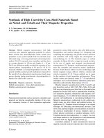
the eigenstates of a molecule, producing time-dependent virtual states as shown in Fig.
2.1 [31]. Since Raman scattering results from a transition between two stationary states
of the molecules, the difference in energy between the vibrational levels is carried off
9
by the scattered photons, and the frequency shift can be observed. Using perturbation
theory and the time-dependent Schrödinger equation, it is predicted that Raman
scattering is weaker than Rayleigh scattering by about three orders of magnitude. In
addition, from the Boltzmann distribution, most of the molecules are initially in the
lowest vibrational state, and therefore Stokes Raman scattering is usually stronger than
anti-Stokes scattering.
|m-1>
|m>
|m+1>
Virtual State
ω
in
ω
sc
ω
st
ω
as
Fig. 2.1 Energy diagram of light scattering. When the initial and final stationary
states are the same (ω
sc
= ω
in
), Rayleigh (elastic) scattering occurs. Stokes
Raman scattering (ω
st
< ω
in
) is a result of molecule vibration transition to a
higher energy level (|m+1>), while anti-Stokes Raman scattering (ω
as
>ω
in
) is
due to a decrease in quantum number, |m> to |m-1>.
Raman spectroscopy has been developed as a powerful tool for chemical
measurements of molecular species [4, 32-35]. It can be used to analyze different kinds
of materials such as gases, vapors, aerosols, liquids and solids. Clinical applications of
Raman spectroscopy and microscopy have been widely demonstrated [36-39], but they
are limited not only by the difficulty in acquiring the inherently weak tissue Raman
signals interacting with a strong fluorescence background, but also by the relatively
too long spectral and imaging acquisition time. Enhancement on weak Raman signals
10
by several orders of magnitude can be realized by coherent Raman technique, of which
coherent anti-Stokes Raman scattering (CARS) is the most popular.
2.1.2 Fundamental theory of CARS
Coherent anti-Stokes Raman scattering (CARS) is a well-known four-wave mixing
process involving a pump, a Stokes and a probe field with frequencies of
p
,
S
,
pr
and
, respectively, interacting with matter to induce a third-order nonlinear
ization
)3(
polar
P
at the anti-Stokes freq
prSp
uency of
as
[8, 27].
Generally, experiments are often performed in a frequency-degenerated manner for
simplicity, that is, the pump and probe beams come from the same urce
(
p
laser so
pr
), atso th
Spas
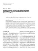
2 . Fig. 2.2(a) shows the energy diagram of the
CARS process [40].
(a)
(b)
|ν=0
|ν=1
Ω
ω
p
ω
s
ω
as
ω
p
k
p
k
p
k
s
k
as
Fig. 2.2 (a) Energy diagram of CARS process, and (b) Phase matching
tio
T
condition in CARS radia n.
he anti-Stokes field (
as
E ) is related with the third-order nonlinear polarization
(
)3(
P
) by the wave equation (assuming an isotropic medium) derived from the
Maxwell’s relations [41],
.
4
)(
)3(
2
2
2
2
P
cc
E
as
asasas
as
(2.2)
11
Here
as
is the dielectric constant, c is the light speed in vacuum and
)3(
P
can be
described by
whe
ear
nti
represents the Ram
an response of the molecular vibrations. It is expressed as
,)(
SppSpp
EEEEEEP
(2.3)
re
p
E and
S
E are the amplitudes of the pump and Stokes fields, respectively;
)3(
is the third-order nonlin susceptibility, which comprises a resonant part,
r)3(
,
and a nonresonant part,
nr)3(
. The resonant part
r)3(
is a complex qua
*)3()3(*)3()3( nrr
ty and
,
)(
)3(
i
A
Sp
where
A is a constant related to the mode density and the R
r
(2.4)
am
an cross-section,
is the vibrational frequency, and
2 is Raman linewidthe th.
The intensity of the CARS signal,
, is written as,
as
I
,
)2/( kl
The last factor )2/(sinc
2
kl in Eq. (2.5) is m ed en the wave vectors of the
pump, the Stokes and the fields,
p
k ,
S
k ,
as
k , respectively, satisfy the
ndition,
)2/(sin
2
2
2
2
4
2
)3()3(
2
kl
EE
c
I
Sp
nrr
as
as
as
(2.5)
aximiz wh
CARS
phase-m
atching
co
lk , where l is the coherent interaction length and
asSp
kkkk 2 is the wave vector mismatch. Because of the strictly-defined phase
relationship, CARS signal can only be coherently generated in a certain direction, as
shown in Fig. Eq. (2.5), obviously, the overall CARS signal is
proportional to
2.2(b). From
2
)3()3( nrr
, and the resonant CARS signal can be greatly enhanced
12
when the resonant condition of
Sp
holds. The nonresonant part
nr)3(
is a
real quantity and is essentially independ f the excitation frequencies. It is
important to realize that the concurrent
nr)3(
is the source of the non
en
y-degener
p
t o
ant
back
re
thi
r n
reson
ground lim
iting the contrast and sensitivity o sonant CARS detection.
The third-order nonlinear susceptibility (
)3(
) is actually a four-rank tensor
containing 81 elements, of which only a few are independent in a symmetrical system.
In the most commonly used frequenc ARS, the components of induced
rd-orde onlinear polarization at
as
f
ated C
S
2 by the pump and Stokes fields at
p
and
S
, respectively, can be written as
,(
*
)3(
pas
).()()(
Spp
EEE
),3)(
)3(
Sas
P
,
p
(2.6)
The subscripts
refer to the Cartesian axis labels and define the polarization
directions of the induced polarization (CARS), the pump, probe and Stokes fields,
respectively. Considering the macroscopic symmetry properties of the medium, the
number of terms in Eq. (2.6) can be further reduced. For isotropic media such as
liquids and gases, it has been shown by symmetry arguments that o ree
susceptibility terms are independent ing r
e z-axis, and “1” and
eptibility com
ponents and is defined as
nly th
“2” indicate the x-
with the fo
()3()3(
s propagate along th
llow
)3
elationship:
.
1221121211221111
(2.7)
Here, supposing the beam
)3(
and y-axis, respectively
.
The ratio between the susc
)3(
1221
)3(
1111
13
the CARS depolarization ratio,
nr
nr
)3(
1111
and
nr)3(
1221
r
r
)3(
1111
(2.8)
Here,
r)3(
1221
nr
and
r
are the nonresonant and resonant CARS depolarization ratios,
respectively. Far from g to Kleinman’ any resonance of the system, accordin s symmetry
conjecture [42], 3/
)3(
1111
)3(
1221
)3(
1212
)3(
1122
nrnrnrnr
holds, and .3/1
nr
2.2 Experimental Instrumentations of CARS Microscopy
CARS imaging provides a new approach to generate chemically selective contrast
based on molecular vibrations, and therefore it has become an attractive technique for
a broad variety of biological and biomedical applications. To establish a robust CARS
microscope, several aspects of strategies should be considered, including the selection
of ultrafast laser sources, excitation geometry and detection schemes, and methods for
nonresonant background suppression.
2.2.1 Laser sources for CARS microscopy
To choose the ideal laser sources for CARS microscopy, several parameters should be
remarked. First is the wavelength range. It has been found that CARS with UV/VIS
wavelength excitation results in large nonresonant background due to the two-photon
resonant interactions [24]. In contrast, NIR excitation minimizes the nonresonant
signals because they are far away from
the two-photon resonance [8, 18]. Another
advantage of NIR excitation is the low absorption and therefore s
mall photodamge to
the samples [43]. In addition, NIR light can penetrate deeper than UV/VIS light in
14
highly scattering samples, which is important for CARS clinical applications. Secondly,
pulsed laser excitation is necessary because CARS signal generation is cubically
dependent on the power of the incident light intensities [41]. The pulse width is
another im
portant parameter that affects the resonant signal to nonresonant background
ratio in CARS imaging. It has been proved that the bandwidth of a several-picosecond
(2~5ps) pulse can match well with the linewidth of most of the Raman resonant
vibr
RS
ation bands (10~20 cm
-1
) for optimizing the CARS signal excitation with improved
spectral resolution and minimized nonresonant background [44].
In the first CARS m
icroscopy built by Ducan and coworkers [24], two
synchronously pum
ped mode-lock pulsed dye lasers were used. Zumbusch et al. [18,
26] used a regeneratively amplified Ti:sapphire laser pumped optical parametric
am
plifier system (Coherent, RegA/OPA). After that, two electronically synchronized
mode-locked Ti:sapphire oscillators with high repetition rate (~80 MHz) and several ps
temporal bandwidth significantly improved the spectral resolution and sensitivity in
CARS microscopy [45]. As a simple and economic approach, one T
i:sapphire fs/ps
laser source and one OPO system can also be used [46]. However, the limitation of this
schem
e is that it can only cover the Raman shift above ~2000 cm
-1
. As the latest CARS
source, Ganikhanov et al. used the signal and idler output from an optical parametric
oscillator (OPO) as the pump and Stokes beam, respectively, for CARS microscopy
and the most recently design can maintain the pump and Stokes pulse trains are
temporally synchronized and spatially overlapped at the output of the OPO [47, 48].
Besides, fib
er laser sources have the potential to develop low cost and portable CA
15