Protein folding quality control in the endoplasmic reticulum in
Bạn đang xem bản rút gọn của tài liệu. Xem và tải ngay bản đầy đủ của tài liệu tại đây (4.85 MB, 190 trang )
PROTEIN FOLDING QUALITY CONTROL
IN THE ENDOPLASMIC RETICULUM
IN BUDDING YEAST
XIE WEI
(B. Sc., USTC)
A THESIS SUBMITTED
FOR THE DEGREE OF DOCTOR OF PHILOSOPHY
TEMASEK LIFE SCIENCES LABORATORY
NATIONAL UNIVERSITY OF SINGAPORE
2010
ii
ACKNOWLEDGEMENT
I would like to express my deepest thanks to my supervisor A/Prof. Davis Ng for his
professional guidance, his valuable insight and his stimulating discussion. I am
extremely grateful for his constant support and encouragement through the course of
study.
Many thanks to my graduate committee members, Drs. Gregory Jedd, Naweed Naqvi
and Yeong Foong May, for their helpful discussions and suggestions on this work.
I also thank all current and previous members of Cell Stress and Homeostasis Group.
Special thanks to Dr. Kazue Kanehara, for her help and contribution in the work in
Chapter 3, and for the opportunity to participate in her exciting work in Chapter 4.
I thank Ms. Wang Songyu and Dr. Ng Kian Hong for their critical readings of this
thesis.
I acknowledge Temasek Holdings for the financial support to my work.
Finally, I would like to thank my family: my father, my mother, and my fiancée, Ms
Yau Wing Tak, for their selfless support, for always being there for me through all these
years.
iii
TABLE OF CONTENTS
Title page i
Acknowledgements ii
Table of contents iii
Summary vi
List of figures ix
List of tables xii
List of abbreviations xiii
List of publications xvi
CHAPTER 1: Introduction 1
1.1 General introduction 1
1.1.1 Quality control in the cell 1
1.1.2 The secretory pathway 2
1.1.3 Quality control in the ER 3
1.1.4 Advantages for studying quality control in yeast 4
1.2 ER quality control machinery 6
1.2.1 Role of N-linked glycosylation in ERQC 6
1.2.1.1 The calnexin/calreticulin cycle 8
1.2.1.2 “Mannose timer” hypothesis 12
1.2.2 ER molecular chaperones 16
1.2.2.1 BiP/Kar2p 16
1.2.2.2 PDI 18
1.3 ER-associated protein degradation 19
1.3.1 ERAD depends on ubiquitin-proteasome system 20
1.3.2 Distinct ERAD complexes 21
1.3.2.1 The Hrd1p complex 22
1.3.2.2 The Doa10p complex 32
iv
1.3.2.3 Mammalian ERAD complexes 35
1.4 Objectives of the thesis 36
CHAPTER 2: Materials and methods 38
2.1 S. cerevisiae strains and genetic methods 38
2.1.1 List of strains used in this study 38
2.1.2 Media for culturing S. cerevisiae 38
2.1.3 Mating and sporulation of S. cerevisiae 38
2.1.4 Transformation of S. cerevisiae 46
2.1.4.1 Low efficiency plasmid transformation 46
2.1.4.2 Preparation of yeast competent cells 47
2.1.4.3 High efficiency DNA fragment transformation 47
2.2 Molecular biology methods 48
2.2.1 List of plasmids used in this study 48
2.2.2 List of oligonucleotide primers used in this study 48
2.2.3 Plasmid construction 48
2.2.4 Yeast genomic DNA extraction 66
2.3 Biochemistry methods 67
2.3.1 Antibody used in this study 67
2.3.2 TCA precipitation of yeast whole cell lysate 67
2.3.3 Western blot of yeast proteins 68
2.3.4 Cycloheximide-chase analysis 69
2.3.5 Cell labeling and immunoprecipitation 69
2.3.6 Yeast microsome preparation and native co-immunoprecipitation 70
2.3.7 Protease sensitivity assay 71
2.3.8 Preparation of yeast proteins for mass spectrometry 72
2.4 Cell biology and microscopy methods 73
2.4.1 Indirect immunofluorescence 73
2.4.2 Confocal microscopy 74
v
CHAPTER 3: Quality control of glycoproteins in the ER 76
3.1 Introduction 76
3.2 Results 78
3.2.1 A bipartite signal targets misfolded glycoproteins to ERAD 78
3.2.2 Local conformational perturbations activate non-signal glycans 89
for ERAD
3.2.3 The CPY ERAD determinant is recognized by the BiP/Kar2p 97
chaperon
3.2.4 Substrate signaling domains act as reporters of protein misfolding 102
3.3 Discussion 111
CHAPTER 4: Quality control of non-glycosylated proteins in the ER 118
4.1 Introduction 118
4.2 Results 120
4.2.1 Novel PrA variants reveal a third substrate class of the yeast 120
Hrd1p complex
4.2.2 The glycan-independent ERAD requires most but not all factors 125
of the Hrd1p complex
4.2.3 ngPrA∆295-331 competes with the glycan-dependent substrate 132
CPY* for degradation
4.2.4 The glycan-independent mode of Hrd1p pathway recognizes 132
distinct degradation signals
4.3 Discussion 138
CHAPTER 5: Conclusions and future directions 143
References 147
vi
SUMMARY
Endoplasmic reticulum (ER) is the first membrane compartment of secretory pathway in
eukaryotic cells. Newly synthesized proteins are translocated into ER lumen, and they
are screened by endoplasmic reticulum quality control (ERQC) system. Only correctly
folded and functional proteins can be sorted out to Golgi and later membrane
compartments. Misfolded proteins are retained in the ER and turned over by a
mechanism conserved from yeast to human known as endoplasmic reticulum-associated
protein degradation (ERAD). While the mammalian system is less understood, the
ERAD mechanism in yeast is explained in more detail, and it is shown to be centered on
two membrane associated E3 ubiquitin ligases: Hrd1p and Doa10p. Previous studies
suggested that Hrd1p ubiquitinates misfolded luminal proteins and membrane proteins
with luminal lesions, while Doa10p targets membrane proteins with misfolded cytosolic
domain. But how exactly the two ERAD E3s detects these lesions remains elusive.
In this thesis, I have used Saccharomyces cerevisiae as a model organism to study the
quality control of two classes of ER luminal proteins – N-linked glycoproteins and
non-glycosylated proteins, both of which are ERAD substrates and degraded by Hrd1p
when misfolded.
In Chapter 3 of this thesis, to study how misfolded N-linked glycoproteins are
recognized by ERQC and ERAD, I started with analyzing two model substrates CPY*
vii
and PrA*. Both of these misfolded ER luminal proteins contain multiple N-linked
glycans, but only one of them is necessary and sufficient for ERAD. Serial deletion
analyses in neither CPY* nor PrA* identified ERAD determinant in the polypeptide
primary sequences, suggesting the determinant might exist in higher order structures. I
inspected the tertiary structure of wild type CPY and found the specific ERAD signal
glycan is positioned on an 11-stranded β-sheet that is arranged mostly in parallel. This
suggests that formation of the local structure adjacent the glycan is dependent on the
overall folding of the polypeptide. Biochemical analysis of CPY* showed that the
polypeptide region adjacent the ERAD signal glycan – termed bipartite ERAD signal, is
tightly bound to Kar2p, a molecular chaperone in the ER lumen and essential
component of the Hrd1p ERAD complex. Indeed the bipartite signal is as simple as a
glycan attached to an unfolded/disordered structure. Consistent with this hypothesis,
lesions introduced throughout CPY to specifically disrupt local structures surrounding
non-ERAD glycans could efficiently report to ERAD through that designated glycan.
Moreover, the position of the bipartite signal on a glycoprotein suggests a possible role
in sensing the overall folding of the polypeptide. Normally the bipartite signal exists in
a stable conformation buried into the tertiary structure of a folded glycoprotein to pass
quality control. However should the protein misfold, the bipartite signal will remain
disordered and exposed to ERQC and ERAD.
In Chapter 4 of this thesis, I described the study in collaboration with Dr. Kazue
Kanehara (experiments done by Dr. Kazue Kanehara are indicated in respective figure
viii
legends) to decipher the mechanism for quality control of non-glycosylated proteins in
the ER lumen. Similar to N-linked glycoproteins, non-glycosylated proteins also subject
to ERQC, but the exact machinery responsible is largely unknown. In this chapter, Dr.
Kazue Kanehara performed a comprehensive analysis to reveal the genetic requirements
for ERAD of misfolded glycoprotein as well as non-glycosylated proteins. Although
both depend on Hrd1p, glycoproteins require additional luminal factors for their
degradation compared to non-glycosylated proteins. By systematic deleting primary
sequence of non-glycosylated PrA* variant, I discovered a signal in the polypeptide
chain both necessary and sufficient for its degradation, suggesting the
glycan-independent route of Hrd1p ERAD pathway also operates in a signal-receptor
based mechanism.
ix
LIST OF FIGURES
Figures Pages
Figure 1.1 Saccharomyces cerevisiae under microscopy 5
Figure 1.2 Synthesis of N-linked oligosaccharide and its 7
transfer to a polypeptide
Figure 1.3 Regulation of calnexin/calreticulin cycle by 10
de-glucosylation and re-glucosylation enzymes
Figure 1.4 Mannosidase-lectin signal-receptor system 13
Figure 1.5 Organization of the Hrd1p and Doa10p E3 complexes 24
for ERAD
Figure 1.6 ERAD of luminal substrates by the Hrd1p complex 27
Figure 1.7 ERAD of membrane substrates by the Hrd1p complex 30
Figure 1.8 ERAD of membrane substrates by the Doa10p complex 33
Figure 3.1 Deletion variants of CPY* and PrA* are degraded 79
efficiently in wild type cells
Figure 3.2 Ribbon diagram of mature CPY 82
Figure 3.3 Signal glycans and adjacent peptide segments are 84
sufficient to signal ERAD
Figure 3.4 Glycan structure alone is not sufficient for ERAD 87
substrate recognition
Figure 3.5 Glycan-proximal lesions are structural disruptive 90
x
Figures Pages
Figure 3.6 Glycan-proximal lesions can generate artificial 92
ERAD determinants
Figure 3.7 Glycan proximity is not a major determinant of 95
substrate recognition
Figure 3.8 The peptide segments adjacent the CPY* signal 98
glycan are recognized by the chaperone BiP/Kar2p
Figure 3.9 The CPY ERAD determinant can detect lesions 103
throughout the polypeptide
Figure 3.10 Intracellular processing of CPY and PrA point mutants 106
Figure 3.11 The CPY and PrA signal glycans mark domains 108
broadly sensitive to structural defects
Figure 3.12 Model of glycoprotein substrate recognition by the 115
Hrd1p complex
Figure 4.1 Specific PrA* variants bypass the Htm1p requirement 121
for degradation
Figure 4.2 ngPrA variants are substrates of the Hrd1p complex 123
Figure 4.3 The Kar2 chaperone is required for glycan-independent 126
ERAD
Figure 4.4 ngPrA∆295-331 degradation requires multiple 130
components of the Hrd1 complex
xi
Figures Pages
Figure 4.5 Glycan-independent and glycan-dependent substrates 133
of ERAD are competitors for degradation
Figure 4.6 PrA contains a distinct determinant for 136
glycan-independent ERAD
xii
LIST OF TABLES
Tables Pages
Table 2.1 List of yeast strains used in this study 39
Table 2.2 List of plasmids used in this study 49
Table 2.3 List of oligonucleotide primers used in this study 53
Table 3.1 Peptide analysis of CPYΔ2-binding protein 101
Table 4.1 Peptide analysis of ngPrAΔ295-331-binding protein 128
Table 4.2 Genetic requirements for the degradation of 131
Hrd1p-dependent substrates
xiii
LIST OF ABBREVIATIONS
aa Amino acid
ATP Adenosine-5’-triphosphate
BiP Immunoglobulin heavy chain binding protein
CFTR Cystic fibrosis transconductance regulator
CHX Cycloheximide
CNX Calnexin
CPY Carboxypeptidase Y
CRT Calreticulin
DAPI 4’,6-diamidino-2-phenylindole
DMSO Dimethyl sulfoxide
DNA Deoxyribonucleic acid
DTT Dithiothreitol
ER Endoplasmic reticulum
ERAD ER-associated protein degradation
Ero1 ER oxidation 1
ERQC ER quality control
GI Glucosidase I
GII Glucosidase II
Glc Glucose
GlcNAc N-acetylglucosamine
xiv
GT Glucosyltransferase
HA hemagglutinin
HMG-CoA 3-hydroxy-3-methylglutaryl-CoA
HMGR HMG-CoA reductase
HRD HMG-CoA reductase degradation
Hsp Heat shock protein
Htm1 Homologous to mannosidase I
kDa kilo Dalton
Man Mannose
MHC Major histocompatibility complex
Mns1 α-mannosidase I
NEF Nucleotide exchange factor
OD Optical density
OST Oligosaccharyltransferase
PBS Phosphate buffered saline
PCR Polymerase chain reaction
PDI Protein disulfide isomerase
PMSF Phenylmethylsulphonylfluoride
PrA Proteinase A
RING Really Interesting New Gene
RNA Ribonucleic acid
rpm Round per minute
xv
RT Room temperature
SC Synthetic complete
SDS Sodium dodecyl sulphate
SDS-PAGE Sodium dodecyl sulphate-polyacrylamide gel electrophoresis
SEM standard error of the mean
SRP Signal-recognition particle
TCA Trichloroacetic acid
TPR Tetratricopeptide repeats
UPS Ubiquitin-proteasome system
Yos9 Yeast osteosarcoma 9
YPD Yeast peptone dextrose
xvi
LIST OF PUBLICATIONS
Xie, W., Kanehara, K., Sayeed, A., and Ng, D.T. Intrinsic conformational determinants
signal protein misfolding to the Hrd1/Htm1 endoplasmic reticulum-associated
degradation system. Molecular Biology of the Cell. 20, 3317-3329. (2009).
Kanehara, K., Xie, W., and Ng, D.T. Modularity of the Hrd1 ERAD complex underlies
its diverse client range. Journal of Cell Biology. 188(5):707-716. (2010).
Xie, W., and Ng, D.T. ERAD substrate recognition in budding yeast. Seminars in Cell
and Developmental Biology. 21(5):533-539. (2010).
1
CHAPTER 1
Introduction
1.1. General introduction
1.1.1. Quality control in the cell
All free living organisms are made of cells, and in every single cell proteins carry out
most cellular functions. Life depends on cells, and on countless proteins to fold
correctly and function as they are designed to. Cells make proteins in a very similar
way as factories manufacture their various products. As factories sometimes make
faulty products, cells also occasionally produce misfolded and malfunctioning
proteins that could cause problems. In order to alleviate potential problems, cells use
specialized processes to promote correctly folded proteins, and most importantly, to
dispose of misfolded proteins, like factories do to their faulty products. This
specialized process is termed protein folding quality control.
Quality control is of vital importance to all organisms, from single cell to higher
eukaryotes. There are plenty of serious human diseases caused, either directly or
indirectly, by defects in quality control system. For example, cystic fibrosis, a severe
genetic disorder, is caused by the rapid turnover of mutated cystic fibrosis
transmembrane conductance regulator (CFTR) (Ward et al., 1995). And defects in
2
coping with excess misfolded proteins could result in diabetes and neurodegenerative
disorders such as Alzheimer’s disease and Parkinson’s disease (Yoshida, 2007).
Therefore, understanding the basic principles underlying cellular quality control
mechanism could lead to promising treatments or even cures for those diseases.
1.1.2. The secretory pathway
The endoplasmic reticulum (ER) is a major site of protein synthesis in eukaryotic
cells, and it is the first membrane compartment of secretory pathway. Proteins are
destined for the secretory pathway by signal sequences, short hydrophobic segments
at the very N-terminus (Milstein et al., 1972). Polypeptides are synthesized first in the
cytosol by ribosomes, and transported through or integrated into ER membrane
through the Sec61p translocon complex (Rapoport, 2007). This process is termed
translocation, and it has two different routes, co-translational or post-translational
translocation, which is dictated by the hydrophobicity of the signal sequence (Ng et
al., 1996). In co-translational translocation, while a polypeptide is still being
synthesized by the ribosome, its signal sequence is recognized by signal-recognition
particle (SRP) and targeted to the ER membrane by SRP receptor (Halic and
Beckmann, 2005). In post-translational mode, only after the synthesis of the
polypeptide is completed and it’s released from ribosome can it be targeted to the ER
membrane, and this process occurs independently of SRP and its receptor but requires
additional membrane complex Sec62p-Sec63p (Deshaies et al., 1991; Huber et al.,
3
2005a; Huber et al., 2005b; Panzner et al., 1995). In either mode, the diameter of the
Sec61p translocon pore limits the translocating polypeptides with α-helixes at most,
and no tertiary structure can be accommodated (Bostina et al., 2005; Haider et al.,
2006; Kowarik et al., 2002; Saparov et al., 2007; Tian and Andricioaei, 2006; Van den
Berg et al., 2004). This means polypeptides enter the ER lumen largely unfolded,
therefore the ER becomes a site in the cell where major folding events occur.
Inside the ER lumen, a wide range of methods are provided to assist the folding of the
newly translocated polypeptides. Covalent modifications such as N-linked
oligosaccharides are added while the polypeptides are still in the translocon (Helenius
and Aebi, 2004). ER resident molecular chaperones directly bind the folding
polypeptide to prevent aggregation and promote their native conformation (Buck et al.,
2007). In the oxidative environment of the ER lumen, free cysteine pairs on the
polypeptide are prone to form disulfide bonds often indispensible for the complete
folding process (Frand and Kaiser, 1998). Eventually, correctly folded and functional
proteins are packed inside COPII vesicles and transported to Golgi and later
membrane compartments of the secretory pathway (Barlowe, 2003).
1.1.3. Quality control in the ER
The high throughput assembly line of ER protein synthesis will inevitably encounter a
population of proteins that fail to acquire their native structure. The ER must employ
4
a censoring system to search and detain these misfolded ones, otherwise allowing the
malfunctioning proteins to slip through would be detrimental to the overall safety of
the cell, sometime even the whole organism. More than three decades ago, evidence
suggesting the existence of such system was already discovered. A mutant form of
α1-antitrypsin causing severe emphasema and liver disease in humans was retained in
the ER of liver cells (Hercz et al., 1978). More evidences were gathered from the
study of another pathogen influenza virus hemagglutinin (HA). Correctly folded HA
subunits assembled into oligomers can exit the ER (Gething et al., 1986), whereas the
misfolded species are bound to ER resident chaperon BiP (immunoglobulin heavy
chain binding protein) and retained (Hurtley et al., 1989). Similar results were found
in the study on vesicular stomatitis virus G protein, and it is during that time de Silva
and coworkers first gave this system its name: endoplasmic reticulum quality control
(ERQC) (de Silva et al., 1990). Now it is established that ERQC is a surveillance
mechanism, conserved in all eukaryotes, that monitors folding status of newly
synthesized secretory proteins entering the ER lumen. Moreover, after ERQC singles
out the misfolded proteins, it also delivers them to a downstream destruction
mechanism termed ER-associated protein degradation (ERAD).
1.1.4. Advantages for studying quality control in yeast
Conserved among all eukaryotes, ERQC and ERAD have been extensively studied in
both mammals and yeast. Saccharomyces cerevisiae, common name “budding yeast”,
5
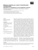
Figure 1.1. Saccharomyces cerevisiae under DIC microscopy
Saccharomyces cerevisiae is a unicellular fungus, generally round in shape and 5-10
μm in diameter, with a doubling time about 2 hours at 30℃. Its reproduction process
starts with the daughter cell emerging as a “bud” from the surface of the mother cell,
hence the common name “budding yeast”. The subsequent asymmetric cell division
results in one bigger mother cell and one smaller daughter.
6
is a unicellular fungus (Figure 1.1). Compared with the mammalians, budding yeast
has a much smaller genome: only 6,000 genes in 12 Mbp, while human has 30,000
genes. Nevertheless it contains all the basic components of ERQC and ERAD,
offering a system to be studied with less complexity. Moreover, the advanced
techniques in yeast genetics, as well as the availability of many mutant strains, makes
the study in all much easier.
1.2. ER quality control machinery
1.2.1. Role of N-linked glycosylation in ERQC
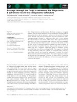
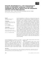
N-linked glycoproteins constitute majority of secretory proteins among all eukaryotes.
The N-linked oligosaccharides are presynthesized on the ER membrane and then
added to proteins all at once (Helenius and Aebi, 2004). The core structure,
Glc
3
Man
9
GlcNAc
2
, is composed of three glucoses, nine mannoses, and two
N-acetylglucosamines (Figure 1.2) (Helenius and Aebi, 2004). Synthesis of the core
oligosaccharide starts first on the cytosolic face with a lipid linkage to the ER
membrane. During the process, an enzyme named Rft1p flips the lipid-linked
oligocaccharide intermediate into the luminal face of the ER membrane (Helenius et
al., 2002), and the synthesis continues. At the last step, an enzyme complex
collectively called oligosaccharyltransferase (OST) transfers the lipid-linked final
oligosaccharide product from the membrane onto the asparagine residue of
7
Figure 1.2. Synthesis of N-linked oligosaccharide and its transfer to a polypeptide
8
Asn-X-Ser/Thr consensus site of the polypeptide (Burda et al., 1999).
One intriguing observation was made: although the glycan is first added to the
polypeptide as Glc
3
Man
9
GlcNAc
2
, protein leaves the ER all bears N-glycans as
Man
8
GlcNAc
2
with all three glucose and one mannose residues trimmed away
(Kornfeld and Kornfeld, 1985). Therefore naturally came the question: why almost all
eukaryotic cells, from yeast to human, have evolved this highly sophisticated
procedure to synthesize a large oligosaccharide, only to trim it down almost right
away in the very same compartment? The logic behind this seemingly energy-wasting
effort is that each trimming product reports to the ER quality control system about the
folding state of the nascent polypeptide chain.
1.2.1.1. The calnexin/calreticulin cycle
As soon as the core Glc
3
Man
9
GlcNAc
2
oligosaccharide is attached to the emerging
polypeptide in the ER lumen, the protein enters the calnexin/calreticulin cycle.
Calnexin (CNX) is a type I transmembrane protein, while calreticulin (CRT) is a
luminal protein, and together these two lectins act as the first stage of the ER quality
control system (Caramelo and Parodi, 2008; Williams, 2006).
Association of the nascent polypeptide chain with the CNX/CRT requires the
sequential trimming of the outmost two glucose residues on branch A of the glycan by
9
glucosidase I and glucosidase II (GI and GII) (Deprez et al., 2005; Hebert et al., 1995)
(Figure 1.3). Interacting with lectins CNX/CRT through the trimmed
Glc
1
Man
9
GlcNAc
2
oligosaccharide protects the emerging polypeptide from forming
aberrant aggregates with other unstructured chains. Thus this association gives the
nascent proteins longer time and better chance to achieve their own native structure.
Besides removing the second glucose residue, GII is also able to cleave the third one,
and this action releases the Man
9
GlcNAc
2
oligosaccharide bearing polypeptide from
CNX/CRT. However, if at this time the polypeptide has not formed a stable structure,
it is allowed to re-associate with CNX/CRT for another folding attempt, and the
re-entry permit is issued by the enzyme glucosyltransferase (GT) (Caramelo et al.,
2004; Pearse et al., 2008; Trombetta et al., 1991). GT adds one glucose residue back
to branch A of the glycan, thus sending it back into the CNX/CRT cycle (Labriola et
al., 1995). The collective de- and re-glucosylation actions of GI, GII and GT ensure
the nascent proteins are retained by CNX/CRT cycle until they are deemed mature
enough to exit, and then other molecular chaperones come into play.
Although the CNX/CRT lectins are absent from the genome of S. cerevisiae, current
knowledge indicates that the use of N-linked oligosaccharide as a signal reporting to
ERQC is conserved among all eukaryotes. In fact, the lack of CNX/CRT cycle in
yeast renders later stages of quality control (for example the “mannose timer”
hypothesis discussed below) in this organism to be more prominent, hence more
detailed insights yielded from the study.