Role of hydrogen sulfide in the pathogenesis of parkinsons disease
Bạn đang xem bản rút gọn của tài liệu. Xem và tải ngay bản đầy đủ của tài liệu tại đây (1.75 MB, 164 trang )
HYDROGEN SULFIDE: A NOVEL NEUROPROTECTIVE
AGENT TO TREAT PARKINSON’S DISEASE
HU LI-FANG
(MD, M.Sci, Nanjing Medical University, China)
A THESIS SUBMITTED
FOR THE DEGREE OF DOCTOR OF PHILOSOPHY
DEPARTMENT OF PHARMACOLOGY
NATIONAL UNIVERSITY OF SINGAPORE
2010
i
ACKNOWLEDGEMENT
I would like to express my deepest gratitude to my supervisor, Prof. Bian Jin-Song, for
giving me the opportunity to work on this research project as a part-time postgraduate
student. I would thank my supervisor for his invaluable comments, enlightening ideas and
continuous encouragement. Without his great support, I would not have made great
progress on my thesis.
I would also thank Prof. Gavin S. Dawe for his guidance in the behaviour study. I
would express my special thanks to Mr. Lu Ming for his kindly help and collaboration in
the animal work.
Sincere appreciation to my colleagues, Neo Kay Li, Pan Tingting, Yong Qian Chen,
Wu Zhiyuan, Ester Khin Sandar Win, Tan Choon Ping, Liu Yihong, Tiong Chi Xin, Xie
Li, and other friends in Prof. Bian‘s lab for their technical support and help in various
aspects over the four years. I would also thank all my friends in Prof. Gavin S. Dawe‘s
lab for their support on my animal work.
I would also extend my deep gratitude to my husband for supporting and inspiring me
to reach my full potential.
ii
TABLE OF CONTENT
PUBLICATIONS vii
ABBREVIATIONS viii
SUMMARY x
Chapter I Introduction 1
1.1 General overview 1
1.2 H
2
S 2
1.2.1 Chemical properties of H
2
S 2
1.2.2 H
2
S toxicity 3
1.2.3 H
2
S biosynthesis and metabolism in mammals 4
1.2.4 Biological roles of H
2
S 10
1.2.4.1 Roles of H
2
S in inflammation 10
1.2.4.2 Role of H
2
S in CNS and CNS diseases 14
1.2.4.3 Roles of H
2
S in cardiovascular system 25
1.2.4.4 Roles of H
2
S in gastrointestinal tract 28
1.2.4.5 Others 30
1.3 PD 31
1.3.1 Epidemiology 32
1.3.2 Risk factors 32
1.3.3 Pathology and pathogenesis 33
1.3.3.1 Pathology 33
1.3.3.2 Pathogenesis 34
1.3.4 Clinical features and diagnosis 38
1.3.5 Treatment 38
1.3.6 Experimental models 39
1.3.6.1 6-OHDA model 40
iii
1.3.6.2 MPTP/MPP
+
model 42
1.3.6.3 Rotenone model 45
1.3.6.4 LPS model 46
1.3.6.5 Other models 49
1.4 Research rational and objectives 52
1.4.1 Rational 52
1.4.2 Objectives 53
Chapter II Anti-inflammatory effects of H
2
S on LPS-stimulated microglia 55
2.1 Introduction 55
2.2 Materials and methods 56
2.2.1 Chemicals and reagents 56
2.2.2 Cell culture 56
2.2.2.1 Microglia cell line culture 56
2.2.2.2 Primary cultured rat cortical microglia and astrocytes preparation 57
2.2.3 NO measurement 58
2.2.4 TNF-α measurement 58
2.2.5 Reverse transcription polymerase chain reactions (RT-PCR) 59
2.2.6 Transfection of CBS and CSE into BV2 cells 59
2.2.7 Western blot analysis 60
2.2.8 Statistical analysis 61
2.3 Results 61
2.3.1 Exogenous H
2
S suppresses LPS-stimulated NO production in microglia 61
2.3.2 The anti-inflammatory effect of H
2
S involves p38 MAPK 63
2.3.3 H
2
S inhibits TNF-
secretion in BV2 cells 65
2.3.4 Endogenous H
2
S regulates NO production in BV2 cells 66
2.3.5 Over-expression of H
2
S synthesis enzyme suppresses NO production in microglia 68
2.3.6 H
2
S suppresses LPS-stimulated NO generation in astrocyte 68
iv
2.4 Discussion 69
Chapter III Anti-inflammatory effects of H
2
S on rotenone-stimulated microglia 73
3.1 Introduction 73
3.2 Materials and methods 74
3.2.1 Chemicals and reagents 74
3.2.2 Cell culture 74
3.2.3 Immunocytochemistry 74
3.2.4 Western blot assays 75
3.2.5 Intracellular ROS assay 75
3.2.6 Extracellular superoxide measurement 75
3.2.7 Microglia-mediated neurotoxicity assay 76
3.2.8 NF-κB activation assay 76
3.2.9 Statistical analysis 77
3.3 Results 77
3.3.1 NaHS inhibits rotenone-stimulated microglia activation 77
3.3.2 NaHS suppresses rotenone-induced intracellular ROS accumulation 79
3.3.3 NaHS inhibits rotenone-induced superoxide release from microglia 80
3.3.4 NaHS attenuates microglia-mediated neurotoxicity 81
3.3.5 NaHS inhibits rotenone-induced p38 MAPK/NF-κB activation 82
3.4 Discussion 86
Chapter IV Anti-apoptotic effect of H
2
S on SH-SY5Y cells 90
4.1 Introduction 90
4.2 Materials and methods 90
4.2.1 Chemicals and reagents 90
4.2.2 Cell culture and treatment 92
4.2.3 Total sulfide measurement 93
4.2.4 Cell viability assay 93
v
4.2.5 Apoptosis quantification 93
4.2.6 Assessment of mitochondrial membrane potential (ΔΨm) loss 94
4.2.7 Western blot analysis 94
4.2.8 Analysis of cytosolic cytochrome c accumulation 94
4.2.9 Caspase-9 activity assay 95
4.2.10 Statistical analysis 95
4.3 Results 95
4.3.1 H
2
S suppresses rotenone-induced cytotoxicity and apoptosis 95
4.3.2 H
2
S inhibits rotenone-induced ΔΨm loss and cytochrome c release 98
4.3.3 H
2
S regulates Bax/ Bcl-2 proteins in SH-SY5Y cells 100
4.3.4 H
2
S suppresses caspase-9/3 activation and PARP cleavage 101
4.3.5 mitoK
ATP
channels contributes to the protective effects of H
2
S 103
4.3.6 Rotenone induces p38/JNK MAPK activation 105
4.3.7 H
2
S inhibits rotenone-induced p38/JNK MAPK activation 107
4.4 Discussion 108
Chapter V Therapeutic effect of H
2
S in rotenone-induced PD model rats 112
5.1 Introduction 112
5.2 Materials and methods 113
5.2.1 Chemicals 113
5.2.2 Animals 113
5.2.3 Behavioural test 113
5.2.4 H
2
S measurement 114
5.2.5 H
2
S-producing activity assay 115
5.2.6 Immunohistochemistry staining 115
5.2.7 NO assay 116
5.2.8 TNF-α assay 116
5.2.9 Western blot analysis 117
vi
5.2.10 Statistical analysis 117
5.3 Results 117
5.3.1 Endogenous H
2
S is reduced in the SN of rotenone-treated rats 117
5.3.2 NaHS alleviates rotenone-induced parkinsonian symptoms in rats 118
5.3.3 H
2
S attenuates rotenone-induced DA neuron loss in the SN 121
5.3.4 NaHS inhibits microglia activation and the subsequent release of inflammatory factors
in the rotenone-induced PD model rats 122
5.4 Discussion 123
Chapter VI General discussion and conclusion 127
6.1 General discussion 127
6.2 Conclusion and perspectives 134
Bibliography 137
vii
PUBLICATIONS
1. Hu LF, Lu M, Tiong CX, Dawe GS, Hu G, Bian JS. Neuroprotective effects of
hydrogen sulfide in Parkinson‘s disease rat models. Aging Cell. 2009; 9(2):135-46.
2. Hu LF, Lu M, Wu ZY, Wong PT, Bian J. Hydrogen sulfide inhibits rotenone-induced
apoptosis via preservation of mitochondrial function. Mol Pharmacol. 2009; 75(1):27-
34.
3. Hu LF, Wong PT, Moore PK, Bian JS. Hydrogen sulfide attenuates
lipopolysaccharide-induced inflammation by inhibition of p38 mitogen-activated
protein kinase in microglia. J Neurochem. 2007; 100(4):1121-8.
4. Hu LF, Pan TT, Neo KL, Yong QC, Bian JS. Cyclooxygenase-2 mediates the
delayed cardioprotection induced by hydrogen sulfide preconditioning in isolated rat
cardiomyocytes. Pflugers Arch. 2008 Mar; 455(6):971-8.
5. Hu LF, Wong PT, Bian J. Hydrogen sulphide: neurophysiology and neuropathology.
Review. Antioxid Redox Signal. 2010 Sep 2.
6. Hu LF, Wong PT, Bian J. Hydrogen sulfide attenuates rotenone-induced
neuroinflammatory responses through down-regulation of NADPH oxidase/ROS
signaling pathways in microglia (in preparation)
7. Tay AS, Hu LF, Lu M, Wong PT, Bian JS. Hydrogen sulfide protects neurons against
hypoxic injury via stimulation of ATP-sensitive potassium channel/protein kinase
C/extracellular signal-regulated kinase/heat shock protein90 pathway. Neuroscience.
2010 Feb 8.
8. Lu M, Choo CH, Hu LF, Tan BH, Hu G, Bian JS. Hydrogen sulfide regulates
intracellular pH in rat primary cultured glia cells. Neurosci Res. 2010; 66(1):92-8.
9. Lu M, Hu LF, Hu G, Bian JS. Hydrogen sulfide protects astrocytes against H(2)O(2)-
induced neural injury via enhancing glutamate uptake. Free Radic Biol Med. 2008;
45(12):1705-13.
10. Lee SW, Hu YS, Hu LF, Lu Q, Dawe GS, Moore PK, Wong PT, Bian JS. Hydrogen
sulphide regulates calcium homeostasis in microglial cells. Glia. 2006; 54(2):116-24.
viii
ABBREVIATIONS
AC
adenylyl cyclase
AD
Alzheimer‘s disease
AIF
apoptosis-inducing factor
AOAA
amino-oxyacetic acid
AP1
activator protein 1
Apaf-1
apoptotic protease activating factor-1
BCA
β-cyanol-l-alanine
CAT
cysteine aminotransferase
CBS
cystathionine-β-synthase
CNS
central nervous system
CSE
cystathionine-γ-lyase
CO
carbon monoxide
COX-2
cyclooxygenase-2
DA
dopamine
ERK
extracellular signal-regulated kinase
GSH
glutathione
GABA
γ-aminobutyric acid
HA
hydroxylamine
H
2
S
hydrogen sulfide
HD
Huntington‘s disease
IL-1β
interleukin 1β
iNOS
inducible nitric oxide synthase
JAK
Janus kinase
JNK
c-Jun N-terminal kinase
K
ATP
ATP-sensitive potassium channel
LPS
lipopolysaccharide
LTP
long-term potentiation
MAO
monoamine oxidase
ix
MAPK
mitogen-activated protein kinase
3-MST
3-mercaptopyruvate sulfurtransferase
mitoK
ATP
mitochondrial K
ATP
channel
MPTP
1-methyl-4-phenyl-1,2,3,6-tetrahydroperidine
NaHS
sodium hydrogen sulfide
NF-κB
nuclear factor kappa-light-chain-enhancer of activated B cells
NMDA
N-methyl-D-aspartic acid
NO
nitric oxide
Nrf-2
Nuclear factor-like 2
NSAIDs
non-steroidal anti-inflammatory drugs
6-OHDA
6-hydroxydopamine
PAG
DL-propargylglycine
PARP
poly (ADP-ribose) polymerase
PD
Parkinson‘s disease
PFA
paraformaldehyde
PGE
2
prostaglandin E
2
PI3K
phosphoinositide 3-kinase
PKA
protein kinase A
PKC
protein kinase C
PLC
phospholipase C
PLP
pyridoxal-5‘-phosphate
PPAR-γ
peroxisome proliferator-activated receptor-γ
ROS
reactive oxygen species
SAM
S-adenosyl-L-methionine
SN
substantia nigra
SNpc
substantia nigra pars compacta
STAT
signal transducers and activators of transcription
TH
tyrosine hydroxylase
TNF-α
tumor necrosis factor-α
VMAT
vesicular monoamine transporter
x
SUMMARY
H
2
S is a novel endogenous gaseous mediator alongside nitric oxide and carbon monoxide.
It serves as an important neuromodulator in regulation of brain functions. PD,
characterized by the progressive loss of DA neurons in midbrain, is the second most
common neurodegenerative disorder among old population. In this thesis, the therapeutic
effect of H
2
S on neurodegeneration and the underlying mechanisms were investigated in
both in vitro and in vivo studies.
Neuroinflammation is one of the main pathological causes/features of PD. In this
thesis, the effect of H
2
S on neuroinflammation was first examined in glial cells. It was
found that both endogenous and exogenous application of H
2
S ameliorated LPS-
stimulated production of nitric oxide and TNF-α, two important pro-inflammatory factors,
in primary cultured microglia and BV2 cells. Similar results were also observed in
primary cultured astrocytes. NaHS, an H
2
S donor, also attenuated rotenone-induced
intracellular reactive oxygen species and extracellular superoxide accumulation in
microglia. This implies that H
2
S plays an anti-inflammatory role in central nervous
system. The underlying mechanisms for the anti-neuroinflammatory role of H
2
S were
demonstrated to be associated with its inhibitory effect on p38 MAPK and NF-κB
signaling pathway. In addition, H
2
S was also found to alleviate inflammation-mediated
neurotoxicity on SH-SY5Y cells. These data suggest that H
2
S may produce
neuroprotective effects via its anti-neuroinflammatory action.
Apart from the indirect neuroprotection, the direct effect of H
2
S on neuronal cells was
also investigated. It was found that H
2
S concentration-dependently suppressed rotenone-
induced cellular injury and apoptotic cell death. NaHS also prevented rotenone-induced
xi
p38- and JNK- MAPK phosphorylation and changes in Bcl-2/Bax levels, ΔΨ
m
dissipation,
cytochrome c release, caspase-9/3 activation as well as PARP cleavage. This effect was
mediated by mitoK
ATP
channels. Therefore, H
2
S may protect neuronal cells against
rotenone-induced apoptosis through preservation of mitochondrial integrity and inhibition
on mitochondrial apoptotic pathways.
The potential neuroprotection of H
2
S was further confirmed in vivo with the rotenone-
induced rat PD model. It was found that both endogenous H
2
S level and its biosynthesis
activity were reduced in rotenone-induced PD rats, implying that the impaired
endogenous H
2
S production may contribute to the development of PD. Interestingly,
NaHS treatment significantly alleviated rotenone-induced behavioral deficits, DA
neuronal loss in substantia nigra, microglial activation as well as elevation of nitric oxide
and TNF-α content in the nigrostriatal tract in rat. These data clearly suggest that H
2
S has
the potential to be developed as a new agent to treat neurodegenerative diseases.
In summary, the present study demonstrates for the first time that H
2
S may serve as a
neuroprotectant to treat and prevent neurotoxin-induced neurodegeneration via anti-
inflammatory and anti-apoptotic mechanisms, and therefore has the potential therapeutic
value for PD treatment.
1
Chapter I Introduction
1.1 General overview
Hydrogen sulfide (H
2
S) poisoning is an uncommon fatal incident that happens now and
then in the world; thus, H
2
S has been known as a toxic gas for more than 300 years.
However, it has recently been referred to as the third gaseous mediator alongside nitric
oxide (NO) and carbon monoxide (CO) because mammalian tissue produces H
2
S with L-
cysteine as a substrate and this gaseous molecule is involved in regulating our body
functions, rather than simply being a toxic pollutant. Two key papers, published in 1996
and 2004, were the first reports to describe the important functions of H
2
S as a novel
neuromodulator and neuroprotectant against oxidative damages in the central nervous
system (CNS) (1, 104). This opens up new directions for exploring H
2
S biology in the
CNS.
Parkinson‘s disease (PD) is a movement disorder characterized by the progressive
loss of dopamine (DA) neurons in the substantia nigra (SN). Its etiology remains elusive
and the mechanisms for initiating and aggravating neuronal death are yet to be defined,
despite years of intensive research. An inflammatory process in CNS, often defined as
neuroinflammation, is recently revealed to play an important role in the cascades of
events leading to DA neuronal loss, and therefore greatly contributes to the pathological
progression of PD. Hence, it is presumed that any strategy aimed at suppressing the
neuroinflammatory process would be potentially effective for PD therapy.
In the peripheral system, H
2
S was recently demonstrated to regulate the inflammatory
process (120, 151, 231). However, its role in neuroinflammation remains unknown.
Given that H
2
S regulates inflammatory processes in various diseases and that H
2
S exists
in brain at relatively high levels, it is hypothesized that H
2
S may regulate
2
neuroinflammatory process, and thus the pathogenesis of PD. Therefore, this thesis was
designed to investigate the potential role of H
2
S in neuroinflammation and DA neuronal
injury. The therapeutic effect of H
2
S on PD was also tested in an animal PD model.
1.2 H
2
S
Similar to NO and CO, H
2
S has historically been known as an industrial pollutant and
environmental toxin; however, it has recently been shown to be an important gaseous
transmitter modulating many physiological and pathological processes. Specifically, H
2
S
exerts important functions in the cardiovascular system and the CNS. It was found to
serve as a vasodilator and a novel neuromodulator. H
2
S is also an endogenous regulator
of inflammatory response, playing both pro- and anti- inflammatory roles in different
systems and situations. Several lines of evidence suggest that H
2
S is an important
biological molecule in mammalian tissues.
1.2.1 Chemical properties of H
2
S
H
2
S is a colourless, flammable gas with a molecular weight of 34.08. It is responsible for
the foul odour of rotten eggs and flatulence. Its structural formula is illustrated as H-S-H,
similar to that of water (H
2
O). It is soluble in water with solubility of 1 g in 242 ml water
at 20 °C. H
2
S is weakly acidic because it easily dissociates into H
+
and HS
-
in solution. It
is noteworthy that both pH and temperature of solution affect H
2
S concentration.
According to a standard Henderson-Hasselbach calculation at 20°C, in solution with pH
at 7.4, H
2
S exists as ~30-33% (one-third) H
2
S and 67-70% (two-thirds) HS
-
, with
negligible S
2-
due to the high pK
a2
(11.96). However, it is not true when temperature
reaches 37°C because pK
a1
at 37°C is 6.755, rather than 7.04 for standard conditions at
20°C. Accordingly, in saline at 37°C and pH 7.4, less than one-fifth of H
2
S exists as the
undissociated form (H
2
S) (82, 166, 192). Because it is unlikely to determine which form
3
of H
2
S (H
2
S, HS
-
, S
2-
, or the mixture) is active, in this thesis the sum of these afore-
mentioned forms will be referred to as ―free H
2
S‖ for the simplicity of conveying ideas.
In some literature, the sum of H
2
S and HS
-
is also denoted as total sulphide. However,
this is somewhat confusing because some chemistry reports also use this term for acid-
labile sulphide (ALS) and dithiothreitol (DTT) -labile sulphide (DLS). Therefore, we
prefer to use free H
2
S to define the sum of H
2
S, HS
-
and S
2
-
in the present dissertation.
1.2.2 H
2
S toxicity
H
2
S has been known as a highly toxic gas for almost 300 years. H
2
S poisoning is a
serious issue due to its widespread environmental and occupational exposure derived
from industrial activities, such as paper pulp mills, petroleum refinery and urban sewers.
H
2
S is a broad-spectrum poison affecting different systems, among which the nervous
system is the most often intoxicated. Acute inhalation of H
2
S (500-1000 ppm) causes
neurotoxic effects such as headache, dizziness, amnesia and even unconsciousness
(‗knockdown‘), due to the direct toxic effect of H
2
S on the brain. These toxic effects are
usually reversible because the sufferers can completely recover from the acute
intoxication if the exposure is rapidly removed. However, if exposure is more pronounced
(over 1000 ppm) or prolonged, fatal respiratory paralysis may occur and even lead to
death, as a result of hypoxia secondary to H
2
S-induced respiratory insufficiency. Death
may come instantly, just like a vivid description ―death comes on like a stroke of
lightening‖, when the concentration of H
2
S is higher than 5000 ppm (150, 163). In a few
cases, acute but nonfatal H
2
S intoxication is followed by brain damage featured by
permanent neurological sequelae.
H
2
S toxicity has been attributed to its ability to inhibit cytochrome c oxidase in a
similar manner to hydrogen cyanide poisoning (197). It suppresses oxygen utilization and
4
results in central respiratory paralysis. Warenycia et al. believed that its inhibition on
monoamine oxidase (MAO) also contributes to the loss of central respiratory drive after
fatal intoxication with H
2
S (208). Recent studies found that generation of excessive ROS
via a CYP450-dependent mechanism also contributed to H
2
S-induced cytotoxicity (50).
Of note, human nose is very sensitive to H
2
S and can detect its unpleasant smell as low as
0.02 ppm (estimated level of H
2
S in normal atmosphere is approximately 0.0001 ppm).
However, it appears to be true only at low concentrations. At higher concentrations (up to
100 ppm), the rotten-egg smell of H
2
S disappears and it emerges as an odourless gas,
greatly increasing the risk of H
2
S poisoning. This implies that disappearance of
unpleasant smell could be a sign for increasing concentrations of H
2
S at sites where
potential exposure of this poison exists.
1.2.3 H
2
S biosynthesis and metabolism in mammals
The desulfhydration of Cys is proposed to be the major source of H
2
S in mammals. This
process is catalyzed via two pyridoxal-5‘-phosphate (PLP)-dependent enzymes CBS and
CSE. In the transsulfuration pathway, Cys is derived from Hcy with CBS catalyzing the
β-replacement reaction of Hcy to yield cystathionine which is then lyzed by CSE into Cys
and α-ketobutyrate (Figure 1.1). It has been shown that CBS can efficiently produce H
2
S
via a β-replacement reaction in which Cys is condensed with Hcy to form cystathionine
and H
2
S and this reaction is far more efficient when compared to β-elimination of Cys
(33). Detailed kinetic analysis performed by Banerjee‘s group demonstrated that CBS
produces H
2
S overwhelmingly from Cys+Hcy (96%) under simulated physiological
conditions, while Cys and Cys+Cys accounts for only 1-3 % (71). Therefore, cysteine and
homocysteine are the preferred substrates of CBS for H
2
S biosynthesis. On the other hand,
CSE produces H
2
S from Cys (70%) or Hcy (γ-elimination, 29%) under normal conditions
5
(10 µM Hcy). Interestingly, when the concentration of Hcy was increased from 10 to 40
and 200 µM to simulate mild and severe hyperhomocysteinemia, the contribution from
Hcy increased from 29% to 63% and 90%, respectively, while contribution from Cys
decreased correspondingly to 37% and 10%, respectively (13). As Vmax for the γ-
elimination of Hcy is twice that for the β-elimination of Cys, this shift may represent a
marked increase in the generation of H
2
S under hyperhomocysteinemic conditions (13).
Therefore, H
2
S production derived from CSE is sensitive to homocysteine (35). Basically,
under normal conditions (10 µM Hcy) CSE represents ~32% of the H
2
S generation by the
transsulfuration pathway, but it increases to ~45% and ~74% under moderate and severe
hyperhomocysteinemia conditions (182). In contrast, CBS is not sensitive to Hcy
concentrations with Cys+Hcy as the predominant substrates (71). For this reason, in
homocystinurics with CBS deficiency, CSE may be the major enzyme to produce H
2
S.
Moreover, the level of H
2
S produced by CSE is predicted to be higher due to the
enhanced accumulation of Hcy (35).
CBS is highly expressed in the brain and thus believed to be the primary physiologic
source of H
2
S in the CNS (1), although both CBS and CSE activities were detected in
different brain regions (9, 201). CBS is a cytoplasm PLP-dependent enzyme. Human CBS
has a complex structure and regulatory mechanisms (134). It contains the N-terminal
heme-binding domain, the catalytic domain, and the C-terminal regulatory domain. Two
other gaseous transmitters, CO and NO, can bind to the heme-binding domain and result
in the inhibition of CBS activity (190, 191). Moreover, the S-adenosyl-L-methionine,
which may bind to the C-terminal domain, can instantaneously activate CBS. At the
transcriptional level, glucocorticoids can stimulate the CBS gene expression whereas
insulin can inhibit it (162).
6
L-Hcy
cystathionine
H
2
S
3-mercaptopyruvate
sulfane sulfur
H
2
S
reducing agents
(DTT, GSH, etc)
Hcy
cys tathionine
GSH
methionine
B12
folate
transsulfurationpathway GSH synthesis pathway
transamination
α-ketobutyrate
L-Cys
Cysteine catabolism pathway
demethylation
serine
homoserine
pyruvate
serine
CBS
CSE
CSE
CAT
3-MST
CBS
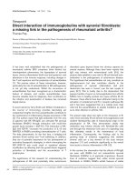
Figure 1.1 Endogenous sources of H
2
S in mammalian. H
2
S is endogenously produced by the action of
CBS and CSE in the transsulfuration pathway. By kinetic simulation, it is found that CBS generates H
2
S
most efficiently from Cys+Hcy, with cystathionine as a byproduct. This reaction contributes >95% of the
net H
2
S production by CBS. On the contrary, the preferred substrates for CSE are Cys and Hcy. Together
they contribute well over 90% of the net H
2
S production by CSE. In addition, the CAT and 3-MST are
components of the Cys catabolism pathway. CAT catalyzes the transamination of Cys to yield 3-
mercaptopyruvate, a substrate of 3-MST to produce pyruvate and sulfane sulfur, which may liberate H
2
S in
the presence of reductants such as dithiothreitol (DTT) and GSH. The transsulfuration pathway is critical
for creating Cys from the essential amino acid methionine, which is first converted to Hcy by demethylation.
CBS condenses serine and Hcy to produce cystathionine, which is converted to Cys and α-ketobutyrate by
CSE. The synthesis of glutathione (GSH) is regulated at the substrate level by Cys. Thus, the
transsulfuration pathway also links to the GSH homeostasis in brain.
The cellular localization of CBS is still controversial. Using immunohistochemical
techniques, Robert et al. showed that CBS protein has a predominantly neuronal
localization in most areas of the brain, especially in hippocampus and cerebellum (166).
In contrast, Enokido et al. later demonstrated that CBS is preferentially expressed in
astrocytes rather than neurons, which is verified by combined biochemical and
histological examination, as well as in situ hybridization (51). This fits with recent
7
findings that CBS mainly localizes to astrocytes (112). Lee et al. demonstrated that the
basal H
2
S level in unstimulated human astrocytes is about 3.0 µmol/g protein, which is
7.9 fold higher than in cultured microglia. More importantly, only astrocytes, instead of
microglia, are strongly immunostained for CBS (112). However, Vitvitsky and his
colleagues showed the incorporation of radiolabel from methionine into glutathione (GSH)
in both cultured human astrocytes and neurons (201). Another group also showed that
inhibition of CSE leads to a significant loss of GSH in adult brain slices (48). Since the
only known route for the transfer of radiolabelled methionine to GSH is via the
transsulfuration pathway involving CBS and CSE, these experiments indirectly justify the
existence of CBS in both astrocytes and neurons. Nevertheless, studies consistently
identified the temporal expression of CBS in developing and adult mouse CNS. During
the embryonic period, CBS protein level is generally low, but it dramatically increases
from late prenatal to early postnatal period (51, 166).
In biomedical studies, small molecule inhibitors, such as hydroxylamine and
aminooxyacetate acid (AOAA), have been used to determine the significance of the
endogenously generated H
2
S. These agents are able to inhibit the biosynthesis of H
2
S
from Cys but they are general inhibitors of all PLP-dependent enzymes and are used quite
to liberate bound PLP for quantitation (92, 184). In addition to heme and PLP,
hydroxylamine also reacts with non-heme iron proteins, e.g. ribonucleotide reductase and
is used to inhibit cell growth. Hence, caution should be taken when interpreting results
obtained from work involving these inhibitors.
In addition to CBS, there is also report showing that CSE plays an important role in
human brain, despite its predominant localization in the cardiovasculature. In fact, CSE is
critical for maintaining GSH homeostasis in brain, which in turn preserves mitochondrial
8
function (48). CSE is the rate-limiting enzyme in the transsulfuration pathway for the
sulfur transfer methionine to Cys, which is a limiting reagent in the synthesis of GSH.
Moreover, CSE mRNA is localized in brain and found to be predominantly present in
neurons by in situ hybridization. The CSE activity in mouse brain was as low as 1% of
the hepatic activity. However, in human brain the activity was 100 times more than that in
mouse brain. Furthermore, an intact transsulfuration pathway in the brain mediated by
both CBS and CSE links to GSH homeostasis, which greatly contributes to the redox-
buffering capacity in brain (201). Nevertheless, the general consensus is that CSE is the
primarily physiological source of H
2
S generation in the peripheral tissues. There is
definitive evidence that CSE knockout mice developed hypertension, which establishes
that H
2
S as a major physiologic signalling molecule regulating vascular tone in mammals.
So far, there is little knowledge about the physiologic relevance of CSE relative to CBS
in brain, in addition to its role in transsulfuration pathway linking to GSH homeostasis.
This issue merits further investigation.
Recently, Kimura‘s group reported another source of H
2
S in the brain homogenates of
CBS-knockout mice (177). They show 3-mercaptopyruvate sulfurtransferase (3-MST) in
combination with Cys aminotransferase (CAT) produces H
2
S from Cys. Like CBS and
CSE, CAT is also a PLP-dependent enzyme, which catalyzes the metabolism of Cys and
α-ketoglutarate to yield 3-mercaptopyruvate as the substrate for 3-MST. 3-MST is
localized to mitochondria and nerve endings. As its name implies, it belongs to the family
of sulfurtransferases, which catalyze the transfer of sulfane sulfur from persulfide or
thiosulfate or mercaptopyruvate to an acceptor, and liberates H
2
S under certain conditions.
Thus, 3-MST does not produce H
2
S by itself. Instead, it produces sulfane sulphur (or
bound sulphur), which in the presence of reductants like dithiothrietol , liberates H
2
S (92).
9
Bound sulfur may be a source of H
2
S in brain and it can immediately release H
2
S in
response to physiologic stimulation (90). This may explain why H
2
S was not depleted in
the brain homogenates of CBS knock mice. Presumably, H
2
S is derived from this pool of
sulfane sulfur under reducing conditions. However, the physiological significance of H
2
S
derived from this source is yet to be determined with 3-MST knockout mice or other
techniques. With respect to development, 3-MST protein expression in the mouse brain is
maintained from embryonic day 16 (E16) to postnatal day 14 (P14) but downregulated
between P28 and P52, and then increased slightly thereafter up to P156 (177).
However, the contributions and differences of CBS and 3-MST with respect to H
2
S
generation under physiological and pathological conditions are still not clearly
understood. As these two enzymes have different cell-type specific expression profiles in
the brain, it is possible that they may have different functions in various
pathophysiological situations. It may be speculated that CBS may relate closely to the
anti-neuroinflammatory role of H
2
S whilst 3-MST may contribute more to the anti-
oxidant action due to their different cellular localization.
In addition to biosynthesis, there are two forms of sulfur stores in mammals, acid-
labile sulfur and bound sulfane sulfur (90). The former store, mainly localized to the iron-
sulfur center of enzymes in mitochondria, releases H
2
S under acidic conditions whilst the
latter store, primarily localized to the cytoplasm, releases H
2
S under reducing conditions.
The physiological importance of H
2
S released by bound sulphur remains unclear.
However, the general consensus is that acid-labile sulfur is not a source of H
2
S under
physiological conditions. Although H
2
S is proposed to undergo various chemical
reactions for its catabolism in mammals, such as oxidation to sulphate, methylation to
10
methanethiol and dimethyl sulphide as well as reaction with methallo- or disulfide-
containing proteins (i.e. haemoglobin) (189), its fate in cells remains elusive.
1.2.4 Biological roles of H
2
S
H
2
S functions as an important molecule mediating various biological effects in mammals
since it could be endogenously produced and regulated by mammalian tissues. Recently,
there is an explosion of papers describing its functions in various systems and conditions.
1.2.4.1 Roles of H
2
S in inflammation
To date, both pro- and anti-inflammatory effects of H
2
S in vitro and in vivo have been
reported. H
2
S is herein positioned as a novel regulator of inflammation.
Pro-inflammation
Most evidence indicating a pro-inflammatory effect of H
2
S comes from various animal
models of inflammatory diseases.
Caecal ligation and puncture-induced sepsis and LPS-induced endotoxemia Hui et al.
first reported that vascular H
2
S markedly elevated in rats with septic and endotoxic shock
(89). Similarly, Zhang et al. found that induction of sepsis by caecal ligation and puncture
(CLP) resulted in substantial upregulation of CSE mRNA in liver, associated with
increased plasma H
2
S level and liver H
2
S production (236). Furthermore, injection of
NaHS upregulated the production of proinflammatory mediators such as interleukin-1β
(IL-1β), IL-6, tumor necrosis factor-α (TNF-α) and macrophage inflammatory protein
(MIP)-2 in lung and liver (234), elevated substance P (SP) generation in lung (233), and
promoted the leukocyte activation and trafficking (235). Administration of PAG (a CSE
inhibitor) produced the opposite effects. These observations strongly suggest that H
2
S
could aggravate the sepsis-associated systemic inflammatory response and thus H
2
S has a
11
pro-inflammatory role in this sepsis model. Supportively, in another systemic
inflammation model LPS-induced endotoxemia, plasma H
2
S level and CSE mRNA as
well as its activity in liver and kidney were also significantly increased compared to that
in control group. Intriguingly, plasma H
2
S concentration was found to be substantially
increased (~3.4 fold) in patients with septic shock compared to healthy controls. NaHS
injection resulted in marked inflammatory damage in lung, as evidenced by a
considerable increase in plasma TNF-α level and myeloperoxidase (MPO) activity in both
lung and liver. Inhibition of endogenous H
2
S by PAG administration alleviated
multiorgan injury caused by endotoxemia (39, 120). More importantly, it appears that
there is an interplay between NO and H
2
S in the pathogenesis of LPS-induced
endotoxemia because NO-releasing flurbiprofen could reduce the H
2
S production and
thus attenuate LPS-induced plasma accumulation of various cytokines (39). All these data
clearly suggest that H
2
S plays a pro-inflammatory role in systemic inflammation, at least
in these two septic shock models.
Acute pancreatitis Acute pancreatitis is a sudden inflammation of pancreas,
commonly occurs in clinical practices. It is often associated with severe complications
and high mortality in severe cases. Both CBS and CSE, two H
2
S -forming enzymes were
found to be highly expressed in pancreatic acinar cells; thus the possible role of H
2
S was
intensively explored in this disease with the mice model induced by caerulein. Bhatia et
al. first noted that blockade of H
2
S formation by PAG, prophylactic as well as therapeutic
treatment, significantly alleviated the caerulein-induced pancreatitis severity and its
associated lung injury (15). Subsequently, researchers from the same group found that
caerulein increased the levels of H
2
S and CSE mRNA but decreased CBS mRNA in
isolated mouse pancreatic acinar cells (187). In addition, PAG treatment not only
12
inhibited endogenous H
2
S formation, but also suppressed the up-regulation of SP and its
receptor neurokinin-1 (NK-1R) as well as the gene expression of preprotachykinin-A, a
precursor of SP, in the caerulein-induced acini. Furthermore, NaHS resulted in a
significant increase of SP and its receptors. These data suggest that H
2
S also exerts a pro-
inflammatory action in acute pancreatitis, possibly mediated by SP-NK-1R related
pathway. Collectively, these in vivo and in vitro findings consistently indicate the pro-
inflammatory effects of H
2
S in acute pancreatitis.
Carregeenan-induced hindpaw edema The possible role of H
2
S in another
inflammatory situation, carrageenan-induced hindpaw oedema was also investigated (14).
An increase of H
2
S synthesis enzyme activity and MPO activity was observed in inflamed
hindpaw. Pretreatment with PAG dose-dependently reduced carrageenan-induced
hindpaw oedema, in which the activities of both H
2
S synthesis enzyme and MPO were
also decreased in a dose-dependent manner. These observations also support a pro-
inflammatory role of H
2
S in this model.
Neurogenic inflammation Neurogenic inflammation is defined as an inflammation
caused by an injurious stimulus of afferent neurons resulting in release of neuropeptide
such as SP and NK A, which may affect vascular permeability and help initiate pro-
inflammatory responses at the site of injury. Since H
2
S is reported to be able to regulate
SP release in several diseases, it is possible that H
2
S may play an important part in the
pathogenesis of neurogenic inflammation. Trevisani et al. reported that NaHS induced the
airway contraction and increased neuropeptide release (196). These effects could be
abolished by co-pretreatment with tachykinin receptor antagonists, and attenuated by the
transient receptor potential vanilloid 1 (TRPV1) antagonist capsazepine. These data
suggest that H
2
S evoked tachykinin-mediated inflammatory reaction in airways, resulting
13
from the stimulation of TRPV1 receptor on afferent nerve endings. Similar effects of H
2
S
on TRPV1-mediated neurogenic inflammation are recently reported in polymicrobial
sepsis (5). These two studies consistently demonstrate a regulatory role of H
2
S in
neurogenic inflammation.
Anti-inflammation
A great body of evidence from both in vivo and in vitro studies mentioned above strongly
supports the pro-inflammatory actions of H
2
S. However, lots of studies also note an anti-
inflammatory role of H
2
S.
H
2
S is demonstrated to alleviate the inflammatory hallmarks including swelling and
pain. Tissue swelling often results from plasma exudation (or ‗edema formation‘) and
neutrophil infiltration, as well as adherence of leukocytes to vascular endothelium. H
2
S-
releasing drug S-diclofenac was shown to inhibit the carrageenan-evoked hindpaw
swelling and neutrophil infiltration, as determined by the decrease of hindpaw MPO
activity (179). Moreover, S-diclofenac produced these effects more potently than its
parental moiety diclofenac. Hence, the enhanced anti-inflammatory effect of S-diclofenac
was proposed to be related to its ability to release H
2
S at the inflamed site. H
2
S donors
were also displayed to induce neutrophil apoptosis and suppress endothelial adhesion
molecules expression. With intravital microscopy, both NaHS and Na
2
S were found to
inhibit aspirin-evoked leukocyte adherence in mesenteric venules via activation of K
ATP
channels (231). In addition, in vitro and in vivo studies have shown that H
2
S inhibits the
generation and release of pro-inflammatory mediators. For example, in cultured murine
RAW264.7 macrophages, H
2
S donors (NaHS and GYY4137, a novel slow-releasing H
2
S
donor) were shown to concentration-dependently inhibit LPS-stimulated release of pro-
inflammatory mediators such as IL-1β, IL-6, TNF-α, NO and PGE
2
but enhance the