Role of poly (ADP ribose) polymerase 1 and copper homeostasis factor, antioxidant protein 1 in the maintenance of genomic integrity
Bạn đang xem bản rút gọn của tài liệu. Xem và tải ngay bản đầy đủ của tài liệu tại đây (3.68 MB, 204 trang )
ROLE OF POLY (ADP-RIBOSE) POLYMERASE 1 AND COPPER
HOMEOSTASIS FACTOR, ANTIOXIDANT PROTEIN 1 IN THE
MAINTENANCE OF GENOMIC INTEGRITY.
LAKSHMIDEVI BALAKRISHNAN
B.SC. (HONS.), NUS
A THESIS SUBMITTED
FOR THE DEGREE OF DOCTOR OF PHILOSOPHY
DEPARTMENT OF PHYSIOLOGY
NATIONAL UNIVERSITY OF SINGAPORE
2010
i
Acknowledgements
“For your thoughtfulness and generosity, from you I have learned much of life’s philosophy.
Thank you sincerely.” - Author Unknown
I would like to express my most sincere gratitude to my supervisor, Associate Professor
M. Prakash Hande, whose attitude to continual learning and many other qualities worthy of
emulating greatly motivated my decision to pursue a doctorate. Thank you for your patience,
mentorship and support over the past 7 years and hope to have your continual guidance in the
years to come.
My heartfelt thanks also go to my fellow lab mates, past and present who made the
difficult times tolerable and the joyous times more memorable. Special thanks to Dr.
Swaminathan Sethu for his critical review of the thesis, Dr. Grace Low for her efforts with PCR,
Mr. Shriram Venkatesan and Ms. Kalpana Gopalakrishnan for their help in experiments. Thanks
also due to friends from the ROS and Tumour laboratory, Cancer and Metastasis laboratory,
Cytokine Biology laboratory, Molecular and Cellular Immunology laboratory, Dr. Taneja’s
laboratory and Dr. Martin Lee’s laboratory for the many occasions they have enabled my
research with equipment, reagents, scientific suggestions and words of support. I would also like
to thank Mr J. Manikandan for all his invaluable help with microarray analysis. Heartfelt thanks
to Mr. Ganesan Arasapam for his efforts in PCRarray and Ms. Cynthia and Mr. Ghee Chong
from the National Cancer Centre who accommodated my multiple requests for radiation time
slots. Sincere thank you to Ms. Lee Shu Ying, Mr. Zhang Jie, Mr. Toh Kok Tee and Ms. Saw
Marlar from the NUMI confocal microscopy and flow cytometry units for their many useful
suggestions that greatly assisted my experiments. Thank you Prof Zhao-Qi Wang and Prof
Jonathan Gitlin for kindly providing the cell lines required for my study.
Special thanks are in also in order to Ms.Yasaswini Sampath Kumar, Mr. Dulesh Peris,
Dr. Peter Pushparaj, Dr. Jude Aarthi, Dr. Pratiba Kurupati and Mr. Gireedhar Venkatachalam for
their invaluable help and support in my project. For their ready support and encouragement for
my graduate studies, my warmest thank you to Dr. Martin Lee and Dr. Deng Yuru. My sincere
appreciation also goes out to Dr. Srividya Swaminathan and Dr. Deng Lih Wen for taking time
out to review my progress as part of the TAC committee. For clearing the many administrative
hurdles, thank you to Ms. Asha Das, Ms. Jeanie Ong, Ms. Kamsitah, Ms. Vasantha Nathan, Ms.
Kumari and Ms. Eileen Kuan.
I cannot thank enough my friend, Dr. Anuradha Poonepalli, who was there for me in so
many ways throughout my doctorate. I am also deeply thankful for the unconditional love,
support and understanding from my parents, sisters, in laws, friends and my better half, Dr.
Vinoth Kumar without whom my PhD would not have been possible.
I thank the examiners for taking time to evaluate my thesis. Last but not least, I thank the
National University of Singapore, Yong Loo Lin School of Medicine and the Department of
Physiology for the opportunity to pursue my doctorate.
ii
Table of Contents
Acknowledgements i
Table of Contents ii
Summary vi
List of Tables viii
List of Figures ix
List of publications xv
List of conference presentations xvii
Chapter 1: Introduction 1
1.1 Review of Literature 1
1.1.1 Genomic Instability 1
1.1.1.1 Telomere mediated genomic instability 2
1.1.1.1.1 Telomeres 2
1.1.1.1.2 Telomere dysfunction and tumourigenesis 6
1.1.1.1.3 DNA repair proteins in telomere
maintenance 8
1.1.2 Inducers of genomic instability 10
1.1.2.1 Oxidative stress 10
1.1.2.2 Arsenic-induced oxidative stress 14
1.1.2.3 Radiation 18
1.1.3 Mechanisms for preventing genomic instability 20
1.1.3.1 Poly (ADP-ribose) polymerase 1 (PARP-1) 21
1.1.3.1.1 Role of PARP-1 at the telomeres 24
1.1.4 Copper metabolism and disease 27
1.1.4.1 PARP-1 and Copper metabolism 29
1.1.5 Copper chaperone, Antioxidant protein 1 (ATOX1) 31
1.2 Rationale and thesis objectives 33
1.3 Significance of the study 36
1.4 Aims 37
iii
Chapter 2: Methods and Materials 38
2.1 Cell lines used in this study 38
2.1.1 Mouse embryonic fibroblasts 38
2.1.2 Human cell lines 38
2.2 Chemicals utilised 39
2.2.1 Sodium arsenite 39
2.2.2 Hydrogen peroxide 39
2.2.3 Gamma radiation 40
2.2.4 Copper pre-treatment 40
2.2.5 Bathocuproine sulphonate pre-treatment 40
2.3 Cytotoxicity Assays 40
2.3.1 Crystal violet assay 40
2.3.2 3-(4, 5-Dimethylthiazol-2-yl)-2, 5 Diphenyltetrazolium assay
(MTT assay) 41
2.4 DNA damage analysis 42
2.4.1 Alkaline Single Cell Gel Electrophoresis Assay (Comet assay) 42
2.4.2 γH2AX foci quantitation 43
2.5 Genotoxicity Assays 44
2.5.1 Chromosomal aberration analysis 44
2.5.1.1 Metaphase preparation 44
2.5.1.2 Peptide Nucleic Acid Fluorescence In Situ Hybridisation
(PNA-FISH) 44
2.5.2 Cytokinesis Blocked Micronucleus Assay (CBMN
assay) 45
2.6 Gene expression analysis 46
2.6.1 Microarray analysis 46
2.6.2 Real Time Reverse Transcriptase Polymerase Chain Reaction
Array 46
2.6.2.1 RNA extraction 47
2.6.2.2 cDNA synthesis 47
2.6.2.3 Real time PCR 47
2.6.2.4 Analysis of PCR data 48
2.7 Superoxide measurement 49
2.8 Cell cycle analysis 49
2.9 Telomere length analysis by Flow-FISH 50
iv
2.10 Bioinformatics analysis 51
2.10.1 Functional gene annotation analysis 51
2.10.2 Identification of ATOX1 consensus sequences and binding
motifs in promoter sequences of potential target genes 52
2.11 Real time Reverse Transcriptase Polymerase Chain Reaction for ATOX1
and PARP-1 53
2.13 Statistical analysis 54
Chapter 3: Results 55
3.1 Role of PARP-1 in regulating telomere-mediated genomic stability
following arsenite-induced oxidative
stress 55
3.1.1 Cells lacking PARP-1 displayed elevated DNA damage 55
3.1.2 Absence of PARP-1 enhances chromosomal instability 58
3.1.3 Arsenite-induced telomere attrition was greater in PARP-1
-/-
mouse
embryonic fibroblasts 63
3.1.4 PARP-1
-/-
MEFs are more sensitive to arsenite-induced cell
death 65
3.1.5 Differential gene expression patterns in PARP-1
+/+
and PARP-1
-/-
cells after arsenite treatment 68
3.1.6 DNA damage and oxidative stress pathway specific analysis of
gene expression profiles in PARP-1 deficient MEFs under
conditions of arsenite-induced oxidative stress 71
3.1.7 Copper containing genes were differentially expressed in PARP-1
deficient MEFs 80
3.2 Role of copper in DNA damage response 84
3.2.1 Copper supplementation reduced levels of double strand breaks
following genotoxic damage in normal MEFs 84
3.2.2 Copper metabolism diseases display increased susceptibility to
DNA double strand breaks 88
3.2.3 Copper supplementation and chelation affected susceptibility of
cells with copper metabolism defects to DNA damage 91
3.2.4 Menkes disease lymphoblastoid cells displayed increased genomic
instability 104
3.3 Role of copper chaperone, antioxidant protein 1 (ATOX1) in DNA
damage response 106
3.3.1 ATOX1 levels are reduced in PARP-1 deficient MEFs 106
3.3.2 ATOX1 deficient MEFs display increased sensitivity to arsenite-
induced DNA damage 108
v
3.3.3 ATOX1 deficient MEFs displayed increased MN formation upon
As
3+
and radiation exposure 111
3.3.4 ATOX1 deficient MEFs display increased levels of chromosomal
aberrations following As
3+
treatment and radiation exposure 116
3.3.5 ATOX1 deficient MEFs sustained increased levels of double
strand breaks as evidenced by increased γH2AX foci formation in
response to DNA damaging agents 121
3.3.6 ATOX1 deficiency is associated with increased superoxide
formation 122
3.3.7 ATOX1 deficiency causes differences in survival upon DNA
damage in MEFs 128
3.3.8 Absence of ATOX1 causes changes in gene expression for genes
in the DNA damage and oxidative stress pathways 131
3.3.8.1 Genes in the antioxidant defense pathway were
differentially expressed between ATOX1 proficient and deficient
cells and following radiation exposure 131
3.3.8.2 Genes involved in DSB repair were significantly up-
regulated upon DNA damage in ATOX1 deficient
cells 132
3.3.9 ATOX1 consensus sequences present in some genes involved in DNA
damage response and antioxidant defense 133
Chapter 4: Discussion 137
4.1 PARP-1 is an important factor in the maintenance of chromosome-genome
stability in response to arsenite-induced damage 137
4.2 Copper homeostasis may affect the response to DNA damaging agents 141
4.3 ATOX1 is important for the maintenance of chromosomal stability in the
presence of DNA damaging agents 145
Chapter 5: Conclusions and future directions 153
Chapter 6: Bibliography 156
vi
Summary
Telomeres are the terminal nucleoprotein structures of chromosomes, protecting
chromosomal ends from nuclease attack and recombination. Dysfunctional telomeres trigger
genomic instability that underlies tumourigenesis. Poly (ADP-Ribose) Polymerase 1 (PARP-1),
an important player in the base excision repair pathway, is a regulator of telomere length
and
telomeric end-capping function. In this study, we wanted to investigate the role of PARP-1 at the
telomeres under conditions of DNA damage. Sodium arsenite, the DNA damaging agent used in
this study, is a potent environmental toxicant and a known inducer of oxidative damage. We
identified that PARP-1 is a critical factor required for mouse cells to withstand arsenite-induced
chromosomal aberrations and cell death. PARP-1 was also observed to have an essential function
in defence against telomere attrition and resultant genomic instability.
Interestingly, our microarray analysis revealed differential expression of copper
metabolism and copper binding proteins following arsenite-induced DNA damage. Additionally,
a link between copper metabolism and PARP-1 has been recently demonstrated where, copper
was able to inhibit PARP-1 activity. Copper is a key component of enzymatic anti-oxidative
defence systems yet under conditions of copper excess, it can be a key inducer of ROS. Defects
in copper homeostasis are implicated in pathophysiologies such as cancer. Gene set enrichment
analysis indicated that genes involved in copper metabolism were significantly differentially
expressed in the absence of PARP-1 and following arsenite treatment. We thus investigated if
copper metabolism may directly have a role in DNA damage response in mammalian cells.
Copper supplementation reduced the levels of double strand breaks induced by genotoxicants in
normal MEFs. Yet, in copper metabolism disease conditions such as Menkes and Wilson’s
vii
diseases, patient lymphoblastoid cells displayed increased levels of DSBs and genomic
instability. These findings reiterate the importance of tight regulation of copper levels in the
cellular milieu for proper biological function.
We then further explored if specific factors in the copper metabolism pathway may affect
the susceptibility to DNA damage. Antioxidant protein 1 (ATOX1), a copper chaperone, was
down regulated in PARP-1 deficient MEFs. Furthermore, ATOX1 was recently established to be
a copper-dependent transcription factor. While the antioxidant effects of ATOX1 have been
demonstrated, its role in DNA damage response or the maintenance of genomic stability has not
been clearly elucidated. We identified that Atox1 mRNA levels rose in response to hydrogen
peroxide and arsenite exposure. Hence, we investigated the effect of ATOX1 deficiency in MEFs
under conditions of genotoxicant-induced DNA damage. Increased DNA damage was observed
in Atox1 deficient MEFs when challenged with sodium arsenite and radiation. The absence of
ATOX1 was also responsible for increased levels of ROS as well as DSB sustained by the cells.
In addition, genes in the DNA damage signalling, oxidative stress and anti-oxidant defence
pathways were differentially expressed in the absence of ATOX1. Given that oxidative processes
are major sources of DNA damage, we propose that the antioxidant properties of ATOX1 may
protect genomic integrity. Although the nature of PARP-1 and ATOX1 interaction has not yet
been elucidated, this study proposes a new paradigm for how copper metabolism impacts cellular
oxidation state and genome stability.
viii
List of Tables
• Table 1: Effect of PARP-1 deficiency on telomere maintenance and chromosome-
genomic instability.
• Table 2: Chromosomal aberrations observed in PARP-1
+/+
and PARP-1
-/-
MEFs
following arsenite treatment.
• Table 3: Differentially expressed genes in the oxidative stress and antioxidant defense
pathway from PARP-1
+/+
and PARP-1
-/-
MEFs after arsenite treatment by microarray.
• Table 4: Differentially expressed genes in the DNA damage signalling pathway from
PARP-1
+/+
and PARP-1
-/-
MEFs after arsenite treatment by microarray.
• Table 5: Expression of genes in the copper metabolism pathway from PARP-1
+/+
and
PARP-1
-/-
MEFs by microarray.
• Table 6: Chromosomal aberrations observed in ATOX1
+/+
and ATOX1
-/-
MEFs
following sodium arsenite treatment by PNA-FISH.
• Table 7: Chromosomal aberrations observed in ATOX1
+/+
and ATOX
-/-
MEFs following
radiation by PNA-FISH.
• Table 8: Differentially expressed genes in the DNA damage signalling pathway from
ATOX1
+/+
and ATOX1
-/-
MEFs after arsenite and radiation treatment by PCRarray.
• Table 9: Differentially expressed genes in the oxidative stress and antioxidant defense
pathway from ATOX1
+/+
and ATOX1
-/-
MEFs after arsenite and radiation treatment by
PCRarray.
• Table 10: Bioinformatics search of Atox1 consensus sequences and response elements in
the promoter of genes involved in DNA damage response and antioxidant defense.
ix
List of Figures
• Figure 1: Telomere structure.
• Figure 2: The telomeric end replication problem.
• Figure 3: Breakage-fusion-bridge cycles.
• Figure 4: Model of telomere-mediated genomic instability.
• Figure 5: ROS levels determine cellular outcomes.
• Figure 6: Hypothesis for induction of oxidative DNA adducts and protein cross-links by
arsenic.
• Figure 7: Induction of DNA damage by radiation.
• Figure 8: Intracellular uptake and transport of copper.
• Figure 9: SYBR Green–stained comets in PARP-1
+/+
and PARP-1
-/-
MEFs following
arsenite treatment by comet assay.
• Figure 10: DNA damage as measured by the comet assay in PARP-1
+/+
and PARP-1
-/-
MEFs following arsenite exposure for:
- (A) 30 minutes
- (B) 24 hours
• Figure 11: Binucleated cells from PARP-1
+/+
and PARP-1
-/-
MEFs following arsenite
treatment by cytokinesis-blocked micronucleus assay.
• Figure 12: Micronuclei induction measured by the cytokinesis-blocked micronucleus
assay in PARP-1
+/+
MEFs and PARP-1
-/-
MEFs following arsenite treatment for:
- (A) 24 hours
- (B) 48 hours
x
• Figure 13: Telomere PNA-FISH analysis on metaphase spreads from PARP-1
+/+
and
PARP-1
-/-
MEFs following arsenite exposure.
• Figure 14: Chromosome aberrations detected by telomere PNA-FISH analysis in PARP-
1
+/+
MEFs and PARP-1
-/-
MEFs following arsenite treatment for:
- (A) 24 hours
- (B) 48 hours
• Figure 15: Telomere length measurements by flow FISH in PARP-1
+/+
and PARP-1
-/-
MEFs with arsenite treatment for:
- (A) 24 hours
- (B) 48 hours
• Figure 16: Cell cycle profile assessed by propidium iodide staining with flow cytometry
in PARP-1
+/+
and PARP-1
-/-
MEFs with arsenite treatment for:
- (A) 24 hours
- (B) 48 hours
• Figure 17: Cell viability assessed by MTT assay in PARP-1
+/+
and PARP-1
-/-
MEFs with
arsenite treatment for:
- (A) 24 hours
- (B) 48 hours
• Figure 18: Genes with differential expression in arsenite treated samples between PARP-
1
+/+
and PARP-1
-/-
MEFs.
• Figure 19: Classification of differentially expressed genes by Gene Ontology according
to the biological process.
xi
• Figure 20: Differentially expressed genes in PARP-1
+/+
and PARP-1
-/-
MEFs from the
oxidative stress and antioxidant defense pathway after arsenite treatment.
• Figure 21: Differentially expressed genes in PARP-1
+/+
and PARP-1
-/-
MEFs from the
DNA damage signalling pathway after arsenite treatment.
• Figure 22A: Real time rtPCR expression of Parp-1 mRNA in PARP-1
+/+
and PARP-1
-/-
MEFs.
• Figure 22B: Differentially expressed copper containing genes in PARP-1
+/+
and PARP-1
-
/-
MEFs after arsenite treatment.
• Figure 23: γH2AX foci staining in MEFs.
• Figure 24: γH2AX foci staining in normal MEFs treated with:
- (A) Various doses of copper prior to arsenite treatment.
- (B) Sodium arsenite with and without 10µM of copper pre-treatment.
- (C) Hydrogen peroxide with and without 10µM of copper pre-treatment.
- (D) Radiation exposure with and without 10µM of copper pre-treatment.
• Figure 25: γH2AX foci staining in copper metabolism disease cells following:
- (A) Sodium arsenite
- (B) Hydrogen peroxide
- (C) Radiation
• Figure 26: γH2AX foci staining with sodium arsenite treatment following copper
supplementation or chelation in:
- (A) Normal human lymphoblastoid cells
- (B) Menkes disease human lymphoblastoid cells
- (C) Wilsons disease human lymphoblastoid cells
xii
• Figure 27: γH2AX foci staining with hydrogen peroxide treatment following copper
supplementation or chelation in:
- (A) Normal human lymphoblastoid cells
- (B) Menkes disease human lymphoblastoid cells
- (C) Wilsons disease human lymphoblastoid cells
• Figure 28: γH2AX foci staining with radiation exposure following copper
supplementation or chelation in:
- (A) Normal human lymphoblastoid cells
- (B) Menkes disease human lymphoblastoid cells
- (C) Wilsons disease human lymphoblastoid cells
• Figure 29: γH2AX foci staining with copper supplementation in copper metabolism
disease cells with
- (A) Sodium arsenite
- (B) Hydrogen peroxide
- (C) Radiation
• Figure 30: γH2AX foci staining with copper chelator supplementation in copper
metabolism disease cells with:
- (A) Sodium arsenite
- (B) Hydrogen peroxide
- (C) Radiation
• Figure 31: Micronuclei induction measured by the cytokinesis-blocked micronucleus
assay in normal, Menkes disease and Wilson’s disease human lymphoblastoid cells
following genotoxic damage with:
- (A) Sodium arsenite
xiii
- (B) Radiation
• Figure 32: Microarray expression of ATOX1 in PARP-1
+/+
and PARP-1
-/-
MEFs with
arsenite treatment.
• Figure 33: Real time rtPCR expression of Atox1 and Parp-1 mRNA in PARP-1
+/+
,
PARP-1
-/-
, ATOX1
+/+
, ATOX1
-/-
MEFs following genotoxic damage.
• Figure 34: Atox1 mRNA expression upon arsenite and hydrogen peroxide treatment.
• Figure 35: Atox1 mRNA expression in primary lymphocytes with radiation exposure.
• Figure 36: DNA damage as measured by the comet assay in ATOX1
+/+
and ATOX1
-/-
MEFs following arsenite exposure.
• Figure 37: Micronuclei induction measured by the cytokinesis-blocked micronucleus
assay in ATOX1
+/+
MEFs and ATOX1
-/-
MEFs following arsenite treatment for:
- (A) 24 hours
- (B) 48 hours
• Figure 38: Micronuclei induction measured by the cytokinesis-blocked micronucleus
assay in ATOX1
+/+
MEFs and ATOX1
-/-
MEFs following radiation exposure.
• Figure 39: Chromosome aberrations detected by telomere PNA-FISH analysis in
ATOX1
+/+
MEFs and ATOX1
-/-
MEFs following arsenite treatment for:
- (A) 24 hours
- (B) 48 hours
• Figure 40: Chromosome aberrations detected by telomere PNA-FISH analysis in
ATOX1
+/+
MEFs and ATOX1
-/-
MEFs following radiation exposure.
• Figure 41: γH2AX foci staining in ATOX1
+/+
MEFs and ATOX1
-/-
MEFs following
genotoxic damage with copper supplementation following:
xiv
- (A) Sodium arsenite
- (B) Hydrogen peroxide
- (C) Radiation
• Figure 42: Superoxide formation observed in ATOX1
+/+
and ATOX
-/-
MEFs following
radiation.
• Figure 43: Cell survival by Crystal violet assay in ATOX1
+/+
MEFs and ATOX1
-/-
MEFs
following arsenite damage with copper supplementation or chelation for:
- (A) 2 hours
- (B) 24 hours
- (C) 48 hours
• Figure 44: Model of how PARP-1 and ATOX1 deficiency may affect tumourigenesis.
xv
List of Publications
• Gurung RL, Balakrishnan L, Bhattacharjee
RN, Jayapal M, Swaminathan S, Hande MP.
(2010) Inhibition of Poly (ADP-Ribose) Polymerase-1 in telomerase deficient mouse
embryonic fibroblasts increases arsenite-induced genome instability. (Genome Integrity,
in press).
• Srikanth P, Banerjee B, Poonepalli A, Balakrishnan L, Low GKM, Hande MP. (2009)
Telomere-mediated genomic instability in cells from Ataxia Telangiectasia patients. Acta
Medica Nagasakiensia. 53:45-48
• Vinoth KJ, Heng BC, Poonepalli A, Banerjee B, Balakrishnan L, Lu K, Hande MP, Cao
T. (2008) Human embryonic stem cells may display higher resistance to genotoxic stress
as compared to primary explanted somatic cells. Stem Cells Dev.; 17(3):599-607.
• Newman JP, Banerjee B, Fang W, Poonepalli A, Balakrishnan L, Low GK,
Bhattacharjee RN, Akira S, Jayapal M, Melendez AJ, Baskar R, Lee HW, Hande MP.
(2008) Short dysfunctional telomeres impair the repair of arsenite-induced oxidative
damage in mouse cells. J Cell Physiol.; 214(3):796-809.
xvi
• Poonepalli A, Balakrishnan L*, Khaw AK, Low GK, Jayapal M, Bhattacharjee RN,
Akira S, Balajee AS, Hande MP. (2005) Lack of poly (ADP-ribose) polymerase-1 gene
product enhances cellular sensitivity to arsenite. Cancer Res.; 65(23):10977-83.
(*Equal contribution; Results from this publication has been included in this thesis)
xvii
List of Conference presentations
• Jayapal M, Sethu S, Low G, Ting A, Sundaram N, Gopalakrishnan K, Balakrishnan L,
Khaw AK and Hande MP. (2009) Environmental genomics: a post genomic approach to
analysing biological responses to environmental toxicants. Vellore Institute of
Technology, Vellore, India.
• Vinoth KJ, Lu K, Liu H, Toh WS, Swaminathan S, Balakrishnan L, Hande MP, Cao T.
(2009) In-vitro Genotoxicity Testing: Human Embryonic Stem cells and Derived
progenies. 2nd Meeting of IADR Pan Asian Pacific Federation (PAPF) and the 1st
Meeting of IADR Asia/Pacific Region (APR), Wuhan, China.
• Balakrishnan L, Jayapal M, Hande MP. (2009) DNA repair deficiency and Telomere
Dysfunction enhances Arsenite-induced Genomic Instability and Cytotoxicity in
Mammalian Cells. Cantoblanco Workshops: Molecular Mechanisms of Genomic
Stability, Madrid, Spain.
• Srikanth P, Banerjee B, Poonepalli A, Balakrishnan L, Low GKM, Hande MP. (2008)
Oxidative damage induced telomere mediated genomic instability in cells from Ataxia
Telangiectasia patients. Keystone Symposia on Molecular and Cellular biology – Stem
cells, cancer and aging. Singapore.
• Balakrishnan L, Gurung RL, Hande MP. (2007) Effect of Combined inhibition of
PARP-1 and telomerase. 5th Anniversary Congress of International Drug Discovery
Science and Technology, Shanghai, China.
• Balakrishnan L, Poonepalli A, Low GKM, Newman, JP, Kashimshetty R, Khaw AK,
Bhattacharjee RN, Akira S, Jayapal M, Balajee AS, Hande MP. (2005) Oxidative
xviii
damage-induced telomere attrition and genomic instability in DNA repair deficient
mammalian cells, 1
st
Asian Congress of Radiation Research, Hiroshima, Japan.
• Newman J, Balakrishnan L, Khaw AK, Poonepalli A, Lee HW, Hande MP. (2005)
Short dysfunctional telomeres impair the repair of arsenite-induced oxidative damage in
mouse cells, Keystone Symposium: Stem Cells, Senescence and Cancer.
• Balakrishnan L., Hande MP. (2005) Protective Effect Of Ocimum Sanctum Against
Arsenite-induced Oxidative Damage In Mammalian Cells. Combined Scientific Meeting
of Singapore Health Services (SingHealth), the National Healthcare Group (NHG) and
the National University of Singapore (NUS).
• Balakrishnan L, Poonepalli A, Khaw AK, Hande MP. (2004) Role of PARP and p53 in
survival against arsenite-induced oxidative stress in mammalian cells, Kyoto University-
NUS International Symposium.
• Balakrishnan L, Hande MP. (2004) Protective Effects Ocimum sanctum Against
Arsenite-induced Oxidative Damage in Mammalian Cells. International Conference of
Complementary and Alternative Medicine.
• Balakrishnan L, Hande MP. (2004) p53 Is Essential for Survival of Arsenite-Induced
Cellular Damage In Mouse Embryonic Fibroblasts. National Undergraduate Research
Opportunities Programme (NUROP) Congress.
• Poonepalli A, Newman J, Ali S, Balakrishnan L, Low GKM, Goh S, Fang W, Khaw
AK, Hande MP. (2004) Oxidative damage and telomere rapid deletion in mammalian
cells. EMBO Workshop/58th Harden Conference - Telomeres and Genome Stability.
1
CHAPTER 1
Introduction
1.1 Review of Literature
1.1.1 Genomic Instability
Although genetic variation is an essential feature of evolution, genomic instability
in normal cells may underlie tumourigenic progression (Hanahan and Weinberg, 2000).
Genomic instability is characterised by the increased tendency of cells to acquire
spontaneous, extensive and progressive changes in the genome (Lengauer et al., 1998).
Genomic instability can be classified into chromosomal instability (CIN), referring to
changes in chromosome number that lead to chromosome gain or loss, micro- and
minisatellite instability (MIN), which leads to repetitive-DNA expansions and
contractions [reviewed in (Aguilera and Gómez-González, 2008)], CpG Island
Methylator phenotypes (CIMP) [reviewed in (Jass, 2007)] and a putative type that has
been suggested to be point mutation instability (PIN) (Bielas et al., 2006). Some
mutations in normal somatic cells predispose cells to further genomic changes,
perpetuating genomic instability and subsequently cancer. These include alterations in
genes regulating DNA repair, cell-cycle control mechanisms, and direct molecular
modifications in dominantly transforming cellular proto-oncogenes [reviewed in
(Aguilera and Gómez-González, 2008)]. Molecular mechanisms maintaining genomic
stability are deficient in cancer, further aggravating the accumulation of genetic
mutations and deficiencies of diverse mechanisms beyond repair [reviewed in (Meeker
and Argani, 2004)].
2
1.1.1.1 Telomere-mediated genomic instability
The view that telomere dysfunction can serve as a potent driving force in the
production of complex chromosomal rearrangements and aneupliody was first shown in
maize where dicentric chromosomes induced prolonged breakage-fusion-bridge (BFB)
cycles (McClintock, 1938, 1941). Telomeres are highly regulated, terminal
ribonucleoprotein structures that shield the ends of linear chromosomes (Blackburn,
1991). Telomeres are composed of guanine-rich hexameric DNA repeats and specific
telomere binding proteins (Figure 1A) (Blackburn, 1991). Telomeres prevent the natural
ends of chromosomes from being recognised as double strand breaks and protect the
chromosomes from degradation and chromosomal fusions (Blackburn, 1991; Greider,
1991)
. They also function in meiotic and mitotic pairing, as well as chromosome
segregation during meiosis and mitosis, (Pandita et al., 2007) with key roles in nuclear
organization and transcriptional silencing (Blackburn, 1991; Greider, 1990, 1991). The
physical structure of the telomeres is believed to be in the form of a telomere loop (T
loop) and displacement loop (D-loop) structure where the C terminal portion of telomeres
folds back on itself to form the large T-loop and the 3' G-strand binds to the double-
stranded telomere repeat sequence of the 5'-end strand, forming a D-loop (Figure
1B)(Greider, 1999). The protective function of telomeres is attributed to this physical
conformation.
1.1.1.1.1 Telomeres
Telomere synthesis and maintenance are mediated by the telomerase enzyme. The
telomerase complex is composed of an RNA component (hTR or hTERC; Figure 1C)
3
(Greider and Blackburn, 1985) with a sequence complementary to the telomeric repeats
(Greider and Blackburn, 1989), and a catalytic component, the telomerase reverse
transcriptase (hTERT) enzyme (Nakamura et al., 1997). With the RNA component as a
template, TERT reverse transcribes and adds hexanucleotide telomeric repeats onto the 3’
end (Greider and Blackburn, 1989; Yu et al., 1990), maintaining telomere length.
Telomerase, however, is expressed only in germ cells, stem cells (Wright et al., 1996)
and re-expressed in majority of cancer cells (Kim et al., 1994). A small proportion of
cancer cells utilise a telomerase independent mechanism termed alternative lengthening
of telomere (ALT) employing homologous recombination for the maintenance of
telomeres (Bryan et al., 1995). In the absence of telomerase, 50-150 bp of DNA is lost
from the telomeres with each cell division (Harley et al., 1990; Hastie et al., 1990; Levy
et al., 1992; Olovnikov, 1971, 1973). This phenomenon, known as the end replication
problem (Figure 2), is due the unidirectional DNA polymerases not being able to fill the
gap left by the primer on the terminal end of the lagging strand (Harley et al., 1990;
Hastie et al., 1990; Levy et al., 1992). The terminal gap is further enlarged by the action
of putative 5' to 3' exonucleases, which degrade 130-210 nucleotides (Hug and Lingner,
2006). The gaps left by the primers at the ends of the chromosome result in net
shortening of the telomeres with each replication cycle (Harley et al., 1990; Hastie et al.,
1990; Olovnikov, 1971, 1973; Watson, 1972) (Figure 2). Telomere loss is further
aggravated by a variety of endogenous and exogenous factors such as oxidative stress,
psychological stress and deficiencies in DNA repair factors (Finkel et al., 2007).
4
A
B
C
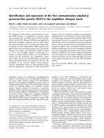
Figure 1: Telomere structure.
The fluorescence image shows the location of a telomere within a chromosome. Mammalian telomeres
consist of TTAGGG repeats with a single-stranded 3’ overhang of the G-rich strand. (A) Specific
protein complexes bind to the double- and single-stranded telomeric DNA. (B) The single-stranded
overhang can invade the double-stranded portion of the telomere, forming protective loops — such as t-
loops and D-loops — at the invasion site. (C) The telomerase complex (which contains the telomerase
RNA template and the reverse transcriptase (TERT) interacts with the overhang and is regulated by
telomeric proteins. Other factors that can interact with telomeres are listed. Bidirectional arrows
indicate interactions (Verdun and Karlseder, 2007).
5
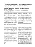
Figure 2: The telomeric end replication problem.
The replication forks move in opposite directions. Since DNA polymerases only elongate in the 5′ to 3′
direction, each fork contains a leading and a lagging strand. Lagging strand synthesis cannot be
completed because the removal of primers thus causing net loss of DNA base pairs from the lagging
strand (Hug and Lingner, 2006).
6
Studies demonstrated the formation of tumours in mice with telomere dysfunction
(Artandi et al., 2000; Blasco et al., 1997; Chin et al., 1999; Rudolph et al., 1999).
Following this, several studies have highlighted the role of telomeres in inducing
genomic instability and thus promoting tumourigenesis (de Lange, 2005; Desmaze et al.,
2003; Meeker and Argani, 2004; Meeker et al., 2004; Murnane, 2006; O'Hagan et al.,
2002). Hence, when telomeres are shortened to a critical length or when the secondary
structure is compromised, the telomeres are unable to effectively protect the terminal
ends. This causes the cell to elicit a DNA damage response, leading to cell cycle arrest
and subsequently cell death (Wright and Shay, 1992). Telomere protection and
maintenance are essential for prevention of genomic imbalances through BFB cycles
[reviewed in (Feldser et al., 2003)], a major cause of structural chromosomal instability.
These BFB cycles permit the accumulation of gross changes in the genome that underlie
tumourigenic progression (Figure 3).
1.1.1.1.2 Telomere dysfunction and tumourigenesis