Anti inflammatory effects of inhibitors of the NF kb pathway in the mouse asthma model
Bạn đang xem bản rút gọn của tài liệu. Xem và tải ngay bản đầy đủ của tài liệu tại đây (10.38 MB, 206 trang )
ANTI-INFLAMMATORY EFFECTS OF INHIBITORS OF THE
NF-κB PATHWAY IN THE MOUSE ASTHMA MODEL
BAO ZHANG
(M.Med.)
A THESIS SUBMITTED FOR THE DEGREE OF DOCTOR OF
PHILOSOPHY
DEPARTMENT OF PHARMACOLOGY
NATIONAL UNIVERSITY OF SINGAPORE
2008
ii
ACKNOWLEDGEMENTS
First and foremost, I would like to deeply thank my supervisor Professor Wong
Wai-Shiu Fred for his guidance and assistance through my Ph.D studies. Without
his help and encouragement, I definitely could not overcome so many obstacles in
the projects. His attitude and discipline will encourage me to continue the research
work in the future.
I would also like to thank Professor Bernard Leung for his invaluable advice and
efforts on my research works.
I am grateful to Amy Lin, Shuhui, Shouping, Ryan, all colleagues in our lab, and
friends who helped me in the experiments, shared me with their experience, and
supported me.
Thanks to National University of Singapore for providing me chances of studying
in Singapore.
Finally, I would like to extend my sincere gratitude to my parents, my wife, my
brother, and sister in law for their endless love, support, and patience all the time.
Bao Zhang
July 2008
iii
TABLE OF CONTENTS
ACKNOWLEDGEMENTS ii
TABLE OF CONTENTS iii
SUMMARY vii
LIST OF TABLES ix
LIST OF FIGURES x
LIST OF ABBREVIATIONS xiii
LIST OF PUBLICATIONS AND CONFERENCE ABSTRACTS xvi
1. INTRODUCTION 1
1.1. Asthma 2
1.1.1. Epidemiology of asthma 2
1.1.2. Susceptibility genes of asthma 3
1.1.3. Pathophysiology of asthma 4
1.1.3.1. Mast cells 8
1.1.3.2. Eosinophils 9
1.1.3.3. T lymphocytes 13
1.1.3.4. B lymphocytes 16
1.1.3.5. Epithelial cells 17
1.1.3.6. Mucus hypersecretion 20
1.1.3.7. Airway hyperresponsiveness 21
1.1.4. Current therapy for asthma 24
iv
1.1.5. New therapy for asthma 27
1.2. NF-κB signaling pathway 30
1.2.1. Introduction of the NF-κB pathway 30
1.2.2. Role of the NF-κB pathway in allergic inflammation 39
1.3. Inhibitors of NF-κB signaling cascades 40
1.3.1. The GSK-3β inhibitor 41
1.3.1.1. The GSK-3 pathway 41
1.3.1.2. GSK-3β and the NF-κB pathway 42
1.3.1.3. GSK-3β inhibitors 44
1.3.2. Andrographolide 48
1.3.2.1. Introduction of andrographolide 48
1.3.2.2. Andrographolide and the NF-κB pathway 51
1.4. The animal model of asthma 52
2. RATIONALE AND OBJECTIVES 55
3. MATERIALS AND METHODS 58
3.1. Materials and reagents 59
3.2. Asthma mouse model 61
3.3. Measurement of AHR 62
3.4. Collection of bronchoalveolar lavage (BAL) fluid from mice 66
3.5. Total and differential BAL fluid cell counts 66
3.6. ELISA 67
v
3.6.1. Cytokines and chemokine levels in BAL fluid 67
3.6.2. Immunoglobulin levels in serum 69
3.7. Histology 70
3.8. Western blotting 72
3.9. Reverse Transcription-Polymerase Chain Reaction (RT-PCR) 73
3.10. Alanine and aspartate aminotransferase assay 74
3.11. NF-κB transcription factor assay 74
3.12. Cell cultures 76
3.12.1. Lymphocyte recall experiments 76
3.12.2. Normal human bronchial epithelial cells 77
3.12.3. A549 cells 77
3.13. Statistical analysis 78
4. ANTI-INFLAMMATORY EFFECTS OF A GLYCOGEN SYNTHASE
KINASE-3Β INHIBITOR, TDZD-8, IN A MOUSE ASTHMA MODEL 79
4.1. Results 80
4.1.1. Effects of TDZD-8 on OVA-induced eosinophil recruitment
in BAL fluid 80
4.1.2. Effects of TDZD-8 on OVA-induced pulmonary cell
infiltration and mucus production 80
4.1.3. Effects of TDZD-8 on cytokine levels in BAL fluid 84
4.1.4. Effects of TDZD-8 on serum IgE levels 84
4.1.5. Effects of TDZD-8 on lung mRNA expression of
inflammatory markers 89
4.1.6. Effects of TDZD-8 on OVA-induced AHR in mice 92
4.1.7. Immunoblot analysis of lung NF-κB p65 92
vi
4.1.8. Effects of TDZD-8 on TNF-α stimulated human bronchial
epithelial cells 95
4.2. Discussions 99
5. ANTI-INFLAMMATORY EFFECTS OF ANDROGRAPHOLIDE IN A
MOUSE ASTHMA MODEL 108
5.1. Results 109
5.1.1. Effects of andrographolide on OVA-induced inflammatory
cell recruitment in BAL fluid 109
5.1.2. Effects of andrographolide on OVA-induced airway cell
infiltration and mucus production 112
5.1.3. Effects of andrographolide on cytokine levels in BAL
fluid 116
5.1.4. Effects of andrographolide on serum Ig levels 116
5.1.5. Effects of andrographolide on antigen recall in bronchial
lymph node cells 120
5.1.6. Effects of andrographolide on lung mRNA expression of
inflammatory markers 124
5.1.7. Effects of andrographolide on OVA-induced AHR in
mice 127
5.1.8. Effects of andrographolide on TNF-α-induced NF-κB
activation in A549 cells 128
5.1.9. Effect of andrographolide on NF-κB DNA-binding activity in
vivo 132
5.1.10. Effects of andrographolide on activities of MAP kinases in
vitro 136
5.2. Discusssion 139
6. CONCLUSION 154
7. REFERENCES 157
vii
SUMMARY
The NF-κB family is a central player in coordinating both innate and
adaptive immunity and is involved in the regulation of a broad array of genes in
response to diverse stimuli. The NF-κB family also plays a key role in the
initiation and development of asthma. Because the NF-κB transcription factors are
central to both normal biological functions and pathological conditions, absolute
inhibition of NF-κB per se may not be a safe approach. Rather, appropriate and
specific inhibition of signaling molecules that regulate NF-κB activity may be an
effective anti-inflammatory strategy for asthma. The objectives of my thesis
project were to examine the potential anti-inflammatory effects of a GSK-3β
inhibitor, namely TDZD-8, and a herbal medicinal, namely andrographolide in a
mouse asthma model and elucidate their mechanisms in the regulation of NF-κB
pathway.
BALB/c mice sensitized and challenged with ovalbumin developed allergic
airway inflammation. Intravenous administration of TDZD-8 significantly (P <
0.05) inhibited ovalbumin-induced increases in total cell counts, eosinophil counts,
IL-5, IL-13, and eotaxin levels in bronchoalveolar lavage fluid, and OVA-IgE in
serum. In addition, TDZD-8 reduced ovalbmuin-induced increase in mRNA levels
of inflammatory molecules, infiltration of inflammatory cells, and mucus
hypersecretion in lungs. TDZD-8 also suppressed airway hyperresponsiveness to
methacholine in mice. Western blotting of the whole lung and human bronchial
viii
epithelial cell showed that TDZD-8 may exert its anti-inflammatory effects by
inhibiting the phosphorylation of p65.
Andrographolide attenuated inflammatory cell counts, IL-4, IL-5, IL-13,
and eotaxin levels in bronchoalveolar lavage fluid, concentration of total IgE,
OVA-IgE, OVA-IgG1 in the serum, and expression of inflammatory molecules in
the lung, in a mouse asthma model. Andrographolide also suppressed OVA-
induced infiltration of inflammatory cells and mucus hypersecretion in the lungs,
and OVA-induced airway hyperresponsiveness to methacholine. Western blotting
and TransAM assay suggested that andrographolide may exert its anti-
inflammatory effects by inhibiting the phosphorylation of IKKβ and suppressing
the DNA-binding activity of p65.
Taken together, these present findings implicate that appropriate and
specific inhibition of signaling molecules that regulate NF-κB pathway may have
therapeutic potential for the treatment of allergic airway inflammation.
ix
LIST OF TABLES
Table Title Page
1.1 Susceptibility genes identified for asthma 5
1.2 Knockout mouse models for the NF-κB pathway 34
3.1 Primer Sets for RT-PCR 75
x
LIST OF FIGURES
Figure Title Page
1.1 Schematic diagram of pathogenesis of asthma 6
1.2 Roles of epithelium on innate and adaptive immunity 18
1.3 Members of NF-κB family 31
1.4 Canonical and non-canonical pathways of the NF-κB family 35
1.5 The functions of NF-κB in hematopoietic differentiation 38
1.6 The Structure of TDZD-8 46
1.7 Proposed binding mode of TDZD-8 to GSK-3β 47
1.8 Andrographis paniculata 49
1.9 Major active components of andrographis paniculata 50
3.1 Aerosol delivery system 63
3.2 The Buxco system 64
3.3 Type of cells found in BAL fluid of mice 68
4.1 Differential cell counts in BAL fluid 81
4.2 Effects of TDZD-8 on BAL fluid cell infiltration 82
4.3 Effects of TDZD-8 on peripheral blood mononuclear cell (PBMC) 83
4.4 A-D,I Effects of TDZD-8 on lung tissue inflammatory cell infiltration 85
4.4 E-H, J Effects of TDZD-8 on mucus production 86
4.5 Effects of TDZD-8 on Th2 cytokines levels in BAL fluid 87
4.6 Effects of TDZD-8 on eotaxin level in BAL fluid 88
xi
4.7 Effects of TDZD-8 on serum IgE production 90
4.8 Effects of TDZD-8 on pulmonary mRNA expression of
inflammatory markers 91
4.9 Effects of TDZD-8 on airway resistance 93
4.10 Effects of TDZD-8 on airway dynamic compliance 94
4.11 Effects of TDZD-8 on NF-κB subunit p65 phosphorylation
in lung tissue 96
4.12 Effects of TDZD-8 on TNF-α-induced phosphorylation of p65 in
normal human bronchial epithelial cells 97
4.13 Effects of TDZD-8 on TNF-α-induced expressions of
proinflammatory cytokines in normal human bronchial epithelial cells 98
5.1 Differential cell counts in BAL fluid 110
5.2 Effects of andrographolide on BAL fluid cell infiltration 111
5.3 Effects of andrographolide on PBMC 113
5.4 A-D, I Effects of andrographolide on lung tissue inflammatory
cell infiltration 114
5.4 E-H, J Effects of andrographolide on mucus production 115
5.5 Effects of andrographolide on Th2 cytokines in BAL fluid 117
5.6 Effects of andrographolide on eotaxin level in BAL fluid 118
5.7 Effects of andrographolide on IFNγ level in BAL fluid 119
5.8 Effects of andrographolide on serum IgE production 121
5.9 Effects of andrographolide on serum IgG production 122
5.10 Effects of andrographolide on OVA-specific response in vitro 123
5.11 Effects of andrographolide on Con-A response in vitro 125
5.12 Effects of andrographolide on pulmonary mRNA expression
of inflammatory markers 126
xii
5.13 Effects of andrographolide on airway resistance 129
5.14 Effects of andrographolide on airway dynamic compliance 130
5.15 Effects of andrographolide on TNF-α induced NF-κB activation in
A549 cells 131
5.16 Effects of andrographolide on TNF-α induced translocation of p65 in
A549 cells 133
5.17 Effects of andrographolide on TNF-α-induced p65 DNA-binding activity
in A549 cells 134
5.18 Effects of andrographolide on p65 DNA-binding activity in lung tissue 135
5.19 Effects of andrographolide on the activities of serum ALT and AST 137
5.20 Effects of andrographolide on TNF-α-induced MEK and ERK activation
in A549 cells 138
xiii
LIST OF ABBREVIATIONS
AHR airway hyperresponsiveness
ALT alanine aminotransferase
AMCase acidic mammalian chitinase
AMV avian myeloblastosis virus
AP alkaline phospatase
AP-1 activator protein-1
APC antigen presenting cell
ASM airway smooth muscle
AST aspartate aminotransferase
ATP adenosine triphosphate
BAL bronchoalveolar lavage
BCA bicinchoninic acid
BCIP 5-bromo-4-chloro-3-indoyl-phosphate
bFGF basic fibroblast growth
BSA bovine serum albumin
cAMP adenosine 3′,5′-cyclic monophosphate
CBP CREB-binding protein
CCR C-C chemokine receptor
Cdyn dynamic compliance
Con-A concanavalin A
COPD chronic obstructive pulmonary disease
COX-2 cyclooxygenase-2
CS inhaled corticosteroid
DC dendritic cells
DEPC diethylpyrocarbonate
DMSO dimethyl sulphoxide
ELISA enzyme-linked immunosorbent assay
ECP eosinophilic cationic protein
ECL enhanced chemiluminescent
ECM extracellular matrix
ERK extracellular signal-regulated kinase
FACS fluorescence-activated cell sorter
FBS fetal bovine serum
FcεRΙ high affinity IgE receptor
FcεRΙΙ low affinity IgE receptor
FITC fluorescein isothiocyanate
GM-CSF granulocyte/macrophage colony-stimulating factor
GR glucocorticoid receptors
GRE glucocorticoid-response elements
GSK-3 glycogen synthase kinase-3
xiv
HDAC histone deacetylase
HEPES 4-(2-hydroxyethyl)-1-piperazineethanesulfonic acid
HRP horseradish peroxidase
ICAM-1 intercellular adhesion molecule-1
ICOS inducible costimulatory protein
IFN interferon
Ig immunoglobulin
IκB inhibitor of NF-κB
IKK inhibitor of NF-κB kinase
IL interleukin
iNOS inducible nitric oxide synthase
JAK janus kinase
JNK c-Jun NH2-terminal kinase
LPS lipopolysaccharide
LT leukotriene
MAPK mitogen-activated protein kinase
MBP major basic protein
MCP-1 monocyte chemoattractant protein
MEK MAP/ extracellular signal-regulated kinase kinase
MHC major histocompatibility complex
MMP matrix metallopeptidase
NBT nitroblue tetrazolium
NEMO nuclear factor-κB essential modulator
NFAT nuclear factor of activated T-cells
NF-κB nuclear factor-κB
Nrf2 nuclear factor-E2-related factor-2
OVA ovalbumin
PAGE polyacrylamide gel electrophoresis
PAF platelet-activating factor
PAMP pathogen-associated molecular pattern
PAS periodic acid-schiff
PBS phosphate-buffered saline
PCR polymerase chain reaction
PDE4 phosphodiesterase type 4
PE phycoerythrin
PMBC peripheral blood mononuclear cells
PVDF polyvinylidene difluoride
RANTES regulated upon activation, normal t-cell expressed, and
secreted
REL v-rel reticuloendotheliosis viral oncogene homolog
RHD rel homology domain
RI lung resistance
xv
RT reverse transcription
STAT signal transducers and activators of transcription
SCF stem cell factor
SDS sodium dodecyl sulfate
SEM standard error of the mean
TCR t-cell receptor
TDZD-8 4-benzyl-2-methyl-1,2,4-thiadiazolidine-3,5-dione
TEMED tetramethylethylenediamine
TGF transforming growth factor
Th2 t helper2
TLR toll-like receptor
TMB 3,3´,5,5´-tetramethylbenzidine
TNF-α tumor necrosis factor-α
TPL2 tumor progression locus 2
Treg regulatory T cell
TSLP thymic stromal lymphopoietin
TTBS tween-20 tris buffered saline
VCAM-1 vascular cell adhesion molecule-1
VEGF vascular endothelial growth factor
VLA-4 very late antigen-4
WNT wingless and int-1
xvi
LIST OF PUBLICATIONS AND CONFERENCE ABSTRACTS
Publications
Bao, Z., Lim, S. M., Liao, W. P., Lin, Y. Z., Thiemermann, C., Leung, B. P., and
Wong, W. S. (2007). Glycogen synthase kinase-3beta inhibition attenuates asthma
in mice. Am J Respir Crit Care Med 176, 431-438.
Lai, W. Q., Goh, H. H., Bao, Z., Wong, W. S., Melendez, A. J., and Leung, B. P.
(2008). The role of sphingosine kinase in a murine model of allergic asthma. J
Immunol 180, 4323-4329.
Liao, W., Bao, Z., Cheng, C., Mok, Y. K., and Wong, W. S. (2008). Dendritic
cell-derived interferon-gamma-induced protein mediates tumor necrosis factor-
alpha stimulation of human lung fibroblasts. Proteomics 8, 2640-2650.
Bao, Z., Guan, S.P., Cheng, C., Wu, S. L., Leung, B. P., and Wong, W. S. The
anti-inflammatory effects of andrographolide in a mouse asthma model. (In
revision 2008)
Conference Abstracts
Bao, Z., Lim, S. H., Thiemermann, C., Wong, W. S. (2006). Anti-inflammatory
effects of glycogen synthase kinase-3 beta inhibitor in a mouse asthma model.
Acta Pharmacologica Sinica 27, 270-270.
xvii
Wong, W. S., Bao, Z., Lim, S. H., Thiemermann, C., (2006). Anti-inflammatory
effects of glycogen synthase kinase-3 beta inhibitor in a mouse asthma model.
Respirology 11, A137-A137.
Bao, Z., Lim, S., Lin, Y., Leung, B. P., Thiemermann, C., Wong, W. S. (2007).
Anti-inflammatory effects of GSK-3B inhibitor TDZD-8 in a mouse model of
asthma. Inflammation Research 56, S416-S416.
Liao, W. P., Bao, Z., Cheng, C., Wong, W. S. (2008). Dendritic cell-derived
interferon-γ-induced protein mediates tumor necrosis factor-α stimulation of
human lung fibroblasts. 1st International Singapore Symposium of Immunology.
1
1. INTRODUCTION
2
1.1. Asthma
1.1.1. Epidemiology of asthma
Asthma is a common chronic disease which affects around 300 million people
of all ages and ethnic backgrounds. The prevalence of asthma is high in
industrialized countries such as United Kingdom (15.3%), New Zealand (15.1%),
Australia (14.7%), and United States (10.9%) when compared with non-
industrialized countries, for instance, Mexico (3.3%), India (3%), and Iran (5.5%)
(Masoli et al., 2004). A dramatic increase in the prevalence of asthma was
reported in many countries from the 1960s to the 1990s (Eder et al., 2006). One
popular theory which explains the rising prevalence of asthma today, especially in
industrialized societies, is the “hygiene hypothesis”. This hypothesis contributes
the rising prevalence of asthma to the decreasing infection rates in children due to
cleaner environments in industrialized countries. It is derived from the observation
that the risk of hay fever varies inversely with family size, and is further
supported by the phenomenon that exposure to microbial products released by
farm animals exerts a protective role against the development of asthma (Strachan,
1989). Although asthma is generally not a life-threatening disease, mortality rates
are still considerable, accounting for about 1 in every 250 deaths worldwide
(Masoli et al., 2004). Furthermore, asthma is the third leading cause of
hospitalization, exceeded only by pneumonia and injuries, among persons under
18 years of age in the United States (Eder et al., 2006). High hospitalization fee,
3
together with high incidence, lead to asthma related costs exceeding those of
tuberculosis and acquired immunodeficiency syndrome (AIDS) combined,
accounting for 1% to 2% of the total health-care budget in industrialized countries
(Braman, 2006). Furthermore, the economic burden of asthma disproportionately
affects uncontrollable asthma patients. In both western and developing countries,
10% to 20% uncontrollable asthma patients are responsible for approximately
50% of direct or indirect costs, whereas 70% of mild asthma patients account for
only 20% of total costs (Beasley, 2002; Braman, 2006). In summary, the rising
prevalence, mortality, and high economic burden of asthma are having huge
effects on the health-care systems worldwide. Therefore, more research should be
done to better understand the pathophysiology of asthma and further explore
potentially effective therapies for this disease.
1.1.2. Susceptibility genes of asthma
Both genetic background (atopy) and environmental factors (allergens, viruses,
and occupational exposures) contribute to the initiation and development of
asthma (Busse and Lemanske, 2001). In developed countries, 30% of the
population is atopic, whereas only 10-12% of the population suffers from asthma,
suggesting that allergic responses to inhaled allergens are considered as risky
factors rather than causative factors of asthma (Hammad and Lambrecht, 2008).
Therefore, it is critical to identify both genetic and environmental factors and their
interactions that might contribute to the development of asthma in a sensitized
4
subject. It has been more than 10 years since the first genome-wide screen for
asthma and atopy susceptibility loci (Ober and Hoffjan, 2006). In a decade, rapid
advances in identifying susceptibility genes for asthma have uncovered numerous
genes which are crucial to the pathogenesis of asthma (Table 1.1) (Vercelli, 2008).
These asthma susceptibility genes are involved in probably all aspects of asthma
including innate immunity and immunoregulation, T helper 2 (Th2) cell
differentiation and effector function, epithelial biology and mucosal immunity,
lung function, airway remodeling, and disease severity (Vercelli, 2008). Despite
the apparent achievements in asthma genetics, there remain huge confusing
discrepancies about the linkage between genotypes and phenotypes of asthma.
Both gene-environment and gene-gene interactions might dramatically change the
impact of a specific gene on the complex phenotypes, as are supported by
epidemiological studies of asthma (Moffatt et al., 2007; Vercelli, 2008). Finally,
understanding of asthma genetics not only helps us unravel the pathogenesis of
this disease, but may also provide information for pharmacogenetic approaches,
leading to individualization of treatments with high efficacy and low side effects
for patients (Hall, 2006).
1.1.3. Pathophysiology of asthma
Asthma is a chronic airway disease which is characterized by airway
inflammation, mucus hypersecretion, and airway hyperresponsiveness (AHR)
(Figure 1.1) (Busse and Rosenwasser, 2003).
5
Table 1.1 Susceptibility genes identified for asthma (Adaped from Vercelli, 2008)
Gene Chromosome Function and pathway
GSTM1 1p13.3 Environmental and oxidative stress-detoxification
FLG 1q21.3 Epithelial barrier integrity
IL10 1q31-q32 Immunoregulation
CTLA4 2q33 T-cell-response inhibition and immunoregulation
IL-13 5q31 Th2 effector function
IL-4 5q31.1 Th2 differentiation and IgE induction
CD14 5q31.1 Innate immunity-microbial recognition
SPINK5 5q32 Epithelial serine protease inhibitor
ADRB2 5q31-q32 Bronchial smooth-muscle relaxation
HAVCR1 5q33.2 T-cell-response regulation-HAV receptor
LTC4S 5q35 Cysteinyl leukotriene biosynthesis, inflammation
LTA 6q21.3 Inflammation
TNF-α 6q21.3 Inflammation
HLA-DRB1 6q21 Antigen presentation
HLA-DQB1 6q21 Antigen presentation
HLA-DPB1 6q21 Antigen presentation
GPRA 7q14.3 Regulation of cell growth and neural mechanisms
NAT2 8p22 Detoxification of drugs and carcinogens
FCERIB 11q13 High-affinity Fc receptor for IgE
CC16 11q12.3-q13.1 Epithelium-derived anti-inflammatory protein
GSTP1 11q13 Environmental and oxidative stress,
detoxification
IL-18 11q22.2-q22.3 Induction of IFNγ and TNF
STAT6 12q13 IL-4 and IL-13 signalling
NOS1 12q24.2-q24.31
Nitric oxide synthesis — cell–cell communication
CMA1 14q11.2 Mast-cell chymotryptic serine protease
IL-4R 16p12.1-p12.2 α-chain of the IL-4 and IL-13 receptors
CCL11 17q21.1-q21.2 Epithelium-derived eosinophil chemoattractant
CCL5 17q11.2-q12 Monocyte, T-cell and eosinophil chemoattractant
ACE 17q23.3 Inactivation of inflammatory mediators
TBXA2R 19p13.3 Smooth-muscle contraction, inflammation
TGFB1 19q13.1 Immunoregulation, cell proliferation
ADAM33 20p13 Cell–cell and cell–matrix interactions
GSTT1 22q11.23 Environmental and oxidative stress,detoxification
6
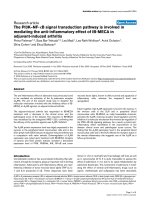
Figure 1.1 Schematic diagram of pathogenesis of asthma. Definition of
abbreviations: ICAM-1 = intercellular adhesion molecule-1; TSLP = thymic
stromal lymphopoietin; VCAM-1 = vascular cell adhesion molecule-1; VLA-4 =
very late antigen-4.
7
Inhaled allergens, often the initiator of the asthma, are taken up by lung dendritic
cells (DC). Then, under the presence of low concentration toll-like receptor (TLR)
agonist within the allergen itself or the presence of proteolytic activity within the
allergen, DCs are activated and migrate to the draining lymph nodes where they
present allergens to naïve CD4+ T cells, promoting the differentiation of naïve
CD4+ T cells into Th2 cells (Hammad and Lambrecht, 2008). Th2 cells have a
central role in the pathogenesis of asthma and produce an array of cytokines such
as interleukin-4 (IL)-4, IL-5, IL-9, and IL-13. IL-4 is mainly responsible for the B
cells isotype switching. Under the presence of IL-4, IL-13, and other molecules, B
cells undergo isotype switching and synthesize IgE which is released into
circulation, eventually binding to high affinity IgE receptors (FcεRΙ) on the
surface of mast cells. Crosslinking of antigens, IgE, and FcεRΙ on mast cells lead
to the degranulation of mast cells and the release of mediators including histamine,
leukotrienes, and cytokines, causing acute bronchoconstriction (Busse and
Lemanske, 2001). IL-5 is the most critical cytokine mediating the differentiation,
activation, and survival of eosinophils, which may contribute to both
inflammation and airway remodeling in asthma (Simon and Simon, 2007). IL-9
could promote the proliferation of mast cells. Furthermore, IL-13 is the most
pivotal effector of all Th2 cytokines, inducing almost all pathophysiological
features of asthma comprising airway inflammation, AHR, mucus oversecretion,
and airway remodeling (Wills-Karp, 2004). In addition, adhesion molecules, their
8
receptors, and chemokines are vital for the transmigration of inflammatory cells
from circulation into inflammatory sites in response to allergic provocation
(Rosenberg et al., 2007). Infiltration of inflammatory cells and the release of Th2
cytokines may lead to transient and reversible AHR, whereas multiple structural
changes in the airway, known as airway remodeling, could contribute to persistent
AHR (Cockcroft and Davis, 2006).
1.1.3.1. Mast cells
Mast cells arise from CD34+ pluripotent stem cells in the bone marrow,
circulate in the blood as precursors, and then undergo tissue-specific maturation.
In tissue, mast cells mature under the influence of stem cell factor (SCF) and its
receptor CD117. In addition to SCF, mast cell growth and differentiation is
manipulated by various cytokines, including IL-3, IL-4, IL-6, IL-9, IL-10, and
nerve growth factor (Brown et al., 2008).
Mast cells are activated by the crosslinking of FcεRΙ or by non-IgE-mediated
pathways through complement receptors or toll-like receptors. Upon activation,
mast cells release an array of mediators, cytokines, and chemokines. The pattern
of mediator release is modulated by cytokines, growth factors, and the
microenvironment (Brown et al., 2008). For instance, IL-4 could augment FcεRΙ-
mediated responses by mast cells (Bischoff et al., 1999), whereas, IL-10 and
transforming growth factor-β (TGF-β), produced by regulatory T cells could
diminish those reactions (Royer et al., 2001).