Proline rich acidic protein 1 in life and death 5
Bạn đang xem bản rút gọn của tài liệu. Xem và tải ngay bản đầy đủ của tài liệu tại đây (1.11 MB, 7 trang )
138
pathways were activated in this caspase-dependent apoptosis induced by the
inhibition of PRAP1 in response to 5-FU treatment.
Collectively, our results indicate that PRAP1 repression enhanced caspase-
dependent apoptosis in cells treated with 5-FU. These results strongly suggest that
PRAP1 knockdown increases the sensitivity of cells to DNA damage by genotoxic
agents and modulates cell fate by increasing cell death via caspase 8- and 9-
dependent apoptosis
3.11 Other m echanisms em ployed b y PRAP1 inhibition is e nhancing cell
death
3.11.1 Effect of PRAP1 on cytoskeleton
3.11.1.1 Effects of PRAP1 on cellular morphology
Our results showed that repression of PRAP1 in p53-/- cells enhanced the
cell death at high dose of 5-FU independent of cell cycle arrest interference,
suggesting the involvement of other mechanism in addition to its role in p21
regulation. As the PRAP1 knockdown cells showed a distinct morphology, we
investigated whether PRAP1 plays a role in the maintenance of cytoskeleton. As
shown in phase contrast pictures of Figure 3.67, the morphology of cells with
perturbed PRAP1 expression was distinctively different from that of their
respective controls. In cells with overexpression of PRAP1, a more elongated
morphology was displayed, and the volume of the cells was much reduced as
compared to control. The cells grew in less-defined clusters, and were more
scattered. The nuclei of cells with overexpression of PRAP1 were also smaller and
disoriented, compared to the nuclei of the control cells that were well-spaced in an
orderly manner. Conversely, in cells with PRAP1 knockdown, a more rounded
139
morphology was observed (Figure 3.67). The cellular volume was also reduced
and the organization of cells in a cluster was less orderly. These results indicated
that perturbation of PRAP1 expression either by overexpression or knockdown
resulted in cellular morphological changes.
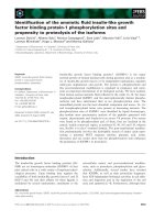
3.11.1.2 Effects PRAP1 on actin filament
To study the effect of PRAP1 on the organization of F-actin, an important
cytoskeletal protein, we stained F-actin with Phalloidin-Alexa Fluor 488. As
shown in Figure 3.67, the untransfected and vector control cells displayed an
extensive network of actin filaments. The actin microfilaments are mainly found
in cortical structures forming characteristic ruffles. There are also numerous long
stress fibers that traversed the cells. Interestingly, in cells with PRAP1
overexpression, there was an obvious reorganization of actin filaments. There was
a significant reduction in the amount of stress fibers formed. F-actin was found to
accumulate at the periphery of the cell.
The organization of actin in the control siRNA treated cells was similar to
that of the untransfected and vector control cells, indicating that transfection of
siRNA did not affect the actin network. Conversely, in cells with PRAP1
knockdown, reorganization of actin filaments were observed. The architecture of
actin network was severely disrupted. There was also a marked reduction in the
number of stress fibers formed. The actin microfilaments were highly
concentrated on the periphery of the cells. Both overexpression and knockdown of
PRAP1 gene resulted in similar effect on the actin network, suggesting that
PRAP1 may act as a biphasic protein. Collectively, these results indicated that
perturbation of PRAP1 expression affects the architecture of actin network,
resulting in a change in cellular morphology.
140
Figure 3.67 Effects of PRAP1 on actin network
Representative immunofluorescence images of F-actin in HCT 116 cells stably expressing PRAP1 (PRAP1) and its empty vector control stable
cell line (Vector), or after transfection of control siRNA and PRAP1 siRNA for 72 hours. F-actin was stained using Palloidine conjugated to
FITC (Green) and nuclear was counter stained with Hoechst (Blue).
141
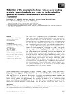
3.11.1.3 Effects of PRAP1 on microtubules
To further study the effect of PRAP1 on cytoskeleton, we also looked at
the other important cytoskeletal network, the microtubules. As shown in Figure
3.68, the vector control cells displayed a very organized network of tubulin with a
distinct microtubule organizing center (MTOC). Similar findings were also
observed with the control siRNA treated cells. Interestingly, in the cells with
overexpression of PRAP1, there was no obvious MTOC found. The intensity of
staining was also much reduced than that of the control cells. The architecture of
the microtubulin network was not well-organized as that of the control cells.
Similar findings were also observed in the cells with PRAP1 knockdown. These
results indicated that PRAP1 affects the organization of microtubules with a major
defect in the formation of MTOC.
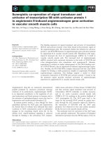
3.11.2 PRAP1 interacts with Hsp 70
In order to study the possible mechanism employed by PRAP1 in
protecting the cells against genotoxic stress, we investigated its potential binding
partners. Briefly, cells were treated with 5-FU or UV and cell lysates were
harvested. Possible binding partners of PRAP1 were pulled down using
glutathione beads conjugated to GST-PRAP1. Proteins bound to GST-PRAP1
were then analyzed on SDS-PAGE gel and visualized using silver staining. Figure
3.69 shows a representative SDS-PAGE gel picture of the pulled down proteins.
Unique bands (indicated in arrow) from GST-PRAP1 beads alone or GST beads
alone were excised and processed for MS-MS analysis. After subjecting these
bands to peptide mass fingerprinting analysis, Hsp 70 was identified as a possible
142
Figure 3.68 Effects of PRAP1 on microtubulin network
Representative immunofluorescence images of microtubules in HCT 116 cells stably expressing PRAP1 (PRAP1) and its empty vector control
stable cell line (Vector), or after transfection of control siRNA and PRAP1 siRNA for 72 hours. Anti-tubulin antibody was used to stain the
microtubulin network (Green) and nuclear was counter stained with Hoechst (Blue).
143
binding partner for PRAP1 (Figure 3.71). To validate our pull-down result,
immunoprecipitation was performed. Briefly, cells were transfected with pcDNA-
PRAP1
Figure 3.69 GST-PRAP1 pull-down
Representative silver staining picture of pull-down assay with GST-PRAP1 using
HCT 116 lysate treated with 5-FU for 24 hours. Lane 1: GST beads alone; Lane 2:
GST-PRAP1 beads with lysate; Lane 3: GST-PRAP1 beads alone; Lane 4: protein
ladder. Unique bands (arrow) as compared to controls (the glutathione beads
conjugated with GST and the GST-PRAP1 bead) were excised for MS-MS
analysis.
144
Figure 3.70 Identification of PRAP1 binding protein by MS-MS
Representative figure showing the identification of PRAP1 binding proteins after
performing MS-MS.
Figure 3.71 PRAP1 physically interacts with Hsp 70
Representative western blot of PRAP1 after immunoprecipitation (IP) with Hsp
70 antibody. Total: Input lysate; IgG: IP with IgG antibody; Hsp 70: IP with Hsp
70 antibody.