Search for new types of deoxyribozymes and development of human topoisomerases inhibitors on the basis of oligonucleotides
Bạn đang xem bản rút gọn của tài liệu. Xem và tải ngay bản đầy đủ của tài liệu tại đây (3.41 MB, 202 trang )
1
PART I: SEARCH FOR NEW TYPES OF
DEOXYRIBOZYMES WITH SELF-CLEAVING
ACTIVITY UNDER HYDROLYSIS PATHWAY
2
CHAPTER 1
Introduction
Nature has spent more than 3 billion years to perfect the many thousands of enzymes
that are utilized in living cells. Yet this long and merciless process of evolution has
given rise to enzymes based on only two molecular formats-protein and RNA. In
opting to build biocatalysts, nature has made two excellent choices. Proteins exploit
the different chemistries offered by their constituent 20 amino acids to form an
incredible array of diverse structures and precisely configured active sites. So together
with their ability to adopt a seemingly endless array of tertiary structures in sequence-
directed fashion, proteins are well suited to serve as enzymes. The splendid catalytic
potential of proteins is exemplified by enzymes, such as orotidine 5’-phosphate
decarboxylase, which coverts substrate to product with a rate enhancement of 17
orders of magnitude over the corresponding uncatalyzed rate [1].
Although RNA’s role in modern biocatalysis might be an “accidental catalysts” from
life’s early evolutionary history [2], catalytic RNAs (ribozymes) are certainly capable
of generating impressive rate enhancements. For example, group I ribozymes catalyze
RNA splicing with a rate enhancement of ~13 orders of magnitude. Only eight classes
of naturally occurring RNA biocatalysts have been identified so far since the 1980s
when Thomas Cech made the first surprising discovery that RNA molecules are
capable of catalyzing reactions in the absence of any protein components [3]. In
contrast to proteins, the diversity of chemical functional groups in RNA is severely
limited, and this fact is widely considered as the most significant determinant that
constrains both the structural and catalytic potentials of RNA. Then why is RNA
3
provided with the catalytic activity of enzymes from evolution? The most important
reason is that RNA is capable of bringing its lesser chemical repertoire to bear on
catalysis by forming surprisingly intricate three-dimensional structures [4-6]. The
versatilities of RNA in forming intricate and functional structures are due to the
combined use of standard Watson-Crick base pairing, non-standard base pairing, and
a variety of non-covalent contacts involving phosphates, ribose oxygen and cationic
metals. A significant component for building complex RNA structures is the 2'
hydroxyl of ribose, as the oxygen atom of this group can act both as a donor and as an
acceptor for hydrogen bonding [7].
1.1 Classes of Ribozymes
Ribozymes, or catalytic RNAs, were first discovered in the laboratory of Tom Cech,
at the University of Colorado, in 1982. They found that the ribosomal RNA precursor
from Tetrahymena thermophila contained an intron, a nonencoding sequence that
interrupts the gene, and was capable of excising itself, in vitro, without any protein or
external energy source [8]. Shortly thereafter, Sidney Altman's group, at Yale
University, showed that the RNA component of RNase P, M1 RNA, from Escherichia
coli was likewise able to process tRNA precursors without any protein factors [9].
This seminal work ushered in a major research activity, and RNA catalysis was soon
found to be widespread in nature, occurring in plants, bacteria, viruses, and lower
eukaryotes.
Seven naturally occurring classes of catalytic RNAs have been identified to date
(hammerhead, hepatitis delta virus, hairpin, Neurospora Varkud satellite, group I
introns, group II introns and ribonuclease P), all of which catalyze cleavage or
4
ligation of the RNA backbone by transesterification or hydrolysis of phosphate groups.
The hammerhead, hepatitis delta virus (HDV), hairpin, and Neurospora Varkud
satellite (VS) ribozymes are small RNAs of 50–150 nucleotides that perform site-
specific self-cleavage [10–16]. The general mechanism of these self-cleavage
reactions is similar to that of many protein ribonucleases in which a 2’-oxygen
nucleophile attacks the adjacent phosphate in the RNA backbone, resulting in
cleavage products with 2’, 3’-cyclic phosphate and 5’-hydroxyl termini. Found in
viral, virusoid, or satellite RNA genomes, these small catalytic RNAs process the
products of rolling circle replication into genome-length strands [17].
5
Base
O
OO
P
O
O
-
O
-
O
O
OHO
HH
Base
H
HO
5'
3'
Base
O
O
P O
-
O
-
O
O
OHO
O
-
Base
5'
3'
O
1
3
O
H
H
4
Base
O
OO
P
O
O
-
O
O
OHO
HO
Base
5'
3'
+
A
B
Base
O
OHO
PO
O
-
O
O
-
O
OHO
Base
5'
3'
Base
O
OH
O
O
OHO
O
Base
5'
3'
+2
Nuc
O
H
1
OH
5
O
H
H
Base
O
OHO
PO
-
O
-
O
O
-
O
OHO
Base
5'
3'
2
3
O
4
Nuc
OH
P OO
O
-
Nuc
2
2
1
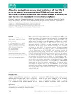
Figure 1-1. Two mechanistic classes of ribozyme phosphodiester cleavage. Circles
represent divalent cations and several ways that they may participate in catalysis.
Although most of the ions are shown participating in the transition state of the
reaction, they may also contribute to ground-state stabilization and substrate binding
(represented by M2, the square labeled 2). (A) Mechanism A (m
A
), observed in metal
catalyzed RNA hydrolysis and RNA cleavage by the hammerhead, hairpin, hepatitis
delta, Neurospora, and tRNA ribozymes. The attack of the 2’-OH group proceeds by
an in-line S
N
2 nucleophilic displacement with inversion of the configuration at
phosphorus [43]. The M2 square represents a divalent cation that promotes catalysis
by the hammerhead ribozyme. In addition to coordinating the nonbridging phosphate
oxygen, M2 may also stabilize the oxyanion nucleophile. (B) Mechanism B (m
B
)
found in catalysis by ribonuclease P, group I and group II intron ribozymes. The
exogenous nucleophile (Nuc), which can be water or hydroxyl functionality, attacks
the phosphorus center by means of S
N
2 displacement [136].The square labeled 3
represents M3, a divalent ion that promotes catalysis by the Tetrahymena ribozyme.
Group I introns, group II introns and ribonuclease P (RNase P) are larger, more
structurally complex ribozymes with several hundred nucleotides in length [18–20].
RNase P cleaves precursor RNA substrates at specific sites to generate functional 5’-
6
termini [21], and group I and II introns catalyze two-step self-splicing reactions [22–
24]. In these large ribozymes, the nucleophile and the labile phosphate are located on
different molecules or are greatly separated in sequence. Thus, the complex folds of
these RNAs serve to orient the nucleophile and phosphate to ensure accurate cleavage
or splicing.
1.1.1 Hammerhead ribozyme structure and catalysis
The hammerhead ribozyme was the first self-cleaving RNA to be discovered [25, 26],
the first ribozyme to be crystallized [27, 28], and has been the focus of more studies
than any other catalytic RNAs. Even so, a complete understanding of its reaction
mechanism still remains elusive [29-31]. Hammerhead and hairpin ribozymes are
found in opposite strands of the same plant virus satellite RNAs, and they catalyze
identical chemical reactions [32]. Nonetheless, the two ribozymes adopt different
structures and have different biochemical features, including different pH and metal-
cation dependencies and different proficiencies for catalyzing RNA ligation [33].
A minimal hammerhead contains three base-paired helices (helices I, II, and III)
around a core of conserved nucleotides. Two crystal structures of non-cleavable
variants of the hammerhead ribozyme have been solved, one with a DNA substrate
strand and the other with an RNA substrate containing a 2’O-methyl group at the
cleavage site [34, 35]. Both showed the three helices are arranged in a Y shape, as
predicted by fluorescence and native gel electrophoresis data [36, 37]. Stems II and III
are essentially coaxial, while helix I lies at a sharp angle to helix II. Backbone
distortions at the junction of helices II and III force nucleotide C17 to stack on stem I
rather than on stem III, placing it in the active site pocket at the three-helix junction.
7
The scissile phosphate on the 3’-side of C17 lies above a hairpin turn of the backbone
formed by the C3-A6 sequence. This CUGA turn is strikingly similar to that found in
the anti-codon loop of tRNA, which serves as a metal binding pocket [38].
Many experiments have been performed to determine the mechanism of hammerhead
self-cleavage and the role of divalent cations in the reaction. Sulfur substitution for
the scissile phosphate oxygens showed that there is inversion of configuration about
the phosphate during the reaction, indicating that the reaction proceeds by in-line
attack of the nucleophile [32, 39-40]. Further, in most of the experiments in which
sulfur was substituted for the pro-R
p
non-bridging oxygen of the scissile phosphate,
the reduced activity was rescued by addition of a thiophilic metal ion, suggesting that
a metal ion directly coordinates this oxygen in the transition state [40-45]. The
reaction rate increases linearly with pH, indicating that the nucleophile is activated by
a hydroxide ion [46]. Either a metal ion hydroxide deprotonates the 2’-hydroxyl
directly, or a metal ion coordinated to the 2’-hydroxyl increases the acidity of the 2’-
oxygen, rendering it susceptible to attack by a hydroxide ion from solution [46, 47].
The ion coordinated to the pro-R
p
phosphate oxygen could perform either of these
functions. If the hammerhead uses a two-metal ion mechanism for self-cleavage, then
an additional directly coordinated ion should stabilize the leaving group oxygen [48,
49]. While sulfur substitution for the leaving group oxygen has failed to identify such
a metal ion, other observations support its existence [47, 50-51]. Thus, there is debate
over the number of divalent cations directly involved in hammerhead catalysis.
Strikingly, recent experiments demonstrate that the ribozyme can function in the
complete absence of divalent cations at extremely high ionic strengths [52]. This
result showed that divalent cations are not essential cofactors in the reaction, though
8
at least one directly coordinated magnesium ion appears to be involved in catalysis
under most conditions in vitro.
1.1.2 Hairpin ribozyme structure and catalysis
Another catalytic RNAs domain found in pathogenic plant virus satellite RNAs is the
hairpin motif. Similar to the other small ribozymes, hairpin catalysts cleave
concatameric precursor molecules into mature satellite RNA during rolling-circle
replication, giving rise to a 2’-3’-cyclophosphate and a free 5’-OH terminus.
Depending on reaction conditions, the hairpin ribozyme may also favour RNA
ligation over cleavage [53]. Up to now, three different hairpin ribozymes have so far
been found in nature, of which the one from satellite RNA associated with tobacco
ring spot virus is the best characterized [54, 55]. The other two hairpin ribozymes,
isolated from different satellite viruses, showed the sequence variations that preserve
the overall structure of the molecules [56].
In naturally occurring hairpin ribozymes, the catalytic entity is a part of a four-helix
junction. A minimal catalytic motif, containing approximately 50 nucleotides, has
been identified that can be used for metal-ion dependent cleavage reactions in trans. It
consists of two domains, each of them harbouring two helical regions separated by an
internal loop, connected by a hinge region. One of these domains results from the
association of 14 nucleotides of a substrate RNA with the ribozyme via base-pairing.
In recent work, crystal structures of hairpin ribozymes bound to a modified, un-
cleavable substrate molecule, a transition state mimic, and a product complex have
been obtained [57,58]. These structures and related biochemical data showed that the
9
active site contains no tightly bound, well ordered metal ions at the site of cleavage.
Catalytic activity of the hairpin ribozyme thus results from distortion and precise
orientation of the substrate RNA and general acid base catalysis by nucleotides in
neighborhood without involvement of metal ions in catalysis [55]. The requirement
for metal ions may be explained by a significant role in the folding process [59, 60]. It
has also been shown that catalytic activity of the hairpin ribozyme can be supported
by spermine, the major polyamine in eukaryotic cells [61].
1.1.3 Hepatitis delta virus and Varkud Satellite ribozymes
The hepatitis delta virus ribozyme was found in a satellite virus of hepatitis B virus, a
major human pathogen [62]. Both the genomic and the antigenomic strands express
cis-cleaving ribozymes of ~85 nucleotides that differ in sequence but fold into similar
secondary structures. A crystal structure of the ribozyme has been determined [63], in
which five helical regions are organized by two pseudoknot structures. There is strong
evidence that the catalytic mechanism of the hepatitis delta virus ribozyme involves
the action of a cytosine base within the catalytic centre as a general acid-base catalyst.
The hepatitis delta ribozyme displays high resistance to denaturing agents like urea or
formamide.
The Varkud Satellite (VS) ribozyme is a 154 nucleotides long catalytic entity that is
transcribed from a plasmid discovered in the mitochondria of certain strains of
Neurospora [64]. The VS ribozyme is the largest of the known nucleolytic ribozymes
and the only one for which there is no crystal structure available to date. The global
structure has been determined by solution methods, particularly Fluorescence
10
Resonance Energy Transfer (FRET), which revealed a formal H shape of the five
helical segments [65].
1.1.4 General properties of introns
Introns, or intervening sequences (IVS), are nonencoding sequences that interrupt the
coding, exon, sequences. These introns must be removed, at the RNA level, in order
for the gene to be expressed functionally. There are five major categories of introns
and splicing mechanisms. These consist of nuclear tRNA introns [66], archaeal
introns [67], nuclear mRNA introns [68], and the group I and group II introns [69, 70].
Of these introns, some members of the group I and group II are clearly capable of
catalyzing their own excision, in vitro, in an RNA-catalyzed fashion.
1.1.4.1 Group I introns
Group I introns are found in the nucleus and organelles of eukaryotes, as well as some
prokaryotes and bacteriophages. These introns are composed of a strictly conserved
core region essential for catalysis and moderately conserved peripheral regions that
enhance their catalytic activity [71-74]. The mechanism of the catalytic reaction with
group I introns involves excision from precursor RNAs through a two-step splicing
reaction.
A prerequisite for splicing is the binding of an exogenous guanosine (exoG) cofactor
to a pocket in the catalytic core of the intron, referred to as G binding site. During the
first step of splicing, the cofactor attacks the 5’-splice site (SS) and attaches to the
intron, resulting in the release of the upstream exon. The exoG leaves the G-binding
site and is replaced by the last nucleotide of the intron, which is always a guanosine
11
(denoted as ωG). The second step is initiated by an attack by the 3’-end of the
released exon on the 3’ SS, which results in ligation of the exons and release of the
intron RNA. Successful catalysis is dependent on the correct folding of the intron [75-
77]. Group I intron splicing requires divalent metal ions for proper folding and
catalysis [77]. Mg
2+
coordination to substrate oxygen atoms activates the nucleophile,
stabilizes the scissile phosphate and stabilizes the developing charge on the leaving
group. The proton of the 2’-OH at the cleavage site is shared between the 2’-and 3’-
oxygens in the transition state [78].
1.1.4.2 Group II introns
Group II introns are found in low frequencies in the mitochondrial genomes of fungi,
sporadically in the organellar genomes of algae. They are numerous in the organellar
genomes of higher plants, and are also surprisingly widespread in the bacterial world
[79]. One of the most interesting features of group II introns is their remarkable
ability to catalyze transposition, and their own migration to specific sites in new
genomes [80]. These large ribozymes are organized into six domains of highly
conserved secondary structure, which folds into a tertiary structure that recognizes 5’-
exon (or oligonucleotide substrates) through two stretches of base pairing.
Furthermore, it has been evidenced that there is a functional inter-exchangeability of
analogous but non-identical domain 1 RNA molecules of group II introns that result
in trans-activation of intron transposition and RNA-based exon shuffling [81]. Group
II introns are true metalloenzymes that require Mg
2+
specifically for folding, and
substrate binding, and for direct coordination to the leaving group during chemical
catalysis [82]. Many studies indicated that domain 5 (D5) forms the active-site centre
of the enzyme, and that individual atoms on D5 contained specific mechanistic roles
12
[82-85]. A variety of biochemical and spectroscopic experiments have shown that a
metal ion binding platform within D5 plays an instrumental role in catalysis.
Available data suggested that a metal ion is directly coordinated to both the 3’-oxygen
and the 2’-OH in the transition state [86].
1.1.5 RNase P
RNase P is a ubiquitous enzyme that acts as an endonuclease to generate the mature
5’-end of tRNA precursors. In bacteria, RNase P exists as a ribonucleoprotein
complex, consisting of a long RNA, typically 300-400 nucleotides in length, and a
small protein of approximately 14 kDa. The discovery that the RNA component of the
enzyme alone still possesses catalytic activity in vitro provided the first example of an
RNA-based catalyst that acts in trans on multiple substrates [87]. RNase P can be
considered to be the only true naturally occurring trans-cleaving RNA enzyme known
to date. For full enzymatic activity under in vivo conditions, however, the protein
component is essential. In human cells, RNase P contains multiple proteinaceous
components and in the absence of protein, the RNA moiety is thought to be
catalytically inactive.
But, in its quest for catalytic perfection, evolution may not have fully exploited all
available molecular formats for enzyme construction. Recently, enzyme engineers
have attempted to construct DNA molecules that have catalytic properties, called
deoxyribozymes. However, the most significant difference between DNA and its
catalytically powerful RNA cousin is the absence of the 2’-oxygen at each nucleotide.
Does the lack of this atom render DNA incapable of forming complex tertiary
structures? The absence of an enchained nucleophile (the 2’-hydroxyl group) at each
13
phosphodiester linkage makes DNA ~100000 fold more stable than RNA under
physiological conditions [88]. Similarly, DNA phosphodiester bonds are 100-fold
more resistant to hydrolytic degradation than the peptide bonds of proteins [89]. This
stability, coupled with the typical double helix structures existing in nature for DNA,
makes DNA an ideal molecule for information storage and transfer. Yet, it is precisely
these characteristics, the ones that are most attractive for building the ideal genetic
molecule, but these must be overcome if DNA is to be turned into powerful enzymes.
DNA can conquer the structurally restrictive effects of its typical duplex structure
simply by abandoning its complementary strand, since single stranded DNA can form
a great diversity of structures by utilizing more exotic secondary and tertiary
interactions including nonstandard base pairs, stabilizing hairpin loops, internal
bulges, multi-stem junctions, pseudoknots, and four stranded G-quadruplex structures
[90]
By using various combinatorial selection strategies, enzyme engineers have created
~100 classes of deoxyribozymes that catalyze nearly a dozen different types of
reaction, including RNA cleavage and DNA modification [94, 95, 97].
1.2 Classes of Deoxyribozymes
While ribozymes consisting of RNA are distributed widely in nature, no DNA
molecules with catalytic activity seem to have evolved. The general impression of
DNA’s potential as an enzyme is shaped by its natural role as a double-helical
molecule used to store genetic information. The repetitive structure of double-helical
DNA is expected to restrict its catalytic potential, just as an antisense oligonucleotide
would inactivate a ribozyme. All deoxyribozymes are in fact single-stranded and
14
therefore, like single-stranded RNAs, have the potential to form higher-ordered
structures.
DNA, by comparison to RNA, is lacking a 2’-hydroxyl group at each nucleotide, and
therefore cannot employ this group to form structure [91, 92]. Without a natural
deoxyribozyme to study, much effort had been spent on establishing DNA’s ability to
form structures or to enhance chemistry relative to either RNA or protein. What is
known about laboratory-created deoxyribozymes indicates that DNA has considerable
potential for folding and catalysis and exhibits true enzyme-like behavior [93]. The
first deoxyribozyme was discovered in 1994 [94] by Ronald R. Breaker and Gerald
Joyce at The Scripps Research Institute in La Jolla, CA. This deoxyribozyme assists
in lead ion dependent RNA cleaving operations. Catalytic amplification was found to
be 100-fold compared to the uncatalysed reaction. All known DNA enzymes have
been created by using in vitro selection process (Figure 1-2). With this method, more
deoxyribozymes have been created to cleave RNA, cleave DNA, cleave N-glycosidic
bonds, modify and ligate DNA, and metalate porphyrin rings [94, 97, 102-103, 106,
134]. Some of these deoxyribozymes can enhance the rate constant for their
corresponding uncatalyzed reactions by 10 billion fold.
15
DNA pool
random dna sequence
Biotin modification
DNA pooldouble stranded DNA
DNA cloning and sequencing
1
2
3
4
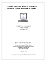
Figure 1-2. In vitro selection process of deoxyribozymes: A population of single
stranded DNA molecules (top) is chemically synthesized with regions of random
DNA sequence (box) and regions of known DNA sequence (plain lines) used for
example as PCR primer binding sites. This population is subjected to the desired
selective conditions (1) during which the portion of the population of different
molecules that can perform the required function, such as self-modification in a
constant-sequence region (filled circle), is allowed to react. Molecules that react
become substrates for PCR amplification (2). Mutations may be introduced at a low
frequency under typical PCR conditions or at a higher frequency with mutagenic PCR
protocols. An enriched population of single-stranded functional sequences is
generated that is identical in format to the starting synthetic pool by the appropriate
choice of PCR primers and other molecular biology methods (3). The enriched
population is subjected to the desired, perhaps more stringent, selective conditions
again (1), and the cycle is repeated until catalytic activity can be detected through
biochemical assay. At this time, the DNA population is cloned and sequenced to
identify individual functional DNAs (4).
1.2.1 RNA-cleaving deoxyribozymes
Most deoxyribozymes created to date promote RNA cleavage by phosphoester
transfer (Figure 1-3). This group of catalytic DNAs highlights the fact that DNA is
capable of forming specific contacts with a number of metal ion or amino acid
cofactors that are essential for function [95]. In addition, the discovery of several
“cofactor less” deoxyribozymes indicates that DNA has the chemical power to
promote RNA transesterification even in the absence of external chemical groups [95,
16
96]. The first report of an RNA-cleaving deoxyribozyme in 1994 revealed that a short
DNA (38 mer) could form a complex structure that enhanced the site-specific,
multiple turnover cleavage of RNA substrates by ~100,000 fold in the presence of
divalent lead [94]. The subsequent research showed not only that DNA could act as a
true catalyst but also that such enzymes could be readily obtained. Some of the more
recently derived examples also showed that deoxyribozymes can be made to have an
effect on biological systems and that they have promise in diagnostic and therapeutic
applications. One RNA-cleaving deoxyribozyme provides a striking illustration of the
impact that the study of catalysis by DNA can have on biology.
17
c
16.2-11 RNA-cleaving
deoxyribozyme
N N N N N N N N A U N N N N N N
II
I
N N N N N N N N
U G N N N N N N
5'
5'
cleavage site
G
U
G
A
C
C
C
C
T
U
G
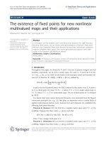
Figure 1-3. RNA cleavage by transesterification and two deoxyribozyme motifs that
catalyze this reaction. (a) RNA strand scission occurs when the oxygen of the 2’
hydroxyl group serves as a nucleophile to attack the phosphorus center of the adjacent
phosphoester linkage in an S
N
2-like reaction, resulting in 2’, 3’-cyclic phosphate and
5’-hydroxyl products. Proposed secondary structures of the 10–23 (b) and 16.2–11 (c)
RNA-cleaving deoxyribozymes.
18
1.2.2 DNA-cleaving deoxyribozymes
The absence of 2’-hydroxyl group makes DNA approximately 105 times more
resistant to hydrolysis compared with the rate constant of the related internal RNA
transesterification reaction [88, 98]. The extrapolated background rate of DNA
hydrolysis is ~10
–12
min
–1
at neutral pH and at 25°C. As a result, even a 100 million
fold rate enhancement during an in vitro selection reaction for DNA hydrolysis would
yield less than 1% cleaved DNA in a 12 hrs incubation. It is expected that
deoxyribozymes that catalyze phosphoester hydrolysis versus RNA transesterification
will require a more sophisticated active site in order to permit water to make an
efficient nucleophilic attack on phosphorus. Therefore, deoxyribozymes that
hydrolyze phosphoester must be exceedingly rare in a given random-sequence
population, and as might be predicted; there have been no reports of hydrolytic self-
cleaving DNAs.
1.2.2.1 DNA self-cleavage reaction by oxidative mechanism
However, there are two alternative mechanisms for DNA cleavage that are exploited
by deoxyribozymes. First, the spontaneous oxidative cleavage of DNA [99] by
hydroxyl radicals can be induced by using redox-active metals such as Cu
2+
and
reducing agents such as ascorbate [100, 101]. In an earlier study, Carmi et al.
identified two classes of DNA enzymes which can self-cleave by using an oxidative
mechanism [102]. Class I self-cleaving deoxyribozymes require Cu
2+
and ascorbate to
promote DNA cleavage, whereas class II DNAs require only the addition of Cu
2+
as a
cofactor. Class II self-cleaving deoxyribozymes [103] appear to catalyze the cleavage
at one particular inter-nucleotide linkage by promoting the attack of a hydroxyl
radical at the 4’ carbon [104], thereby initiating a series of rearrangements [105] that
19
lead to loss of a nucleotide base and chain cleavage. However, hydroxyl radical-
induced cleavage can occur at several positions along the DNA chain by variations of
this mechanism, leading to a diversity of cleavage products [104]. Its structure is also
notable in that there is evidence to support a triplex tertiary interaction with substrate
(Figure 1-4), which has no precedent in nucleic acid enzymes.
Figure 1-4. DNA cleavage reaction by oxidation strategies. (a) Directed attack of a
hydroxyl radical on the C4’ position induces rearrangements that lead to chain
cleavage. (b) Proposed structural model of the class II DNA cleaving deoxyribozyme
includes two base-paired regions and a triplex interaction (dots) with substrate
confirmed by compensatory mutational analysis.
1.2.2.2 DNA self-cleavage reaction by depurination process
A second mechanism of DNA scission involves cleavage following a site-specific
depurination event [106]. The “10–28” deoxyribozyme functions as an N-glycosylase
enzyme by catalyzing the depurination of a specific guanosine residue (Figure 1-5A).
Amines or other agents that can serve as general bases will then accelerate the
cleavage of the abasic DNA linkage. In 2 mM Ca
2+
, the “10–28” enzyme catalyzes
20
depurination of a separate substrate oligonucleotide with a pseudo-first-order rate
constant of 0.2 min
–1
. This corresponds to a rate enhancement for depurination of ~1
million fold over the corresponding uncatalyzed reaction. The mechanism could be an
S
N
1 type, where protonation at the N7 position of guanine allows the base to depart
and thereby permits water to attack the 1’ carbon. The resulting abasic deoxyribose
site in the DNA can undergo ring opening that would make the linkage susceptible to
β-elimination, which would release the pentose while breaking the DNA strand to
yield 3’- and 5’-phosphate terminated fragments. The intermediate abasic site,
released guanine, and phosphate-terminated fragments have all been detected
experimentally for this enzyme system.
21
Figure 1-5. DNA cleavage reaction by depurination process. (a) The proposed
mechanism of the 10–28 deoxyribozyme involves protonation at the N7 position of
guanine, which facilitates the departure of the base moiety and permits subsequent
attack by a water molecule at the resulting C1’ carbocation of deoxyribose. (b)
Proposed secondary structure of the 10–28 N-glycosylase deoxyribozyme.
Although deoxyribozymes with DNA-cleaving activity have been created by an
oxidative mechanism or depurination process, a nucleotide will be eliminated from
the target DNA chain during these cleavage processes, and these elimination
processes will in turn result in the loss of the encoded information in sequence. In
contrast to oxidative DNA cleavage, which results from the facile attack of a hydroxyl
radical on the base or on deoxyribose moieties, direct hydrolysis of phosphate esters is
likely to be far more difficult for a deoxyribozyme to employ, because phosphate ester
hydrolysis in particular is believed to be one of the most challenging objectives
22
considering the extraordinarily slow background rate of hydrolysis of DNA [107]. Yet,
this mechanism may be the most attractive for a DNA-cleaving deoxyribozyme as no
loss of genetic information occurs upon DNA strand cleavage. In view of the
accomplishments of ribozymes, we could believe that DNA molecule could possess
such catalytic capabilities too in nature.
1.3 DNA Cleavage throuth Direct Hydrolysis of Phosphodiester
How can DNA, with its poor complement of chemical functionalities, be catalytic
without outside help? How about cofactors? Protein based enzymes use an array of
organic cofactors to provide external sources of functional groups, hydrogen atoms
and electrons [109]. There are examples of ribozymes that use protein cofactors for
folding or stabilizing their structures [110]. There are many examples of the
nonspecific hydrolysis of RNA using a variety of small peptides [111-113] and
organic molecules [114-116]. In the past years, Roth and Breaker [95] have described
a DNAzyme that strictly requires the imidazole group of histidine for catalysis.
Furthermore, DNAzymes can overcome these functional limitations by utilizing
divalent metal ions cofactors for catalysis [117-118]. Metal ions would function in
two ways. First, they would partially neutralize the negative-charge density on the
backbones of single-stranded DNA, and promote its folding into complex shapes.
Second, water molecules coordinated to divalent cations such as Mg
2+
, Ca
2+
, Zn
2+
or
Pb
2+
would carry out the acid/base catalysis useful for phosphodiester bond hydrolysis.
Selection for deoxyribozymes that catalyze the direct hydrolysis of a phosphoester
linkage is expected to be challenging. However, deoxyribozymes that catalyze DNA
hydrolysis might yet be amenable to isolation by taking advantage of substrate
23
molecules that carry analogues of the DNA phosphodiester linkage. For example,
replacement of the 3’-or 5’-oxygen atom of a DNA phosphodiester with an amine
group yields an analogue DNA linkage that is far more susceptible to hydrolytic
cleavage (due to the nature of the leaving group). The 5’-modified version of this
linkage analogue has already been used as a substrate to select for deoxyribozymes
that cleave phosphoramidate-containing DNAs [108]. The phosphoramidate
modification changes only the leaving group, whereas the geometry and chemical
nature of the transition state remain largely unchanged. Thus, the catalytic
mechanisms that allow an enzyme to cleave this modified DNA might also be
applicable to normal DNA albeit at a much slower rate. Perhaps mutagenized
populations of phosphoramidate-cleaving deoxyribozymes would make an opportune
starting point for the isolation of variant deoxyribozymes that have the catalytic power
to efficiently cleave DNA by hydrolysis. And it is expected that if deoxyribozymes
that catalyze phosphoester hydrolysis versus RNA transesterification really exist in
nature, a more sophisticated active site will be required to permit water molecule to
make an efficient nucleophilic attack on phosphorus.
1.4 G-quadruplexes
DNA can adopt several secondary structures besides the well-known canonical B-type
duplex. DNA sequences that contain four or more closely spaced guanine-tracts can
fold to form intramolecular quadruplexes, which consist of stacked G-quartets [Figure
1-6] that are linked by three loops between the four G-strands [119]. These structures
are stabilized by monovalent cations, especially potassium ion [120], and can adopt a
variety of different folding patterns depending on the relative orientation of the
strands and the position of the loops.
24
Figure 1-6. The guanine tetrad motif and its hydrogen bonding scheme.
For intramolecular quadruplexes, the four G-tracts are separated by loops. These are
of various lengths and can be as short as a single nucleotide [121]. The loops can be
arranged in several different ways [Figure 1-7]; external (propeller) loops link two
adjacent parallel strands [122], while edgewise or diagonal loops link two antiparallel
strands [123]. Some structures contain both edge-wise and propeller loops [124-126].
It is known that loop length and sequence affect quadruplex stability and structure
[127]. Sequences with single nucleotide loops between the guanine-tracts only adopt a
parallel structure, while longer loops can also adopt an antiparallel arrangement of the
strands. Quadruplex stability is also affected by the sequence of the loops [128], and
the bases that flank the quadruplex [129].
Figure 1-7. Strand connectivity alternatives for intramolecular G-quadruplex
structures. (A) All three loops run edgewise and connect adjacent-adjacentadjacent.
(B) One diagonal and two edgewise loops that connect adjacent-diagonal-adjacent. (C)
An example of a loop that runs on the outside of the guanine tetrad core.
25
There is considerable variation in quadruplex structures, depending on the DNA
sequence and the ionic conditions [121]. The biological function of quadruplexes may
well depend on the folded conformation that is adopted, especially if this involves
interaction with specific proteins. Such an effect has been suggested for the NHE
element of the c-myc promoter, which can in principle adopt multiple conformations.
Since the loops can have a considerable effect on quadruplex folding and stability,
many experiments have been designed to examine how changes in loop length affect
quadruplex properties. One very stable intramolecular quadruplex contains four
guanine-tracts that are linked by single T residue [130-132] and this is known to be an
inhibitor of HIV integrase.
1.5 Guanine Quartets as Structural Components for Some DNA Aptamers and
DNA Enzymes
Natural DNA serves almost without exception as an informational macromolecule
that exists primarily as a base-paired duplex. Single- tranded DNAs are rarely
encountered in nature, and even less frequently do these single-stranded DNAs form
shapes (other than the ususal duplex) that serve a functional role. Yet a number of
compelling arguments can be made to support the notion that certain DNAs are
capable of forming structures that mimic those of folded RNAs, including ribozymes.
The G-quartet motif has been identified or implicated in a number of DNA aptamers
and deoxyribozymes, although it is not unique to DNA, and is at the top of the
putative list of recurrent DNA structural motifs. The primary role for the guanine-
quartet in folded DNAs might be to serve as a stable center around which other
structural domains can be assembled. The first example of a protein-specific DNA