The role of paxillin superfamily members hic 5 and leupaxin in b cell antigen receptor signaling 2
Bạn đang xem bản rút gọn của tài liệu. Xem và tải ngay bản đầy đủ của tài liệu tại đây (636.7 KB, 11 trang )
Leupaxin Negatively Regulates B Cell Receptor Signaling
*
Received for publication, June 5, 2007, and in revised form, July 16, 2007 Published, JBC Papers in Press, July 19, 2007, DOI 10.1074/jbc.M704625200
Valerie Chew and Kong-Peng Lam
1
From the Laboratory of Immune Regulation, Biomedical Sciences Institutes, Agency for Science, Technology and Research and
Singapore Immunology Network, Singapore 138673, Singapore
The role of the paxillin superfamily of adaptor proteins in B
cell antigen receptor (BCR) signaling has not been studied pre-
viously. We show here that leupaxin (LPXN), a member of this
family, was tyrosine-phosphorylated and recruited to the
plasma membrane of human BJAB lymphoma cells upon BCR
stimulation and that it interacted with Lyn (a critical Src family
tyrosine kinase in BCR signaling) in a BCR-induced manner.
LPXN contains four leucine-rich sequences termed LD motifs,
and serial truncation and specific domain deletion of LPXN
indicated that its LD3 domain is involved in the binding of Lyn.
Of a total of 11 tyrosine sites in LPXN, we mutated Tyr
22
, Tyr
72
,
Tyr
198
, and Tyr
257
to phenylalanine and demonstrated that
LPXN was phosphorylated by Lyn only at Tyr
72
and that this
tyrosine site is proximal to the LD3 domain. The overexpression
of LPXN in mouse A20 B lymphoma cells led to the suppression
of BCR-induced activation of JNK, p38 MAPK, and, to a lesser
extent, Akt, but not ERK and NF
B, suggesting that LPXN can
selectively repress BCR signaling. We further show that LPXN
suppressed the secretion of interleukin-2 by BCR-activated A20
B cells and that this inhibition was abrogated in the Y72F LPXN
mutant, indicating that the phosphorylation of Tyr
72
is critical
for the biological function of LPXN. Thus, LPXN plays an inhib-
itory role in BCR signaling and B cell function.
Engagement of the B cell antigen receptor (BCR)
2
on B cells
by antigen triggers first the activation of the Src family kinase
Lyn (1–4), which is known to phosphorylate the immunorecep-
tor tyrosine-based activation motifs within the cytoplasmic
domains of the Ig-
␣
and Ig-

subunits that are part of the BCR
complex (5). The phosphorylation of the immunoreceptor
tyrosine-based activation motif then leads to the activation of
the tyrosine kinase Syk (6, 7), which leads in turn to the phos-
phorylation of various downstream proteins such as BLNK,
phospholipase C
␥
2, and Btk (8). As a consequence of the acti-
vation of these signaling proteins, numerous second messengers
and intermediate signal-transducing proteins are activated, and
together, they lead to the activation of several key transcription
factors that regulate new gene expression in B lymphocytes and
that drive unique B cell physiological responses such as prolifera-
tion, cytokine secretion, and differentiation either to memory B or
antibody-producing plasma cells (9).
Because BCR signaling can lead to the activation of B lym-
phocytes, there exist several mechanisms to down-regulate or
modulate BCR signaling to prevent the overt or inappropriate
activation of B cells. Several phosphatases such as the mem-
brane-bound CD45 and intracellular SHP-1 and SHIP-1 are
known to dephosphorylate and hence deactivate key signal
transduction molecules in the BCR signaling pathway (10).
Recent studies also revealed that, in addition to its well estab-
lished role in BCR signal initiation, Lyn can play a negative role
in down-modulating BCR signaling (11, 12). Indeed, despite
showing defects in B cell development, Lyn-deficient mice are
also susceptible to autoimmune diseases, and Lyn-deficient B
cells are hyper-responsive to BCR ligation (13–15).
Another class of signal transduction molecules known as the
adaptor proteins has also been shown to play critical roles in
lymphocyte signal transduction. These proteins do not have
enzymatic activities but mediate protein-protein and protein-
lipid interactions to provide spatiotemporal modulation of BCR
signaling (16). Some of these adaptors are positive regulators of
signal transduction, and they facilitate the assembly of activat-
ing signaling complexes. For example, BLNK has been widely
established as an adaptor protein that couples Syk and Btk to
activate phospholipase C
␥
2 upon BCR ligation, and this subse-
quently triggers downstream calcium fluxes and inositol 1,4,5-
trisphosphate production (17). On the other hand, other adap-
tors such as the Csk-binding protein and the Dok family
members Dok-1, 2, and 3 are known to play a negative role in
immunoreceptor signaling (18). Most of these inhibitory adap-
tors recruit additional inhibitory effectors to the vicinity of pos-
itive regulator of signaling to shut down the signal transduction
processes, e.g. Csk-binding protein is known to recruit Csk,
which inhibits the activation of the Src family tyrosine kinases
(19), whereas Dok-3 is known to recruit SHIP, which dephos-
phorylates activated signaling molecules (20, 21).
Given that certain adaptor proteins such as BLNK (22),
Dok-1 (23), and Dok-3 (24) have been demonstrated to play key
roles in the regulation of BCR signaling, it is conceivable that
other adaptor proteins that have not been studied in the context
of immunoreceptor signaling may also play a critical role in
BCR signal transduction. The paxillin family of adaptor pro-
teins could be one such example. Paxillin and its related family
members Hic-5, leupaxin, and PaxB have not been demon-
* This work was supported by grants from the BiomedicalResearch Council of
the Agency for Science, Technology and Research, Singapore. The costs of
publication of this article were defrayed in part by the payment of page
charges. This article must therefore be hereby marked “advertisement”in
accordance with 18 U.S.C. Section 1734 solely to indicate this fact.
1
To whom correspondence should be addressed: Singapore Immunology
Network, Lab 6-15, 61 Biopolis Dr., Proteos, Singapore 138673, Singapore.
Tel.: 65-6586-9649; Fax: 65-6478-9477; E-mail: lam_kong_peng@immunol.
a-star.edu.sg.
2
The abbreviations used are: BCR, B cell antigen receptor; JNK, c-Jun N-termi-
nal kinase; MAPK, mitogen-activated protein kinase; IL-2, interleukin-2;
ERK, extracellular signal-regulated kinase; HA, hemagglutinin; LPXN, leu-
paxin; BSA, bovine serum albumin; PBS, phosphate-buffered saline; ELISA,
enzyme-linked immunosorbent assay.
THE JOURNAL OF BIOLOGICAL CHEMISTRY VOL. 282, NO. 37, pp. 27181–27191, September 14, 2007
© 2007 by The American Society for Biochemistry and Molecular Biology, Inc. Printed in the U.S.A.
SEPTEMBER 14, 2007 • VOLUME 282 •NUMBER 37 JOURNAL OF BIOLOGICAL CHEMISTRY 27181
at NATIONAL UNIVERSITY OF SINGAPORE on September 7, 2007 www.jbc.orgDownloaded from
strated to play a role in BCR signaling so far. Paxillin is a focal
adhesion adaptor protein that plays an important role in growth
factor- and integrin-mediated signaling pathways (25, 26).
Despite its ability to bind Pyk2 and PTP-PEST, which are mol-
ecules known to play a role in BCR signaling, a previous report
had indicated that paxillin is not tyrosine-phosphorylated in
activated B cells (27). Thus, the role of paxillin in BCR signaling
remains to be confirmed. Another family member (Hic-5) was
reported to be largely absent in lymphocytes (28), hence mini-
mizing the possibility of its participation in BCR signaling.
On the other hand, leupaxin, which is most homologous to
paxillin and detectable as a 45-kDa protein, is preferentially
expressed in hematopoietic cells, including B cells (29). Sim-
ilar to the other paxillin superfamily members, leupaxin con-
tains multiple N-terminal Leu- and Asp-rich sequences (LD
domains) and LIM domains (26, 30). Both LD and LIM
domains had been shown to be important for protein-pro-
tein interactions, and in addition, LIM domains have also
been shown to play a role in the focal adhesion targeting of
paxillin superfamily members (31). Recent works also estab-
lished a role for leupaxin in the function of osteoclasts (32) and in
the migration of prostate cancer cells (33). Leupaxin is known to
interact with Pyk2 (29); Src (34); PEST domain tyrosine phospha-
tase (PEP) (35); and PTP-PEST, pp125
FAK
, and the ADP-ribosyla-
tion factor (ARF) GTPase-activating protein p95
PKL
(32); and
some of these proteins are known to be expressed in B cells.
In this study, we examined the possible role of leupaxin in
BCR signaling. We found that leupaxin was phosphorylated
upon BCR engagement in human BJAB cells. We show that
leupaxin bound Lyn via its LD3 domain and that Lyn phospho-
rylated Tyr
72
of leupaxin. In addition, we demonstrate that leu-
paxin inhibited the JNK and p38 MAPK signaling pathways
downstream of BCR signaling and suppressed the production
of interleukin-2 (IL-2) by activated mouse A20 B cells and that
the inhibition of cytokine secretion by leupaxin required the
phosphorylation of Tyr
72
. Thus, leupaxin plays an inhibitory
role in BCR signaling and B cell function.
EXPERIMENTAL PROCEDURES
Plasmid Construction—The cDNAs encoding Lyn, paxillin,
and Hic-5 were cloned from a murine spleen cDNA library by
PCR. FLAG-tagged wild-type leupaxin was generated from the
cDNA encoding wild-type leupaxin (provided by Dr. A. Gupta,
University of Maryland, Baltimore, MD) (34). FLAG-tagged
leupaxin deletion mutants (⌬LD1, ⌬LD1–2, ⌬LD1–3, ⌬LD1–4,
and ⌬LD3) and tyrosine-to-phenylalanine mutants (Y22F, Y72F,
Y198F, and Y257F) were generated by PCR. All wild-type and
mutated cDNAs were verified by DNA sequencing (data not
shown).
Cells and Transfections—HEK293T cells were grown in Dul-
becco’s modified Eagle’s medium supplemented with 10% fetal
bovine serum, 2 m
ML-glutamine, and penicillin/streptomycin
and transiently transfected using Effectene transfection rea-
gent (Qiagen Inc.). BJAB and A20 cells were grown in RPMI
1640 medium supplemented with 10% fetal bovine serum, 0.05
m
M 2-mercaptoethanol, 2 mML-glutamine, and penicillin/
streptomycin. For transfection of A20 B cells, 1 ϫ 10
7
cells were
mixed with 20
g of plasmid DNA in 500
l of RPMI 1640
medium and electroporated in a 0.4-cm cuvette at 950 micro-
farads and 300 V using a Gene Pulser (Bio-Rad). Transfection
efficiency was assessed with the pEGFP-N2 vector at 36 h post-
transfection by flow cytometry and was determined to be
between 30 and 40%.
Isolation of Subcellular Fractions—BJAB cells were lysed on
ice for 20 min in hypotonic buffer containing 15 m
M Tris-HCl
(pH 7.5), 5 m
M KCl, 1.5 mM MgCl
2
, 0.1 mM EGTA, 0.2 mM
Na
3
VO
4
, and protease inhibitor mixture (Roche Applied Sci-
ence). Cell lysates were homogenized through a 26-gauge nee-
dle and centrifuged at 500 ϫ g. The supernatant was transferred
to polycarbonate tubes and ultracentrifuged at 20,800 ϫ g for
1 h at 4 °C. The supernatant containing the cytosol fraction was
recovered, and the pellet containing the plasma membrane
fraction was solubilized in 150 m
M NaCl, 15 mM Tris-HCl (pH
7.5), 5 m
M EDTA, 1% Triton X-100, 0.2 mM Na
3
VO
4
, and pro-
tease inhibitors.
Antibodies—F(abЈ)
2
fragments of goat anti-mouse IgG and
goat anti-human IgM were purchased from Jackson Immu-
noResearch Laboratories (West Grove, PA). Monoclonal anti-
bodies against human leupaxin (283C and 315G) were obtained
from Dr. A. Gupta (32) and ICOS Corp. (Bothell, WA). The
following commercial antibodies were also used: anti-phos-
pho Akt (Ser
473
/Thr
308
), anti-Akt-1, anti-ERK2, anti-I
B
␣
,
anti-JNK1, anti-Lyn, anti-phospho-ERK, anti-p38, and anti-

-
tubulin (Santa Cruz Biotechnology, Inc.); anti-phospho-stress-
activated protein kinase (SAPK)/JNK (Thr
183
/Tyr
185
) and anti-
phospho-p38 (Thr
180
/Tyr
182
) (Cell Signaling Technology);
anti-FLAG polyclonal and anti-hemagglutinin (HA) mono-
clonal (Sigma); horseradish peroxidase-coupled anti-phos-
photyrosine (4G10; Upstate Biotechnology); and Alexa 546-
conjugated goat anti-mouse and Alexa 488-conjugated
chicken anti-rabbit (Molecular Probes).
Cell Stimulation, Western Blotting, and Immunoprecip-
itations—Cells were resuspended in RPMI 1640 medium at 2 ϫ
10
6
cells/200
l and serum-starved at 37 °C for 1 h prior to
stimulation with anti-Ig antibodies. BJAB cells were stimulated
with 10
g/ml anti-human IgM F(abЈ)
2
fragment, and A20 cells
with 15
g/ml anti-mouse IgG F(abЈ)
2
fragment. After stimula-
tion, cells were lysed on ice for 10 min in lysis buffer containing
1% Nonidet P-40, 10 m
M Tris-HCl (pH 8.0), 150 mM NaCl, 1 mM
EDTA, 0.2 mM Na
3
VO
4
, and protease inhibitor mixture and
sonicated. Cell homogenates were centrifuged at 13,000 rpm
for 15 min at 4 °C, and supernatants were recovered for protein
quantification using a BCA protein assay kit (Pierce). Proteins
were electrophoresed on 10% SDS-polyacrylamide gel and
transferred onto polyvinylidene difluoride immunoblot mem-
branes (Bio-Rad). The membranes were blocked with 5% non-
fat milk in Tris-buffered saline containing 0.1% Tween 20 for
1 h at room temperature and incubated separately with various
antibodies recognizing the different molecules studied. Protein
bands were visualized using horseradish peroxidase-coupled
secondary antibodies and the enhanced chemiluminescence
ECL detection system (Amersham Biosciences). For immuno-
precipitations, cell lysates were precleared with protein A/G
Plus-agarose (Santa Cruz Biotechnology, Inc.) for1hat4°C.
For immunoprecipitation of endogenous proteins, anti-leu-
paxin (LPXN) monoclonal antibody 315G or anti-Lyn antibody
Inhibitory Role of Leupaxin in BCR Signaling
27182 JOURNAL OF BIOLOGICAL CHEMISTRY VOLUME 282•NUMBER 37•SEPTEMBER 14, 2007
at NATIONAL UNIVERSITY OF SINGAPORE on September 7, 2007 www.jbc.orgDownloaded from
was coupled overnight to protein A/G Plus-agarose at 4 °C and
washed twice with lysis buffer before overnight incubation with
precleared cell lysates at 4 °C. For other immunoprecipitations,
agarose beads were covalently coupled with anti-phosphoty-
rosine or anti-FLAG antibody and washed twice with lysis
buffer before incubation with precleared cell lysates. Beads
were then pelleted and washed three times with lysis buffer
before boiling in loading buffer (1% SDS, 1%

-mercaptaetha-
nol, 15% glycerol, and 0.01% bromphenol blue) for 5 min. The
released proteins were resolved on SDS-polyacrylamide gels.
Confocal Microscopy—BCR-stimulated BJAB cells were
washed twice with cold 1% bovine serum albumin (BSA) in
phosphate-buffered saline (PBS) and fixed for 20 min on ice
with 4% paraformaldehyde in PBS. After permeabilization at
room temperature for 10 min with 0.2% saponin and 0.03
M
sucrose in 1% BSA-containing PBS, cells were washed twice
before being deposited onto slides. Cells were blocked with 5%
normal goat serum in 1% BSA-containing PBS at room temper-
ature for 1 h before overnight incubation with primary antibod-
ies at 4 °C. The slides were washed three times with 1% BSA in
PBS and incubated at room temperature for 1 h with Alexa
546-conjugated goat anti-mouse or Alexa 488-conjugated
chicken anti-rabbit antibody to reveal the respective primary
antibodies. Slides were washed three times with 1% BSA in PBS,
mounted, and viewed under a Radiance 2000 confocal laser
scanning microscope (Bio-Rad).
Measurement of BCR-triggered IL-2 Production—10
5
trans-
fected A20 cells in 200
l of culture medium were stimulated
for 24 h at 37 °C in 96-well plates in the presence or absence of
10
g/ml anti-mouse IgG F(abЈ)
2
fragment. The resulting pro-
duction of IL-2 was measured by enzyme-linked immunosor-
bent assay (ELISA) using a mouse IL-2 ELISA kit (BD Bio-
sciences) according to the manufacturer’s protocol. All
cytokine secretion assays were performed in triplicate and
repeated three times. To measure BCR-induced activation of
the IL-2 promoter, A20 cells (10 ϫ 10
6
) were transfected with
an IL-2 promoter-luciferase plasmid as described previously
(36). Briefly, cells were electroporated with 15
g of IL-2 pro-
moter-luciferase plasmid together with 10
g of the indicated
plasmids and 1.5
g of pRL-TK (Renilla) plasmid (to standard-
ize for transfection efficiency). After 40 h, 2 ϫ 10
6
cells were
stimulated for 6 h with 10
g of IgG F(abЈ)
2
fragment. Cells were
harvested, and cell pellets were solubilized in passive lysis buffer
(Promega Corp.) and incubated on a Spiramix roller mixer for 15
min at room temperature. Cell lysate (90
l) was assayed for both
firefly and Renilla luciferase activities using the Dual-Luciferase
reporter assay system (Promega Corp.), and the relative light units
were measured in a TD-20/20 single tube luminometer (Turner
BioSystems, Sunnyvale, CA). Luciferase activity was calculated as
increments (n-fold) in IgG F(abЈ)
2
fragment-induced activity over
basal activity obtained with unstimulated cells.
RESULTS
Leupaxin Is Activated upon BCR Engagement in Human
BJAB B Cells—It was shown previously that LPXN is preferen-
tially expressed in hematopoietic cells (29). However, the role of
LPXN in BCR signaling is not known. We first observed that
LPXN is highly expressed in human BJAB B lymphoma cells
(Fig. 1A), suggesting that it might have a role in some aspects of
B cell physiology. It is known from various studies that the
engagement of BCR on B cells with anti-IgM antibodies or anti-
gens leads to the tyrosine phosphorylation and hence activation
of several downstream signaling proteins such as the tyrosine
kinase Btk and the adaptor protein BLNK (37–39). LPXN is
known to contain 11 tyrosine residues. Therefore, to determine
whether LPXN is involved in BCR signaling, we examined
whether LPXN is tyrosine-phosphorylated upon the engage-
ment of BCR on BJAB cells. As shown in Fig. 1A, treatment of
BJAB cells with 10
g/ml anti-human IgM F(abЈ)
2
fragment led
to the tyrosine phosphorylation of LPXN as shown by immu-
noprecipitating LPXN and immunoblotting it with anti-phos-
photyrosine antibody 4G10. The phosphorylation of LPXN
occurred as early as 1 min after BCR stimulation of BJAB cells
and appeared to peak at the 5- and 15-min time points before
returning to the basal level at the 30-min time point. Con-
versely, immunoprecipitating total cellular phosphotyrosine
proteins from BCR-stimulated BJAB cells with antibody 4G10
followed by immunoblotting with anti-LPXN antibody also
revealed a similar pattern in which LPXN was highly activated
between 5 and 15 min, as LPXN was maximally immunopre-
cipitated at these time points (Fig. 1B). As a comparison, the
tyrosine kinase Lyn (a known component of the BCR signal
transduction system) was also shown to be tyrosine-phospho-
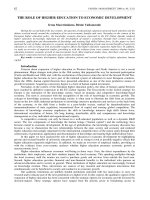
FIGURE 1. LPXN is phosphorylated andrecruitedtotheplasmamembraneof
B cells upon BCR ligation. A, tyrosine phosphorylation of LPXN in BJAB cells.
Cells were stimulated with 10
g/ml anti-human IgM F(abЈ)
2
fragment for various
time points as indicated, and LPXN was immunoprecipitated (IP) and probed
with anti-phosphotyrosine antibody 4G10 or anti-LPXN monoclonal antibody
283C, respectively. B, BJAB cells were stimulated with 10
g/ml anti-human IgM
F(abЈ)
2
fragment for various time points, and cells lysates were subjected to
immunoprecipitation with anti-phosphotyrosine antibody and immunoblotted
(IB) with anti-LPXN and anti-Lyn antibodies. C, membrane recruitment of LPXN
upon BCR stimulation. BJAB cells were stimulated with anti-human IgM F(abЈ)
2
fragment for various time points, and their plasma membrane and cytoplasmic
fractions were immunoblotted with anti-LPXN, anti-Lyn (used as a loading con-
trol for the plasma membrane fraction), and anti-

-tubulin (used as a loading
control for the cytoplasmic fraction) antibodies.
Inhibitory Role of Leupaxin in BCR Signaling
SEPTEMBER 14, 2007 • VOLUME 282 •NUMBER 37 JOURNAL OF BIOLOGICAL CHEMISTRY 27183
at NATIONAL UNIVERSITY OF SINGAPORE on September 7, 2007 www.jbc.orgDownloaded from
rylated in BCR-activated BJAB cells. However, in contrast to
LPXN, the phosphorylation of Lyn seemed to be more pro-
longed and was extended to 30 min after BCR ligation (Fig. 1B).
This might indicate that the activation of LPXN and hence its
involvement in BCR signaling could be more transient com-
pared with Lyn. The tyrosine phosphorylation of LPXN also
indicated that it could be a target of a protein-tyrosine kinase
that was activated downstream of the BCR signaling pathway.
Besides the tyrosine phosphorylation of LPXN, we also
observed the recruitment of LPXN to the plasma membrane of
B cells upon BCR ligation. Several proteins in the BCR signaling
pathway, e.g. the adaptor protein BLNK and the tyrosine kinase
Btk, are known to locate to the plasma membrane and espe-
cially to the lipid raft fraction of B cells following BCR activation
(17). Our results also indicated that LPXN was enriched in the
membrane faction beginning at 5 min after anti-human IgM
F(abЈ)
2
fragment treatment of BJAB cells (Fig. 1C). LPXN could
still be found in the membrane fractions 15 min after BCR liga-
tion and was subsequently sequestered back to the cytoplasm
beginning 30 min after activation. This is consistent with the
tyrosine phosphorylation profile of LPXN as shown in Fig. 1 (A
and B). Lyn, which is known to be constitutively present in the
membrane fraction of B cells (40, 41), was used as a loading
control for the membrane fractions, whereas

-tubulin was
used as a control for the cytoplasmic fractions (Fig. 1C). Thus,
taken together, the data indicate that LPXN is tyrosine-phos-
phorylated and recruited to the plasma membrane following
BCR cross-linking in B cells, suggesting that LPXN could play a
role in BCR signaling.
Leupaxin Interacts with Lyn during BCR Signaling—Previous
studies indicated that members of the paxillin superfamily of
adaptors can interact with members of the Src family of tyro-
sine kinases, e.g. paxillin was shown to bind Src either directly
or via Pyk2 (31), Hic-5 can bind Fyn (42), and LPXN can inter-
act with Src in osteoclasts (34). Because we demonstrated that
LPXN was tyrosine-phosphorylated upon BCR cross-linking
and it is known that Lyn is the predominant Src family tyrosine
kinase found in B cells (43), we investigated whether LPXN can
physically interact with Lyn.
To accomplish this, FLAG-tagged paxillin superfamily mem-
bers (LPXN, Hic-5, and paxillin) were coexpressed with HA-
tagged Lyn in HEK293T cells, and whole cell lysates from the
transfectants were subjected to immunoprecipitation with
anti-FLAG antibody-agarose beads. The immunoprecipitates
were subsequently immunoblotted with anti-Lyn antibody. As
shown in Fig. 2A, all three members of the paxillin superfamily
interacted with Lyn, with Hic-5 co-immunoprecipitating a
larger amount of Lyn, followed by LPXN and finally paxillin.
Used as a negative control, the FLAG tag alone did not show any
nonspecific interaction with Lyn.
As LPXN bound Lyn in overexpression studies in HEK293T
cells, we next examined whether endogenous interaction of
LPXN and Lyn can occur in B cells upon BCR activation. BJAB
cells were treated with 10
g/ml anti-human IgM F(abЈ)
2
frag-
ment for various time points, and cell lysates were immunopre-
cipitated with anti-LPXN antibody and immunoblotted with
anti-Lyn antibody. As shown in Fig. 2B, the interaction between
LPXN and Lyn was detected as early as 1 min and seemed to
peak between 5 and 15 min after BCR ligation in BJAB cells. The
binding of LPXN to Lyn appeared to occur in response to BCR
cross-linking, as LPXN and Lyn could no longer be co-immu-
noprecipitated at the 30-min time point. We were able to show
that an equivalent amount of LPXN (used as a control) was
immunoprecipitated at all time points examined. Thus, Lyn
binding to LPXN appears to be induced by BCR signaling.
To visualize the endogenous interaction of LPXN and Lyn,
we performed immunofluorescence studies in BJAB cells.
LPXN (which stained red) was found to be evenly distributed in
the cytoplasm of unstimulated BJAB cells (Fig. 2C, upper pan-
els). Upon ligation of BCR on BJAB cells, LPXN was recruited to
the plasma membrane at the 5-min time point and remained
there until after 15 min. However, by 30 min, LPXN was seques-
tered back to the cytoplasm (Fig. 2C, upper panels). This obser-
vation was consistent with our membrane fractionation data
shown in Fig. 1C. On the other hand, Lyn (which stained green)
(Fig. 2C, middle panels) was constitutively present in the mem-
brane fractions, as has been reported in a previous study (17).
Interestingly, the merging of the two panels revealed a pattern
of BCR-induced co-localization of LPXN and Lyn upon cell
activation (Fig. 2C, lower panels, yellow). The co-localization of
LPXN and Lyn was clearly visible in the plasma membrane of
BJAB cells at the 5-min time point following BCR ligation
and was slowly diminished from the 15-min time point
onward as LPXN was slowly recruited back to the cytoplasm.
By the 30-min time point, the co-localization of LPXN and
Lyn was minimal as LPXN was sequestered mostly away
from the membrane and predominantly found localized in
the cytoplasm. Taken together, the data in Fig. 2 support the
finding that LPXN interacts with Lyn and that the interac-
tion likely occurs in the plasma membrane of B cells and is
induced upon BCR activation.
Leupaxin Interacts with Lyn through Its LD3 Domain—Be-
cause LPXN is a multidomain adaptor protein containing four
leucine-rich LD motifs and four LIM domains, we were inter-
ested in determining the specific domain within LPXN that is
responsible for mediating the interaction with Lyn. It has been
shown previously that the LD2 domain of paxillin can bind
several proteins, including kinases such as Src, focal adhesion
kinase, and Pyk2 (25). The interaction of paxillin with Src has
been shown to be either direct (near the proline-rich N termi-
nus) or indirect (via focal adhesion kinase or Pyk2 near the LD2
domain) (31). More relevant to our study, a recent report also
indicated that the LD2 domain of LPXN can interact with Src
(34). Thus, we examined the specific LD domain(s) of LPXN
that interact with Lyn.
To determine which LD domain(s) of LPXN are involved in
binding Lyn, we constructed a series of FLAG-tagged trunca-
tion and deletion mutants of LPXN as shown in Fig. 3A. The
FLAG-tagged ⌬LD1, ⌬LD1–2, ⌬LD1–3, ⌬LD1–4, and ⌬LD3
deletion mutants of LPXN were overexpressed in HEK293T
cells together with HA-tagged Lyn. Western blot analyses (Fig.
3B) indicated that all FLAG-tagged LPXN deletion mutants and
HA-tagged Lyn proteins were expressed in the transfected cells.
The various FLAG-tagged LPXN mutants were thus immuno-
precipitated with anti-FLAG antibody and immunoblotted
with anti-Lyn antibody. As shown in Fig. 3B, wild-type LPXN
Inhibitory Role of Leupaxin in BCR Signaling
27184 JOURNAL OF BIOLOGICAL CHEMISTRY VOLUME 282•NUMBER 37•SEPTEMBER 14, 2007
at NATIONAL UNIVERSITY OF SINGAPORE on September 7, 2007 www.jbc.orgDownloaded from
(lane 2) and both the ⌬LD1 and ⌬LD1–2 LPXN deletion
mutants (lanes 3 and 4) were able to bind Lyn, whereas the
⌬LD1–3 and ⌬LD1–4 mutants could not (lanes 5 and 6). This
suggested that the potential binding
domain of LPXN for Lyn could be
the LD3 domain. To further con-
firm that the LD3 domain of LPXN
binds Lyn, we specifically generated
the ⌬LD3 mutant, in which only the
LD3 domain of LPXN was deleted.
Indeed, as shown in Fig. 3B (lane 7),
deleting the LD3 domain of LPXN
abolished the interaction of LPXN
and Lyn, as the two proteins could
no longer be co-immunoprecipi-
tated. We therefore concluded that
the LD3 domain of LPXN is the
domain responsible for interaction
with Lyn.
Leupaxin Is Phosphorylated at
Tyrosine 72 by Lyn—LPXN contains
11 potential tyrosine phosphoryla-
tion sites and has been shown to be
a substrate of tyrosine kinases
(29). Because LPXN was able to
interact with Lyn (Figs. 2 and 3),
we examined whether LPXN can
be a substrate of and be phospho-
rylated by Lyn. FLAG-tagged Lyn
was hyperphosphorylated and con-
stitutively active when transfected
into HEK293T cells (Fig. 4A, upper
panel, lane 2). When FLAG-tagged
LPXN was coexpressed with FLAG-
tagged Lyn in HEK293T cells, it was
tyrosine-phosphorylated by Lyn, as
shown by immunoblotting of whole
cell lysates with anti-phosphoty-
rosine antibody 4G10 (Fig. 4A, lane
3). However, without the coexpres-
sion of FLAG-tagged Lyn, LPXN
was not phosphorylated (Fig. 4A,
upper panel, lane 1). Interestingly,
Lyn was also able to phosphorylate
paxillin and Hic-5 (Fig. 4A, lanes 4
and 5), suggesting that Lyn can
potentially interact with and phos-
phorylate all paxillin family mem-
bers. As control immunoblotting
with anti-FLAG antibody indicated
that all transfected proteins were
equivalently expressed in HEK293T
cells (Fig. 4A, lower panel).
Because Lyn could bind and
phosphorylate LPXN, we next ex-
amined the tyrosine residue(s) in
LPXN that can be phosphorylated
by Lyn. Among the 11 tyrosine resi-
dues in LPXN, 6 were identified as potential sites for phospho-
rylation by kinases using the NetPhos 2.0 program, which pre-
dicts serine, threonine, and tyrosine phosphorylation sites in
FIGURE 2. Interaction of LPXN and Lyn. A, binding of LPXN by Lyn. HEK293T cells were transiently transfected
with plasmids expressing various FLAG-tagged paxillin superfamily members and HA-tagged Lyn. Anti-FLAG
immunoprecipitates (IP) were subjected to immunoblotting (IB) with anti-Lyn antibody (upper panel) to exam-
ine co-immunoprecipitation and hence interaction of LPXN and Lyn. Whole cell lysates were immunoblotted
with anti-FLAG or anti-HA antibody (middle and lower panels) to examine the expression of the transfected
plasmids. B, interaction of endogenous LPXN and Lyn in BJAB cells. Cells were stimulated with 10
g/ml
anti-human IgM F(abЈ)
2
fragment for the indicated time points, and cell lysates were subjected to immunopre-
cipitation with anti-LPXN antibody and immunoblotted withanti-Lynand anti-LPXN (used as a loading control)
antibodies. C, recruitment of LPXN to the plasma membrane upon BCR activation. BJAB cells were stimulated
with 10
g/ml anti-human IgM F(abЈ)
2
fragment for various time points, cytospunontoglass slides, and stained
with anti-LPXN (red) and anti-Lyn (green) antibodies. Co-localization of the two proteins (as indicated in yellow)
was evident by merging the two panels.
Inhibitory Role of Leupaxin in BCR Signaling
SEPTEMBER 14, 2007 • VOLUME 282 •NUMBER 37 JOURNAL OF BIOLOGICAL CHEMISTRY 27185
at NATIONAL UNIVERSITY OF SINGAPORE on September 7, 2007 www.jbc.orgDownloaded from
eukaryotic proteins. Of these 6 tyrosine residues, Tyr
22
, Tyr
72
,
Tyr
198
, and Tyr
257
were the sites with the highest potential for
phosphorylation.
To determine which tyrosine residue in LPXN is phospho-
rylated by Lyn, we mutated individually the 4 tyrosine residues
to phenylalanine to generate the FLAG-tagged Y22F, Y72F,
Y198F, and Y257F mutants. These LPXN mutants were
cotransfected with HA-tagged Lyn into HEK293T cells, and
whole cell lysates were immunoblotted with anti-phosphoty-
rosine antibody 4G10. All four tyrosine-to-phenylalanine
mutants were expressed equivalently in the transfected cells
(Fig. 4B, lower panel). Of the four LPXN tyrosine mutants
examined, Y22F, Y198F, and Y257F remained phosphorylated
in the presence of Lyn. Interestingly, the tyrosine phosphoryl-
ation of LPXN was completely abolished in the Y72F mutant
(Fig. 4B, upper panel), suggesting that Lyn specifically phospho-
rylates Tyr
72
and that this is the only tyrosine-phosphorylated
site in LPXN.
It was possible that by generating the Y72F mutant, we had
disrupted the interaction between LPXN and Lyn. To test this
possibility, the various LPXN mutants and Lyn were co-immu-
noprecipitated from the transfected cells. As shown in Fig. 4C,
all four tyrosine-to-phenylalanine mutants (and Y72F, in par-
ticular) co-immunoprecipitated with Lyn, suggesting that these
mutants can still bind Lyn. We therefore concluded that the
lack of phosphorylation of LPXN by Lyn is not due to its inabil-
ity to interact with Lyn and that the binding and phosphoryla-
tion of LPXN by Lyn are two separable events. Furthermore, as
shown in the schematic map of LPXN in Fig. 4D, Tyr
72
is prox-
imal to the LD3 motif, which we had demonstrated above to be
the domain of LPXN responsible for its interaction with Lyn
(Fig. 3, A and B).
Leupaxin Selectively Inhibits JNK, p38 MAPK, and Akt Sig-
naling in Mouse A20 B Cells—We have so far shown that LPXN
was phosphorylated upon BCR engagement and that Lyn, a
critical kinase in BCR signaling, bound and phosphorylated
LPXN. Thus, it is likely that LPXN plays a role in some aspects
of BCR signaling. BCR engagement in B cells is known to acti-
vate three major signaling pathways downstream of tyrosine
kinase activation, and these include the phospholipase C
␥
2/
protein kinase C/calcium, phosphoinositide 3-kinase/Akt, Vav/
Rac, and Ras/Raf/MAPK pathways (39). The phospholipase
C
␥
2/protein kinase C/calcium pathway further triggers the
activation of NF
B (44).
To elucidate the role of LPXN in BCR signal transduction, we
overexpressed LPXN in mouse A20 B lymphoma cells (Fig. 5A)
and examined the effect of LPXN overexpression on the activa-
tion of MAPKs, Akt, and NK
B upon BCR ligation (Fig. 5,
B–D). First, we examined the relative level of ectopically
expressed LPXN versus the endogenous protein. A20 cells were
transiently transfected with either FLAG vector or FLAG-
LPXN, and whole cell lysates were immunoblotted with anti-
LPXN antibody. As shown in Fig. 5A (left panels), the level of
endogenous LPXN in A20 B cells was rather low, and LPXN
expression in A20 cells was significantly enhanced upon trans-
fection, thus making A20 cells ideal for assessing the effect of
LPXN on BCR signaling.
A20 B cells transfected with FLAG vector or FLAG-LPXN
(Fig. 5A, right panel) were stimulated with 15
g/ml goat anti-
mouse IgG F(abЈ)
2
fragment for various times, and cell lysates
were immunoblotted with specific antibodies recognizing the
phosphorylated and hence activated forms of JNK1, JNK2, p38
MAPK, ERK1, and ERK2 (Fig. 5B). Our results indicate that A20
B cells overexpressing LPXN showed a decreased in the phos-
phorylation of JNK1, JNK2, and p38 MAPK at the 3-, 10-, and
30-min time points after BCR stimulation compared with con-
trol FLAG vector-transfected A20 cells. However, the phospho-
rylation of ERK was largely unaffected upon the overexpression
of LPXN, as at all three time points examined, the extent of ERK
phosphorylation was comparable between FLAG- and FLAG-
LPXN-transfected A20 B cells. We thus conclude that LPXN
plays a negative role in the activation of JNK and p38 MAPK,
but not ERK.
We next examined the effect of the overexpression of LPXN
on the activation of Akt upon BCR ligation. As shown in Fig. 5C,
the phosphorylation of Ser
473
and Thr
308
in Akt, which is indic-
FIGURE 3. LPXN interacts with Lyn via its LD3 domain. A, schematic illus-
tration of the truncation and deletion mutants of LPXN domains. ϩ, positive
binding; Ϫ, no binding with Lyn. B, co-immunoprecipitation of Lyn with
mutant LPXN. HEK293T cells were transiently transfected with plasmids
expressing various FLAG-tagged mutants of LPXN and HA-tagged Lyn. Anti-
FLAG immunoprecipitates (IP) of mutant LPXN were immunoblotted (IB) with
anti-Lyn antibody to examine interaction and with anti-FLAG antibody to
control for the expression of various mutants of LPXN. Whole cells lysates
were also immunoblotted with anti-HA antibody to control for Lyn expres-
sion. WT, wild-type.
Inhibitory Role of Leupaxin in BCR Signaling
27186 JOURNAL OF BIOLOGICAL CHEMISTRY VOLUME 282•NUMBER 37•SEPTEMBER 14, 2007
at NATIONAL UNIVERSITY OF SINGAPORE on September 7, 2007 www.jbc.orgDownloaded from
ative of Akt activation, was slightly reduced at the 10- and
30-min time points in BCR-stimulated A20 B cells overexpress-
ing LPXN compared with A20 cells transfected with the control
FLAG vector. However, the inhibitory effect of the overexpres-
sion of LPXN on Akt activation was not as drastic as that on
JNK and p38 MAPK activation. By contrast, LPXN did not
appear to inhibit the activation of NF
B, as the degradation of
I
B
␣
was largely unaffected in A20 cells overexpressing LPXN
(Fig. 5D). Thus, LPXN appears to play an inhibitory role in BCR
signaling and seems to negatively regulate the activation of JNK,
p38 MAPK, and, to a lesser extent, Akt, but not ERK and NF
B.
Leupaxin Inhibits IL-2 Produc-
tion in A20 B Cells—Because LPXN
appeared to inhibit the activation of
JNK and p38 MAPK during BCR
signaling, we were interested to
determine the effect of LPXN ex-
pression on B cell function. Previous
studies indicated that the overex-
pression of the inhibitory adaptor
Dok-3 in A20 B cells also reduces
JNK phosphorylation and that this
leads to a decrease in the production
of IL-2 in BCR-stimulated A20 cells
(20, 24). Because the overexpression
of LPXN also inhibited JNK activa-
tion, we investigated whether the
overexpression of LPXN could
affect IL-2 secretion by activated
A20 cells.
A20 cells were transiently trans-
fected with FLAG vector and
increasing amounts of FLAG-LPXN
and stimulated with 10
g/ml goat
anti-mouse IgG F(abЈ)
2
fragment at
37 °C for 24 h before culture super-
natants were recovered and assayed
for IL-2 production via ELISA. As
shown in Fig. 6A, A20 B cells trans-
fected with LPXN secreted less IL-2,
and there was a dose-dependent
reduction in IL-2 production with
increasing amounts of LPXN trans-
fected and expressed. Thus, LPXN
inhibited IL-2 secretion by activated
A20 cells in a dose-dependent man-
ner. The total amount of FLAG-
tagged LPXN transfected into A20 B
cells was verified by immunopre-
cipitation with anti-FLAG antibody
and immunoblotting with anti-
FLAG antibody. Increasing amounts
of LPXN were shown to be trans-
fected into A20 cells (Fig. 6A).
To reaffirm our finding, we also
performed an IL-2 promoter-
driven luciferase reporter assay
with A20 cells overexpressing
LPXN. Compared with the ELISA, the IL-2 promoter-driven
luciferase reporter assay provided a more sensitive way to
measure the effect of LPXN overexpression on BCR signal-
ing in A20 cells. As shown in Fig. 6B, control FLAG vector-
transfected A20 cells up-regulated the production of IL-2
when stimulated via BCR as reflected by an increase in IL-2
promoter activity in the luciferase reporter assay. By con-
trast, A20 cells overexpressing LPXN did not show any
increase in IL-2 production when measured in a similar
manner. Thus, the overexpression of LPXN inhibits the pro-
duction of IL-2 by activated B cells.
FIGURE 4. LPXN is phosphorylated at Tyr
72
by Lyn. A, Lyn phosphorylates all members of the paxillin super-
family of adaptors. HEK293T cells were transiently transfected with plasmids expressing various FLAG-tagged
paxillin superfamily members and Lyn. The whole cells lysates were immunoblotted (IB) with anti-phosphoty-
rosine antibody 4G10 and anti-FLAG antibody. B, LPXN is phosphorylated at Tyr
72
. HEK293T cells were tran-
siently transfected with plasmids expressing various FLAG-tagged tyrosine-to-phenylalanine mutants of LPXN
and HA-tagged Lyn. Cell lysates were immunoblotted with anti-phosphotyrosine antibody 4G10 to examine
the phosphorylation of LPXN and with anti-FLAG and anti-HA antibodies to control for the expression of LPXN
mutants and Lyn, respectively. C, the Y72F mutant of LPXN can still bind Lyn. HEK293T cells were transfected as
indicated, immunoprecipitated (IP) with anti-FLAG antibody, and immunoblotted with anti-HA antibody to
examine the binding of mutant LPXN by Lyn. D, Schematic map depicting the 11 tyrosine residues of LPXN. WT,
wild-type.
Inhibitory Role of Leupaxin in BCR Signaling
SEPTEMBER 14, 2007 • VOLUME 282 •NUMBER 37 JOURNAL OF BIOLOGICAL CHEMISTRY 27187
at NATIONAL UNIVERSITY OF SINGAPORE on September 7, 2007 www.jbc.orgDownloaded from
Tyrosine 72 of Leupaxin Is Important for Its Inhibitory
Function—As shown above (Fig. 4B), Tyr
72
of LPXN was the
only tyrosine site phosphorylated by Lyn. Because LPXN was
phosphorylated upon BCR signaling, Tyr
72
could be important
for the biological function of LPXN. To examine whether this
tyrosine site is important for the function of LPXN, we overex-
pressed wild-type LPXN and various tyrosine-to-phenylalanine
mutants in A20 cells and analyzed their effect on BCR-induced
IL-2 production. First, we examined whether the Y72F LPXN
mutant can be tyrosine-phosphorylated upon BCR stimulation.
A20 cells were transiently transfected with FLAG vector or
FLAG-tagged wild-type LPXN or mutant Y22F or Y72F and
stimulated via BCR. The total cell lysates were subjected to
immunoprecipitation with anti-FLAG antibody-agarose beads
and immunoblotted with anti-phosphotyrosine antibody 4G10.
As shown in Fig. 7A (upper panel, lanes 2 and 3), Y72F was not
tyrosine-phosphorylated upon BCR stimulation. The Y22F
FIGURE 5. Overexpression of LPXN inhibits the phosphorylation of JNK,
p38 MAPK, and Akt in A20 B cells upon BCR ligation. A, expression of
ectopically expressed LPXN versus endogenous protein in A20 cells. A20 cells
were transiently transfected with FLAG or FLAG-LPXN, and whole cell lysates
were immunoblotted (IB) with anti-LPXN antibody and subsequently with
anti-actin antibody as a loading control (left panels) or immunoprecipitated
(IP) with anti-FLAG antibody and immunoblotted with anti-LPXN antibody
(right panel). B, LPXN inhibits JNK and p38 MAPK, but not ERK, during BCR
signaling. Transfected A20 cells were stimulated with 15
g/ml anti-mouse
IgG F(abЈ)
2
fragment for various time points, and cell lysates were immuno-
blotted with antibodies that recognize the specific signaling proteins or their
phosphorylated forms as indicated. C, overexpression of LPXN reduces Akt
activation during BCR signaling. Transfected A20 cells were stimulated as
described above and examined for Akt activation using antibodies that rec-
ognize phosphorylated Ser
473
and Thr
308
in Akt as well as total Akt-1. D, nor-
mal degradation of I
B
␣
in BCR-stimulated A20 cells overexpressing LPXN.
Whole cell lysates from transfected A20 cells were examined for the degrada-
tion of I
B
␣
in response to BCR stimulation. The p38 blot was included as a
control for the loading of whole cell lysates.
FIGURE 6. Overexpression of LPXN suppresses IL-2 production in BCR-
stimulated A20 B cells. A, ELISA showing the reduction in IL-2 production by
A20 B cells overexpressing LPXN. A20 B cells were transiently transfected with
different amounts of FLAG-LPXN and stimulated with 10
g/ml anti-mouse
IgG F(abЈ)
2
fragment at 37 °C for 24 h. IL-2 secretion was assayed by ELISA. The
data shown are averages of triplicates and are representative of three inde-
pendent experiments. Western blot data show the relative amounts of LPXN
expressed in A20 cells transfected with various amounts of plasmids. B, IL-2
promoter-driven luciferase reporter assay showing the inhibition of IL-2 pro-
duction by LPXN overexpression. A20 cells were transiently transfected
with IL-2 promoter-luciferase and pRL-TK (Renilla) plasmids and with
either FLAG or FLAG-LPXN and stimulated with 10
g/ml anti-mouse IgG
F(abЈ)
2
fragment at 37 °C. Luciferase activity was measured. All assays
were done in triplicate and repeated three times. Statistical analysis was
done using Student’s t test: *, p Ͻ 0.05; **, p Ͻ 0.01; ***, p Ͻ 0.005. IP,
immunoprecipitation; IB, immunoblot.
Inhibitory Role of Leupaxin in BCR Signaling
27188 JOURNAL OF BIOLOGICAL CHEMISTRY VOLUME 282•NUMBER 37•SEPTEMBER 14, 2007
at NATIONAL UNIVERSITY OF SINGAPORE on September 7, 2007 www.jbc.orgDownloaded from
mutant and wild-type LPXN (used as positive controls) were
tyrosine-phosphorylated upon BCR stimulation (Fig. 7A, upper
panel, lanes 5, 6, 8, and 9). Immunoblotting with anti-FLAG
antibody showed that equivalent amounts of various LPXN
proteins were immunoprecipitated (Fig. 7A, lower panel). Thus,
the data indicate that Tyr
72
is the tyrosine phosphorylation site
of LPXN upon BCR stimulation in A20 cells, consistent with
our earlier results in 293T cells (Fig. 4).
Next, A20 cells were transiently transfected with FLAG alone
or with FLAG-tagged wild-type LPXN and mutants Y22F and
Y72F (Fig. 7B) and stimulated with 10
g/ml goat anti-mouse
IgG F(abЈ)
2
fragment at 37 °C for 24 h before culture superna-
tants were recovered and assayed for IL-2 production via
ELISA. A20 cells transfected with wild-type LPXN showed
inhibition of BCR-induced IL-2 production (Fig. 7B), consistent
with our earlier results (Fig. 6A). A20 cells transfected with the
Y22F LPXN mutant also showed similar repression of BCR-
induced IL-2 production compared with wild-type LPXN. By
contrast, the Y72F LPXN mutant showed no significant repres-
sion of IL-2 production and produced IL-2 at a level compara-
ble with that produced by A20 cells transfected with the control
FLAG vector (Fig. 7B). These findings indicate that Tyr
72
of
LPXN, which was shown above to be phosphorylated by Lyn
(Fig. 4B), is important for LPXN-mediated repression of BCR-
induced IL-2 production.
Similar results were also obtained when we measured IL-2
promoter-driven luciferase activity in A20 cells transfected
with wild-type or mutant LPXN. As demonstrated in Fig. 7C,
IL-2 promoter-driven luciferase activity was significantly sup-
pressed in A20 cells transfected with wild-type and Y22F
LPXN, whereas it was largely unaffected in cells transfected
with the Y72F mutant. Used as a control, FLAG-transfected
A20 cells also had high levels of IL-2 promoter-driven luciferase
activity. Thus, this independent set of experiments further con-
firmed our hypothesis that Ty
72
plays an important role in the
function of LPXN and could be critical for mediating the inhib-
itory role of LPXN in repressing BCR-induced IL-2 production
in A20 B cells.
DISCUSSION
We have demonstrated that LPXN, a member of the paxillin
superfamily, was phosphorylated and recruited to the plasma
membrane upon BCR ligation in human BJAB B lymphoma
cells, indicating that LPXN can potentially play a role in B cell
activation. Previous studies had shown that members of the
paxillin family can interact with members of the Src kinase fam-
ily (31, 34, 42), and indeed, we also established an interaction
between LPXN and Lyn, a Src family tyrosine kinase that plays
a critical role in BCR signal transduction. Although we have
shown that Lyn also interacted with paxillin and another
related family member, Hic-5 (Fig. 2 A), these interactions
might not be physiologically significant in B cells compared
with the interaction between Lyn and LPXN. This is because
the roles of paxillin and Hic-5 in BCR signaling remain contro-
versial, as paxillin was not shown to be phosphorylated upon
BCR ligation (27), whereas Hic-5 is largely absent in lympho-
cytes (28). On the other hand, LPXN is preferentially expressed
in hematopoietic cells, including B cells, and we have shown
here that it could be phosphorylated in B cells upon BCR acti-
vation. Indeed, we further established an interaction between
endogenous LPXN and Lyn upon BCR ligation in BJAB cells.
The induced nature of their interaction further strengthened
our hypothesis that LPXN plays a role in BCR signaling. Inter-
estingly, the kinetics of the interaction between LPXN and Lyn,
FIGURE 7. Tyr
72
of LPXN is important for its inhibition of IL-2 production
in BCR-stimulated A20 B cells. A, Tyr
72
is important for LPXN phosphoryla-
tion in A20 cells. A20 cells were transiently transfected with FLAG-tagged
wild-type LPXN and various mutants and stimulated via BCR. Wild-type LPXN
and the mutants were immunoprecipitated (IP) with anti-FLAG antibody and
immunoblotted (IB) with anti-phosphotyrosine antibody 4G10 to examine
their phosphorylation status. B, ELISA showing the lack of inhibition of IL-2
production in BCR-stimulated A20 cells overexpressing the Y72F mutant of
LPXN. A20 cells were transiently transfected with various FLAG-tagged LPXN
mutants and stimulated for 24 h with 10
g/ml anti-mouse IgG F(abЈ)
2
frag-
ment at 37 °C. Western blot data show the amounts of LPXN expressed in A20
cells transfected with various plasmids. C, IL-2 promoter-driven luciferase
reporter assay showing induction of IL-2 promoter activity in A20 B cells over-
expressing the Y72F mutant, but not wild-type (WT) LPXN or theY22F mutant.
A20 cells were transiently transfected with the IL-2 promoter-luciferase and
pRL-TK (Renilla) plasmids and with FLAG-tagged mutant LPXN and examined
for luciferase activity after BCR stimulation. All assays were done in triplicate
and repeated three times. Statistical analysis was done using Student’s t test:
*, p Ͻ 0.05; **, p Ͻ 0.01; ***, p Ͻ 0.005.
Inhibitory Role of Leupaxin in BCR Signaling
SEPTEMBER 14, 2007 • VOLUME 282 •NUMBER 37 JOURNAL OF BIOLOGICAL CHEMISTRY 27189
at NATIONAL UNIVERSITY OF SINGAPORE on September 7, 2007 www.jbc.orgDownloaded from
as well as LPXN recruitment to the plasma membrane, corre-
sponded with LPXN phosphorylation by Lyn (Figs. 1 and 2). We
further speculate that LPXN can be recruited to the plasma
membrane by a yet unknown mechanism, where it would inter-
act with plasma membrane-located Lyn and be phosphoryla-
tion by Lyn.
We further determined the specific domain of LPXN that
interacts with Lyn. In previous studies, the LD2 domain of pax-
illin was reported to be the domain responsible for its interac-
tion with Src (31), whereas the LD2 domain of LPXN was dem-
onstrated to bind Src in osteoclasts (34). However, using
different truncations of the LD domains of LPXN, we found
LD3 to be the domain of LPXN that binds Lyn. The reason for
the discrepancy between our finding and that published previ-
ously is unclear. However, a possible explanation could be the
involvement of different tyrosine kinases with different LD
domains. In the two previous studies, Src was bound by the LD2
domains of paxillin and LPXN. In our study, we examined the
interaction between Lyn and LPXN, and perhaps, Lyn can bind
only to the LD3 domain of LPXN. Thus, additional experiments
may be needed to examine whether the LD3 domain of paxillin
can also bind Lyn.
Using the NetPhos 2.0 phosphorylation prediction software,
we identified six potential tyrosine phosphorylation sites
among the 11 tyrosine residues in LPXN. We chose to mutate 4
tyrosine residues (Tyr
22
, Tyr
72
, Tyr
198
, and Tyr
257
) to assess
their importance in the function of LPXN. Among these, Tyr
22
was predicted to be the critical tyrosine phosphorylation site, as
it corresponds to Tyr
31
of paxillin, which has been shown to be
important for its activation and function (31, 45). However, our
results show that Tyr
72
is the tyrosine phosphorylation site
important for the activation of LPXN. The phosphorylation of
Y72F mutant LPXN was completely abolished even in the pres-
ence of Lyn (Fig. 4B). This apparent unexpected result marks a
difference between LPXN and paxillin in terms of the tyrosine
phosphorylation sites critical for their biological function. This
may indicate that the function of paxillin and LPXN can be very
different. At this moment, we cannot rule out the possibility
that the other tyrosine sites can also be phosphorylated, per-
haps by tyrosine kinases other than Lyn and in different cell
types and in response to different receptor signaling.
Our biochemical studies also established a role for LPXN
in BCR signaling. The overexpression of LPXN in mouse A20
B lymphoma cells led to a decrease in JNK and p38 MAPK
phosphorylation, whereas the activation of ERK was largely
unaffected. The specific inhibition of JNK and p38 activation
indicates a specific role of leupaxin in BCR signaling. The
upstream kinase mitogen-activated protein kinase/extracel-
lular signal-regulated kinase kinase kinase (MEKK) is likely
to be influenced by LPXN (46), and this will be addressed in
future work. The possible effect of LPXN on the activity of a
downstream target of JNK, AP-1, also remains to be studied (47,
48). Besides the phosphorylation of JNK and p38, the phospho-
rylation of Akt was also reduced in BCR-stimulated A20 B cells
overexpressing LPXN. On the other hand, NF
B activation was
unaffected upon overexpression of leupaxin. Thus, LPXN
appears to function as an inhibitor of specific BCR signaling
pathways.
Consistent with our biochemical analyses suggesting that
LPXN can selectively inhibit certain BCR signaling pathways,
our functional studies showed that A20 B cells overexpressing
LPXN secreted much less IL-2 when activated via their BCR
compared with control cells. The reduced production of IL-2
could be the result of the inhibition of JNK, p38 MAPK, and Akt
signaling in A20 B cells overexpressing LPXN. Indeed, previous
reports correlated IL-2 production to JNK signaling in Jurkat T
cells (47, 48), and there is also reduced JNK phosphorylation
and IL-2 production in A20 B cells overexpressing another
inhibitory adaptor, Dok-3 (20, 24). In addition, it has also dem-
onstrated that the inhibition of p38 MAPK and Akt can affect
IL-2 production in T cells (49–51) and in B cells (52, 53). There-
fore, we speculate that LPXN regulates IL-2 production in B
cells via regulating JNK, p38 MAPK, and Akt activities and that
Tyr
72
of LPXN is critical for the inhibition of BCR-induced IL-2
production (Fig. 7).
The precise mechanisms governing the negative regulatory
function of LPXN in BCR signaling are still largely unknown.
LPXN as a substrate of Lyn may affect the downstream signal-
ing pathways shown previously to be initiated by Lyn. Despite
the established positive role of Lyn in BCR signal initiation,
Lyn-deficient mice are susceptible to autoimmune disease, and
Lyn-deficient B cells are hyper-responsive to BCR ligation (13–
15). Analyses of Lyn-deficient primary B cells showed increases
in MAPK and Akt activation as well as enhanced calcium sig-
naling (54). Lyn has since been described as having both a pos-
itive and a negative regulatory role in BCR signaling (11–14,
54). On the basis of these studies, we speculate that LPXN may
play a role in enhancing the negative regulatory role of Lyn. A
negative signaling pathway regulated by Lyn during BCR signal-
ing involves the phosphatase SHIP-1. Our preliminary data
indicated that the phosphorylation of SHIP-1 was normal in
cells overexpressing LPXN (data not shown), hence ruling out
the possibility of LPXN acting on the SHIP-1 negative regula-
tory pathway. It is also possible that LPXN may function in a
novel pathway downstream of Lyn to enhance its negative reg-
ulatory role in BCR signaling. It had been shown previously that
members of the paxillin superfamily interact with the phospha-
tase PTP-PEST (31, 33, 55). Thus, it is possible that LPXN may
exert its negative regulatory role via PTP-PEST, and this
remains to be determined. Alternatively, members of the pax-
illin superfamily (in particular, paxillin and Hic-5) have been
shown to interact with Csk, which down-regulates Lyn activity
in human and murine platelets (56). Again, LPXN could poten-
tially exert its negative function via Csk. Our preliminary data
suggested that LPXN could interact with Csk (data not shown),
but this awaits further experimentation and confirmation. In con-
clusion, we have established a previously unknown involvement of
LPXN, a member of the paxillin superfamily, in BCR signal trans-
duction and demonstrated a novel inhibitory role for LPXN in
BCR signaling and B cell function.
Acknowledgments—We greatly appreciate the gift of anti-leupaxin
antibodies from Dr. A. Gupta and ICOS Corp. We also thank Chee-
Hoe Ng and Joy En-Lin Tan for technical assistance and members of
the Lam laboratory for critical comments regarding this project.
Inhibitory Role of Leupaxin in BCR Signaling
27190 JOURNAL OF BIOLOGICAL CHEMISTRY VOLUME 282•NUMBER 37•SEPTEMBER 14, 2007
at NATIONAL UNIVERSITY OF SINGAPORE on September 7, 2007 www.jbc.orgDownloaded from
REFERENCES
1. Yamamoto, T., Yamanashi, Y., and Toyoshima, K. (1993) Immunol. Rev.
132, 187–206
2. Burkhardt, A. L., Brunswick, M., Bolen, J. B., and Mond, J. J. (1991) Proc.
Natl. Acad. Sci. U. S. A. 88, 7410–7414
3. Yamanashi, Y., Fukui, Y., Wongsasant, B., Kinoshita, Y., Ichimori, Y.,
Toyoshima, K., and Yamamoto, T. (1992) Proc. Natl. Acad. Sci. U. S. A. 89,
1118–1122
4. Wechsler, R. J., and Monroe, J. G. (1995) J. Immunol. 154, 3234 –3244
5. DeFranco, A. L., Richards, J. D., Blum, J. H., Stevens, T. L., Law, D. A.,
Chan, V. W., Datta, S. K., Foy, S. P., Hourihane, S. L., and Gold, M. R.
(1995) Ann. N. Y. Acad. Sci. 766, 195–201
6. Hutchcroft, J. E., Harrison, M. L., and Geahlen, R. L. (1992) J. Biol. Chem.
267, 8613–8619
7. Burg, D. L., Furlong, M. T., Harrison, M. L., and Geahlen, R. L. (1994)
J. Biol. Chem. 269, 28136 –28142
8. Dal Porto, J. M., Gauld, S. B., Merrell, K. T., Mills, D., Pugh-Bernard, A. E.,
and Cambier, J. (2004) Mol. Immunol. 41, 599–613
9. Tsubata, T., and Wienands, J. (2001) Int. Rev. Immunol. 20, 675–678
10. Plas, D. R., and Thomas, M. L. (1998) J. Mol. Med. 76, 589–595
11. Lowell, C. A. (2004) Mol. Immunol. 41, 631– 643
12. Nishizumi, H., Horikawa, K., Mlinaric-Rascan, I., and Yamamoto, T.
(1998) J. Exp. Med. 187, 1343–1348
13. Chan, V. W., Lowell, C. A., and DeFranco, A. L. (1998) Curr. Biol. 8,
545–553
14. Chan, V. W., Meng, F., Soriano, P., DeFranco, A. L., and Lowell, C. A.
(1997) Immunity 7, 69–81
15. Nishizumi, H., Taniuchi, I., Yamanashi, Y., Kitamura, D., Ilic, D., Mori, S.,
Watanabe, T., and Yamamoto, T. (1995) Immunity 3, 549 –560
16. Kurosaki, T. (2002) Nat. Rev. Immunol. 2, 354 –363
17. Kabak, S., Skaggs, B. J., Gold, M. R., Affolter, M., West, K. L., Foster, M. S.,
Siemasko, K., Chan, A. C., Aebersold, R., and Clark, M. R. (2002) Mol. Cell.
Biol. 22, 2524 –2535
18. Yamasaki, S., and Saito, T. (2004) Semin. Immunol. 16, 421– 427
19. Xu, S., Huo, J., Tan, J. E L., and Lam, K P. (2005) Mol. Cell. Biol. 25,
8486–8495
20. Robson, J. D., Davidson, D., and Veillette, A. (2004) Mol. Cell. Biol. 24,
2332–2343
21. Ng, C H., Xu, S., and Lam, K P. (2007) Blood 110, 259–266
22. Kurosaki, T., and Tsukada, S. (2000) Immunity 12, 1–5
23. Yoshida, K., Yamashita, Y., Miyazato, A., Ohya, K., Kitanaka, A., Ikeda, U.,
Shimada, K., Yamanaka, T., Ozawa, K., and Mano, H. (2000) J. Biol. Chem.
275, 24945–24952
24. Lemay, S., Davidson, D., Latour, S., and Veillette, A. (2000) Mol. Cell. Biol.
20, 2743–2754
25. Schaller, M. D. (2001) Oncogene 20, 6459–6472
26. Tumbarello, D. A., Brown, M. C., and Turner, C. E. (2002) FEBS Lett. 513,
114–118
27. Davidson, D., and Veillette, A. (2001) EMBO J. 20, 3414 –3426
28. Zhang, J., Zhang, L. X., Meltzer, P. S., Barrett, J. C., and Trent, J. M. (2000)
Mol. Carcinog. 27, 177–183
29. Lipsky, B. P., Beals, C. R., and Staunton, D. E. (1998) J. Biol. Chem. 273,
11709–11713
30. Brown, M. C., Curtis, M. S., and Turner, C. E. (1998) Nat. Struct. Biol. 5,
677–678
31. Brown, M. C., and Turner, C. E. (2004) Physiol. Rev. 84, 1315–1339
32. Gupta, A., Lee, B. S., Khadeer, M. A., Tang, Z., Chellaiah, M., Abu-Amer,
Y., Goldknopf, J., and Hruska, K. A. (2003) J. Bone Miner. Res. 18, 669 –685
33. Sahu, S. N., Nu´n˜ez, S., Bai, G., and Gupta, A. (2007) Am. J. Physiol. 292,
C2288–C2296
34. Sahu, S. N., Khadeer, M. A., Robertson, B. W., Nu´n˜ez, S. M., Bai, G., and
Gupta, A. (2007) Am. J. Physiol. 292, C581–C590
35. Watanabe, N., Amano, N., Ishizuka, H., and Mashima, K. (2005) Mol. Cell.
Biochem. 269, 13–17
36. Northrop, J. P., Ullman, K. S., and Crabtree, G. R. (1993) J. Biol. Chem. 268,
2917–2923
37. Mohamed, A. J., Nore, B. F., Christensson, B., and Smith, C. I. (1999)
Scand. J. Immunol. 49, 113–118
38. Fu, C., Turck, C. W., Kurosaki, T., and Chan, A. C. (1998) Immunity 9,
93–103
39. Kurosaki, T. (1997) Curr. Opin. Immunol. 9, 309 –318
40. Rajendran, L., and Simons, K. (2005) J. Cell Sci. 118, 1099–1102
41. Simons, K., and Toomre, D. (2000) Nat. Rev. Mol. Cell Biol. 1, 31–39
42. Ishino, M., Aoto, H., Sasaski, H., Suzuki, R., and Sasaki, T. (2000) FEBS
Lett. 474, 179 –183
43. DeFranco, A. L., Chan, V. W., and Lowell, C. A. (1998) Semin. Immunol.
10, 299–307
44. Weil, R., and Israel, A. (2004) Curr. Opin. Immunol. 16, 374–381
45. Romanova, L. Y., Hashimoto, S., Chay, K. O., Blagosklonny, M. V., Sabe,
H., and Mushinski, J. F. (2004) J. Cell Sci. 117, 3759 –3768
46. Hagemann, C., and Blank, J. L. (2001) Cell. Signal. 13, 863–875
47. Jhun, B. S., Lee, J. Y., Oh, Y. T., Lee, J. H., Choe, W., Baik, H. H., Kim, S. S.,
Yoon, K. S., Ha, J., and Kang, I. (2006) Biochem. Biophys. Res. Commun.
351, 986–992
48. Kaminuma, O., Deckert, M., Elly, C., Liu, Y. C., and Altman, A. (2001) Mol.
Cell. Biol. 21, 3126–3136
49. Perchonock, C. E., Fernando, M. C., Quinn, W. J., III, Nguyen, C. T., Sun,
J., Shapiro, M. J., and Shapiro, V. S. (2006) Mol. Cell. Biol. 26, 6005– 6015
50. Yu, T. C., Liu, Y., Tan, Y., Jiang, Y., Zheng, X., and Xu, X. (2004) Acta
Biochim. Biophys. Sin. 36, 741–748
51. Cantrell, D. (2002) Semin. Immunol. 14, 19–26
52. Motoda, K., Takata, M., Kiura, K., Nakamura, I., and Harada, M. (2000)
Immunology 100, 370 –377
53. Gary-Gouy, H., Harriague, J., Dalloul, A., Donnadieu, E., and Bismuth, G.
(2002) J. Immunol. 168, 232–239
54. Xu, Y., Harder, K. W., Huntington, N. D., Hibbs, M. L., and Tarlinton,
D. M. (2005) Immunity 22, 9–18
55. Nishiya, N., Iwabuchi, Y., Shibanuma, M., Cote, J. F., Tremblay, M. L., and
Nose, K. (1999) J. Biol. Chem. 274, 9847–9853
56. Rathore, V. B., Okada, M., Newman, P. J., and Newman, D. K. (2007)
Biochem. J. 403, 275–281
Inhibitory Role of Leupaxin in BCR Signaling
SEPTEMBER 14, 2007 • VOLUME 282 •NUMBER 37 JOURNAL OF BIOLOGICAL CHEMISTRY 27191
at NATIONAL UNIVERSITY OF SINGAPORE on September 7, 2007 www.jbc.orgDownloaded from