Development and application of bioinformatics tools for discovery disease markers and disease targeting antibodies
Bạn đang xem bản rút gọn của tài liệu. Xem và tải ngay bản đầy đủ của tài liệu tại đây (1.77 MB, 226 trang )
DEVELOPMENT AND APPLICATION OF
BIOINFORMATICS TOOLS FOR DISCOVERING
DISEASE MARKERS AND DISEASE TARGETING
ANTIBODIES
TANG ZHIQUN
(B. Eng & M.Med, HUST)
A THESIS SUBMITTED
FOR THE DEGREE OF DOCTOR OF PHILOSOPHY
DEPARTMENT OF PHARMACY
NATIONAL UNIVERSITY OF SINGAPORE
2007
Acknowledgements
I
ACKNOWLEDGMENTS
The realization of this thesis was achieved due to the support of a large number of
people, all of which contributed in various ways; without them this research
would not have been possible.
First and foremost, I would like to express my sincere and deep gratitude to my
supervisor, Professor Chen Yuzong, who provides me with the excellent guidance
and invaluable advices and suggestions throughout my PhD study in National
University of Singapore. I have tremendously benefited from his profound
knowledge, expertise in scientific research, as well as his enormous support,
which will inspire and motivate me to go further in my future professional career.
I am grateful to our BIDD group members for their insight suggestions and
collaborations in my research work: Dr. Yap Chunwei, Dr Han Lianyi, Dr. Lin
Honghuang, Dr Zheng Chanjuan, Ms Cui Juan, Mr Ung Choong Yong, Mr Xie
Bin, Ms Zhang Hailei, Dr Wang Rong and Ms Jia Jia. I thank them for their
valuable support and encouragement in my work.
Finally, I owe my gratitude to my parents, husband and daughter for their love,
constant support, understanding and encouragement throughout my life.
Table of contents
II
TABLE OF CONTENTS
ACKNOWLEDGMENTS I
TABLE OF CONTENTS II
SUMMARY IIV
LIST OF TABLES VII
LIST OF FIGURES IIX
LIST OF SYMBOLS X
1 Introduction 1
1.1 Overview of disease markers and therapeutic molecules 1
1.2 Current progress in disease marker discovery 3
1.2.1 Introduction to disease differentiation 3
1.2.2 Approaches of disease marker discovery 4
1.2.3 Brief introduction to microarray technology 7
1.2.4 The problems of current marker selection methods 15
1.3 Current progress in disease targeting molecule prediction, antibody as a
case study 17
1.3.1 Overview of disease-targeting molecule 17
1.3.2 Introduction to therapeutic antibody 23
1.3.3 The need for development of antibody-antigen interaction
databases 27
1.3.4 Current progress in antibody-antigen interaction prediction 30
1.4 Scope and research objective 31
2 Methodology 34
2.1 Support Vector Machines 34
2.1.1 Theory and algorithm 34
2.1.2 Performance evaluation 40
2.2 Methodology for gene selection from microarray data 42
2.2.1 Preprocessing of microarray data 42
2.2.2 Gene selection procedure 44
2.2.3 The development of therapeutic target prediction system 49
2.3 Methodology for therapeutic molecule prediction 53
2.3.1 Database development 53
2.3.2 Predictive system development 60
3 Colon cancer marker selection from microarray data 63
3.1 Introduction 63
3.2 Materials and methods 67
3.2.1 Colon cancer microarray datasets 67
3.2.2 Colon cancer gene selection procedure 68
3.2.3 Performance evaluation of signatures 69
3.3 Results and discussion 70
3.3.1 System of the disease marker selection 70
3.3.2 Consistency analysis of the identified disease markers 71
3.3.3 The predictive performance of identified markers in disease
Table of contents
III
differentiation 87
3.3.4 Hierarchical clustering analysis of samples 93
3.3.5 Evaluation of sample labels 94
3.3.6 The function of the identified colon cancer markers 97
3.3.7 Hierarchical clustering analysis of the identified markers 99
3.3.8 Therapeutic target prediction 101
3.4 Summary 104
4 Lung adenocarcinoma survival marker selection 106
4.1 Introduction 106
4.2 Materials and Methods 109
4.2.1 Lung adenocarcinoma microarray datasets and data preprocess 109
4.2.2 Survival marker selection procedure 110
4.2.3 Performance evaluation of survival marker signatures 111
4.3 Results and discussion 113
4.3.1 System of the lung adenocarcinoma survival marker selection 113
4.3.2 Consistency analysis of the identified markers 113
4.3.3 The predictive ability of identified markers 120
4.3.4 Patient survival analysis using survival markers 126
4.3.5 Hierarchical clustering analysis of the survival markers 132
4.3.6 Therapeutic target prediction of survival markers 135
4.4 Summary 138
5 The development of bioinformatics tools for disease targeting antibody
prediction 140
5.1 Introduction 140
5.2 The development of antibody information database 142
5.2.1 The objective of the AAIR development 142
5.2.2 The collection of related information 143
5.2.3 The construction of AAIR database 144
5.2.4 The interface of the AAIR database 146
5.3 Statistic analysis of disease targeting antibody information database 152
5.3.1 Distribution pattern of antibody-antigen pairs 152
5.3.2 Statistical analysis of sequence specificity of antibody-antigen
recognition 158
5.4 Prediction performance of disease targeting antibody prediction system161
5.4.1 Overview of the prediction system 161
5.4.2 Prediction performance 161
5.5 Conclusion 165
6 Conclusion and future works 167
BIOBLIOGRAPHY 170
APPENDICES 194
LIST OF PUBLICATIONS 214
Summary
IV
SUMMARY
Thanks to the rapid progress on the research of genomics and genetics, our
knowledge on the molecular basis of diseases has been significantly enhanced,
which has greatly contributed to the discovery of disease markers for disease
differentiation, and to the design of disease-targeting molecules like
small-molecule agents or antibodies for disease treatment. The key disease
markers determine the characteristics of disease, therefore could be further
analyzed the possibility of these markers severing as targets for disease targeting
molecule design. The main objective of this dissertation is to develop a disease
marker discovery system from microarray data and a bioinformatics tool for
disease-targeting molecule prediction.
It is of crucial essence to find the marker genes responsible for disease initiation
and progress. The marker genes may benefit early disease diagnosis and correct
prediction of prognosis. The expression level of such markers presents potential
therapeutic drug targets and may give suggestions to proper treatment regime.
Microarray can measure the expression level of thousand of genes at one time,
presenting the most important platform for disease diagnosis, disease prognosis
and disease marker discovery. Current microarray data analysis tools provided
good predictive performance. However, the markers produced by those tools have
been found to be highly unstable with the variation of patient sample size and
combination. The patient-dependent nature of the markers diminishes their
application potential for diagnosis and prognosis. To solve this problem, we
developed a novel gene selection method based on Support Vector Machines,
Summary
V
recursive feature elimination, multiple random sampling strategies and multi-step
evaluation of gene-ranking consistency. The as-developed program can be utilized
to derive disease markers which present both good prediction performance and
high levels of consistency with different microarray dataset combinations.
After program implementation, two different cases were tested: colon cancer
marker discovery by using a well-studied 62-sample colon-cancer dataset and lung
adenocarcinoma survival marker discovery by using an 86-sample lung
adenocarcinoma dataset. In the first case, the derived 20 colon cancer marker
signatures are found to be fairly stable with 80% of top-50 and 69%~93% of all
markers shared by all 20 signatures. The shared 104 markers include 48
cancer-related genes, 16 cancer-implicated genes and 52 previously-derived colon
cancer markers. The derived signatures outperform all previously-derived
signatures in predicting colon cancer outcomes from an independent dataset. The
possibility of the markers as therapeutic target was exploited by a therapeutic
target prediction system. Six known targets and 18 potential targets were
identified by this system. In the second case, 21 lung adenocarcinoma survival
markers were shared by 10 marker signatures. 5 known and 7 novel targets were
predicted as therapeutic targets. These results suggested the effectiveness of our
system on deriving stable disease markers and discovering therapeutic target.
One major application of marker discovery is the finding of disease targeting
molecules for disease prevention and treatment. For this purpose, therapeutic
antibodies, a class of effective disease-targeting molecules, were employed to
develop a therapeutic antibody prediction system based on antibody-antigen
Summary
VI
sequence recognition information. Eventually, an antibody antigen information
resource (AAIR) database, which provides information of sequence-specific
antibody-antigen recognition and their immunological relevance, was developed.
Three classes of information are included in the database. The first class is antigen
information consisting of antigen name, sequence, function and source organism.
The second class is antibody information containing antibody isotype, source
organism, molecular and structural type of antibody. The third one is disease and
therapeutic information composed of disease class, targeted disease, diagnosis and
therapeutic indication. Currently, AAIR contains 2,777 antibody-antigen pairs
covering 159 disease conditions, 2,035 antibody heavy chain sequences, 1,701
antibody light chain sequences, 619 distinct antigen sequences (584
proteins/peptides and 35 other molecules), 254 antigen epitope sequences, and 157
binding affinity constants for antigen-antibody pairs from various viruses, bacteria,
tumor types, and autoimmune responses.
The potential application of the data in AAIR for the study of antibody-antigen
recognition was demonstrated by applying machine learning models to predict
antibody from antigen sequence. It can be concluded from the performance of
machine learning models that the information in AAIR is capable of producing
comparable and reasonable preliminary results to characterize pair-wise
interaction between antibody and antigen, and would be useful for antibody and
antigen design.
List of tables
VII
LIST OF TABLES
Table 1-1 A list of public microarray databases 10
Table 1-2 US FDA-approved molecule targeting drugs (small molecules) 19
Table 1-3 US FDA-approved therapeutic antibody drugs 25
Table 1-4 Public antibody and antigen databases. 29
Table 2-1 List of some popular used support vector machines softwares 40
Table 2-2 Relationships among terms of performance evaluation 41
Table 2-3 Entry ID list table 57
Table 2-4 Main information table 57
Table 2-5 Data type table 57
Table 2-6 Reference information table 57
Table 2-7 Logical view of the database 58
Table 3-1 Statistics of the colon cancer gene signatures for differentiating colon
cancer patients from normal people by 10 different studies that used
the same microarray dataset 65
Table 3-2 Distribution of the selected colon cancer genes of the 10 studies in
Table 3-1 with respect to different cancer-related classes 66
Table 3-3 Gene information for colon cancer genes shared by all of the 20
signatures 74
Table 3-4 Statistics of the selected colon cancer genes from a colon cancer
microarray dataset by class-differentiation systems 85
Table 3-5 Overall accuracies of 500 training-test sets on the optimal SVM
parameters 86
Table 3-6 Average colon cancer prediction accuracy and standard deviation of
500 SVM class-differentiation systems constructed by 42 samples
collected from Stanford Microarray Database 87
Table 3-7 Average colon cancer prediction accuracy and standard deviation of
500 SVM class-differentiation systems constructed by using Alon’s
colon cancer microarray dataset 90
Table 3-8 List of colon cancer genes shared by all 20 signatures 99
Table 3-9 Prediction results from therapeutic target prediction system 102
Table 4-1 Statistics of lung adenocarcinoma survival marker signatures from
references 109
Table 4-2 Statistics of the lung adenocarcinoma survival markers by
class-differentiation systems 115
Table 4-3 Gene information for lung adenocarcinoma survival markers shared
by all of 10 signatures 116
Table 4-4 Average survivability prediction accuracy of 500 SVM
class-differentiation systems on the optimal SVM parameters for
lung adenocarcinoma prediction 120
Table 4-5 Average survivability prediction accuracy of the 500 SVM
class-differentiation systems constructed by 84 samples from
independent 122
List of tables
VIII
Table 4-6 Average survivability prediction accuracies of the 500 PNN
class-differentiation systems constructed by 84 samples from
independent 123
Table 4-7 Average survivability prediction accuracy of 500 SVM
class-differentiation systems constructed by 86 samples from Beer’s
lung adenocarcinoma dataset 125
Table 4-8 Average survivability prediction accuracies of the 500 PNN
class-differentiation systems constructed by 86 samples from Beer’s
lung adenocarcinoma dataset 126
Table 4-9 Comparison of the survival rate in clusters with other groups, by
using different signatures and Beer’s microarray dataset 128
Table 5-1 Antibody-antigen pair ID table 145
Table 5-2 Antibody-antigen pair main information table 145
Table 5-3 Antibody-antigen pair data type table 145
Table 5-4 Protein information table 145
Table 5-5 Protein data type table 146
Table 5-6 Reference information table 146
Table 5-7 Distribution pattern of antibody-antigen pairs involved in different
disease classes 153
Table 5-8 Distribution pattern of antibody-antigen pairs involved in different
disease types 154
Table 5-9 Distribution pattern of antigen in different Pfam 157
Table 5-10 Distribution of antigens of different sequence variations that can be
selectively recognized by antibodies in which the VH-VL differ by
one to 208 amino acids 160
Table 5-11 Performance evaluation of SVM prediction system of
antibody-antigen pairs involved in cancer, influenza, HIV infection
and allergy by using five-fold cross validation 162
Table 5-12 Performance evaluation of SVM prediction system of
antibody-antigen pairs for antigens from four different protein
domain families, Keratin high sulfur B2 protein, Adenovirus E3
region protein CR1, Hemagglutinin and Transglycosylase SLT
domain by using five-fold cross validation 164
Table 5-13 Performance evaluation of SVM prediction system of
antibody-antigen pairs 165
List of figures
IX
LIST OF FIGURES
Figure 1-1 Procedure of microarray experiment 8
Figure 1-2 Filter method versus wrapper method for feature selection 14
Figure 2-1 Margins and hyperplanes 36
Figure 2-2 Architecture of support vector machines 40
Figure 2-3 Overview of the gene selection procedure 45
Figure 2-4 Architecture of therapeutic target prediction system 50
Figure 2-5 Flowchart of database design 53
Figure 2-8 Architecture of disease targeting antibody prediction system 61
Figure 3-1 The system of colon cancer genes derivation and colon cancer
differentiation 71
Figure 3-2 Hierarchical clustering analysis of 62 samples from the gene
expression profile of 104 selected genes. 95
Figure 3-3 Hierarchical clustering analysis of 56 samples and 104 genes on
colon cancer microarray 96
Figure 3-4 Classes of genes involved in oncogenic transformation 98
Figure 4-1 Architecture of neural networks 112
Figure 4-2 System for lung adenocarcinoma survival marker derivation and
survivability prediction 114
Figure 4-3 Hierarchical clustering analysis of the 21 lung adenocarcinoma
survival markers from Beer’s microarray dataset (350). The tumor
samples were aggregated into three clusters. Substantially elevated
(red) and decreased (green) expression of the genes is observed in
individual tumors. 129
Figure 4-4 Kaplan-Meier survival analysis of the three clusters of patients from
Figure 4-3 130
Figure 4-5 Hierarchical clustering analysis of the 21 lung adenocarcinoma
markers from Bhattacharjee’s microarray dataset 131
Figure 4-6 Kaplan-Meier survival analysis of the three clusters of patients from
Figure 4-5 132
Figure 5-1 Structure of AAIR 144
Figure 5-2 The interface displaying a research result on AAIR 149
Figure 5-3 Interface displaying the detailed information of an antibody-antigen
pair in the AAIR 150
Figure 5-4 Interface displaying the detailed information of an antibody entry in
AAIR 151
List of symbols
X
LIST OF SYMBOLS
Ab-Ag: antibody-antigen
Ab: antibody
Ag: antigen
ALL: acute lymphoblastic leukemia
AML: acute myeloid leukemia
ANN: artificial neural networks
cAMP: cyclic adenosine monophosphate
cDNA: complementary DNA
CH: the constant region of the heavy chain variable sequence
CL: the constant region of the light chain variable sequence
DNA: deoxyribonucleic acid
EST: expressed sequence tag
FDA: food and drug administration
FN: false negative
FP: false positive
HLA: human leukocyte antigen
IG: immunoglobulin
KEGG: Kyoto encyclopedia of genes and genomes database
KNN: k-nearest neighbors
LS: least square method
MHC: major histocompatibility complex
MIAME: minimum information about a microarray experiment
ML: machine learning
NCBI: national center for biotechnology information
NSCLC: non-small cell lung cancer
NPV: negative predictive value
NSP: the number of non-survivable patients
PCA: principal component analysis
PDB: protein databank
Pfam: protein family
PNN: probabilistic neural networks
PPV: positive predictive value
Q: overall accuracy
RFE: recursive feature elimination
RNA: ribonucleic acid
SAGE: serial analysis of gene expression
SCLC: small cell lung cancer
SE: sensitivity
SMD: Stanford Microarray Database
SMO: sequential minimal optimization
SP: specificity
SP: the number of survivable patients
SQL: structured query language
STDEV: standard deviation
SV: support vector
SVM: support vector machines
List of symbols
XI
TCR: T-cell receptor
TN: true negative
TP: true positive
TTD: therapeutic target database
VH-VL: the variable region of the heavy chain sequence and the variable
region of the light chain variable sequence
VH: the variable region of the heavy chain sequence
VL: the variable region of the light chain variable sequence
WHO: world health organization
Chapter 1 Introduction
1
1 Introduction
Functional genomics has been widely applied in determining disease
mechanisms and identifying disease markers. The possibility of the marker as a
good therapeutic target can be evaluated by how well therapeutic molecules, such
as small molecules or antibodies, can target them. However, the disease marker
selection, which is critical for disease diagnosis, prognosis, treatment and
disease-targeting molecule design, can be a difficult task since human genome
contains approximately 25,000 genes (1), which are expressed at different time
and are cooperated as an integrated team. The discovery of the disease markers
can facilitate disease target identification and disease targeting molecule design.
The first section (Section 1.1) of this chapter gives an overview of disease markers
and therapeutic molecules. The following two sections of this chapter introduce
the current progress in disease marker discovery (Section 1.2) and therapeutic
molecules prediction (Section 1.3). The motivation of this work and outline of the
structure of this document are presented in Section 1.4.
1.1 Overview of disease markers and therapeutic molecules
Knowing the origin of a disease is the first step in understanding the entire
abnormal course of the disease and helping the treatment of the disease.
Sometimes it is very easy to determine the cause of certain diseases, such as
infectious diseases which are generally caused by virus, bacteria or parasites.
However, the sources of some diseases may not be easily identified, especially
some genetic diseases resulting from an accumulation of inherited and
Chapter 1 Introduction
2
environmentally-induced changes or mutations in the genome, such as cancer
(2-6), diabetes (7, 8), cardiovascular disorders (9, 10) and obesity (11). For
accurate disease diagnosis and proper treatment selection, it is very important to
identify the gene markers responsible for disease initiation. Moreover, the
discovery of the markers responsible for disease progress is critical because such
markers can be used to identify disease stages, subtypes and prognosis effect in an
accurate manner. As such, proper treatment regime can be applied and the
survivability of the patients can be ultimately extended (12).
The completion of human genome sequencing (1, 13), and the new, cheap, and
reliable methods in functional genomics such as gene expression analysis present
the potential for disease marker discovery. Most of the markers show significantly
different expression profiles between healthy people and patients, or among the
patients with different progress stages/subtypes/outcomes, characterizing disease
at the molecule level and for diagnosis and prognosis prediction. They can be
further analyzed as the potential disease targets which normally play key roles in
disease initiation (14) or disease progress (15, 16). The disease targets can be used
in developing disease targeting molecules such as antibodies and small molecules
based on the antibody-antigen interaction and protein-small molecule interaction
(17).
Disease targeting molecule design aims to identify small molecules or antibodies
that bind strongly to the disease targets (15, 16). The understanding of the
interaction of targets and therapeutic molecules are crucial for disease targeting
molecule design. The rapid progress in human genome project and functional
Chapter 1 Introduction
3
genomics provides an ever-increasing number of potential therapeutic targets, and
the computational analysis of protein-protein interaction or ligand-protein
interaction should facilitate the therapeutic molecule design.
1.2 Current progress in disease marker discovery
1.2.1 Introduction to disease differentiation
Generally genetic diseases such as cancer are differentiated according to their
gross morphological appearance of the cells and the surrounding tissues. However,
such a differentiation criterion has some limitations. First, it relies on a subjective
review of the tissue, which depends on the knowledge and experience of a
pathologist, and may not be consistent or reproducible (18, 19). Second, this
method provides discrete, rather than continuous classification of disease into
broad groups with limited ability to determine the treatment regime of individual
patients. Third, disease with identical pathology may have different origins and
respond differently to treatment (20). Last but not the least, current pathology
reports offer little information about the potential treatment regime which a
disease will respond to. Therefore, new disease differentiation method is needed
for accurate diagnosis and treatment.
Fortunately, disease differentiation based on molecular profile of diseases can
overcome those limitations (6, 21-24). Microarray technology, which is capable of
providing the expression profile information on thousands of genes
simultaneously, has become a very important component of disease molecular
differentiation. The gene expression profiles can be applied to identify markers
Chapter 1 Introduction
4
which are closely associated with early detection/differentiation of disease, or
disease behavior (disease progression, response to therapy), and could serve as
disease targets for drug design (25). This strategy is widely used in cancer
research for the identification of cancer markers, and provide new insights into
tumorigenesis, tumor progression and invasiveness (5, 6, 26-29).
1.2.2 Approaches of disease marker discovery
1.2.2.1 Traditional gene discovery method
Two approaches, the candidate gene approach and positional cloning
approach, have traditionally been used to discover genes underlying human
diseases.
Candidate gene method is based on prior biochemical knowledge about the genes,
such as putative functional protein domain of genes and tissues in which genes are
expressed (30, 31). Genes underlying familial hypertrophic cardiomyopathy (32),
Li-Fraumeni syndrome (33), retinitis pigmentosa (34, 35), hereditary prostate
cancer risk (31), metastasis of hepatocellular carcinoma (36), and breast cancer
risk (37) were discovered in this manner. However very limited well-characterized
genes are currently available (30), and most genes can not be analyzed in this
manner due to the limitation of biochemical knowledge.
In contrast to candidate gene method, positional cloning identifies genes without
any prior knowledge about gene function. This method is performed in patients
and their family members using DNA polymorphisms. Alleles of markers that are
Chapter 1 Introduction
5
in close proximity to the chromosome location of the disease genes can be
determined by genetic linkage analysis, and critical region can be defined by
haplotype analysis. The candidate genes residing in the critical regions can be
identified (9, 30). This method was applied in identifying genes related with
asthma (38), cardiovascular disorders (9, 10), and diabetes mellitus (8). However,
the nature of positional cloning limits its resolution to relatively large regions of
the genome (30). The candidate genes within a certain critical region need to be
filtered from the relatively large regions of the genome by identifying mutations in
genes that segregate with the disease (30).
1.2.2.2 Proteomics method
Most recent developed proteomics offers the most direct approach to
understanding disease and its molecular markers (39-41). Proteomics refers to the
systematic analysis of protein, protein complexes, and protein-protein interactions
(42). This approach provides complementary information that can be useful in
studying disease processes, such as cardiomyopathies (43), autosomal recessive
malignant infantile osteopetrosis (44-46), lung cancer (40) and prostate cancer
(47). However, this newly-developed and immature method makes limited data
available for comparison and analysis.
1.2.2.3 Genomics method
Genomics method is another new gene discovery method. Two kinds of
technology, phylogenetic profiles and global profiles of gene expression, are
widely used in this approach.
Chapter 1 Introduction
6
Based on sequencing technology, phylogenetic profiles is a powerful
computational strategy that infers gene function from the completed genome
sequences (48-51). This technology assumes that function-related genes are
evolving in a correlated way, so that they are more likely to share homologs
among organisms. Six possible Bardet-Biedel syndrome genes were identified by
this technology (52, 53).
Currently the most important method for disease gene discovery is global profiles
of gene expression based on genomic knowledge. This method discovers disease
genes from the expression level of a set of genes in particular tissues or cell types.
Serial analysis of gene expression (SAGE) (54) is a method which produces a
snapshot of mRNA population in a sample by a sequence-based sampling
technique. Another technology is the newly-developed microarray technology.
Probably as the richest source of gene expression data, microarray data is used in
this study for gene selection. Microarray measures the expression profiles of
thousands of genes at the same time and have been explored for deriving disease
genes or disease markers (5, 26, 55-62), elucidating pathogenesis of disease (55,
60, 63-66), deciphering mechanism of drug action (67-69), determining
treatment-strategies (70, 71), and characterizing genomic activity during various
cellular processes (72-75). The markers in colorectal tumors (76) and
non-Hodgkin’s lymphoma (77), and prognostic markers of acute myeloid
leukemia (78) were identified by using microarray technology.
Chapter 1 Introduction
7
1.2.3 Brief introduction to microarray technology
1.2.3.1 Introduction to microarray experiments
Microarray technology, also known as DNA chip, gene ship or biochip, is one
of the indispensable tools in monitoring genome wide expression levels of genes
in a given organism. Microarrays measure gene expression in many ways, one of
which is to compare expression of a set of genes from cells maintained in a
particular condition A (such as disease status) with the same set of genes from
reference cells maintained under conditions B (such as normal status).
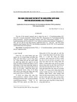
Figure 1-1 shows a typical procedure of microarray experiments (79, 80). A
microarray is a glass substrate surface on which DNA molecules are fixed in an
orderly manner at specific locations called spots (or features). A microarray may
contain thousands of spots, and each spot may contain a few million copies of
identical DNA molecules (probes) that uniquely correspond to a gene. The DNA
in a spot may either be genomic DNA (81), or synthesized oligo-nucleotide
strands that correspond to a gene (82-84). This microarray can be made by the
experimenters themselves (such as cDNA array) or purchased from some suppliers
(such as Affymetrix GeneChip). The actual microarray experiment starts from the
RNA extraction from cells. These RNA molecules are reverse transcribed into
cDNA, labeled with fluorescent reporter molecules, and hybridized to the probes
formatted on the microarray slides. At this step, any cDNA sequence in the sample
will hybridize to specific spots on the glass slide containing its complementary
sequence. The amount of cDNA bound to a spot will be directly proportional to
the initial number of RNA molecules present for that gene in both samples.
Following, an instrument is used to read the reporter molecules and create
Chapter 1 Introduction
8
microarray image. In this image, each spot, which corresponds to a gene, has an
associated fluorescence value, representing the relative expression level of that
gene. Then the obtained image is processed, transformed and normalized. And the
analysis, such as differentially expressed gene identification, classification of
disease/normal status, and pathway analysis, can be conducted.
Figure 1-1 Procedure of microarray experiment
1.2.3.2 Public repository for microarray data
Thanks to the variety of journals and funding agencies which have established
Microarray making Hybridization
+
mRNA reverse transcription
Fluorescentlabeling
Sample A Sample B
RNA extraction
RNA sample A RNA sample B
Microarray hybridization
Microscope glass slides
DNA molecules
amplified by PCR
Spotting
microarray
Image acquisition and analysis
Identification of
differentially
expressed genes
Classification Other analyses (e.g.
pathway analysis)
Cy3 labeled sample A
(green)
Cy5 labeled sample B
(red)
Chapter 1 Introduction
9
and enforced microarray data submission standards, currently, a wealth of
microarray data is now available in different databases such as the Stanford
Microarray Database (SMD) (85), Gene Expression Omnibus (GEO) (86), and
Array Express (EBI) (87). Table 1-1 gives a list of public available microarray
databases. Many of those databases require a minimum information about a
microarray experiment (MIAME)-compliant manner in order to interpret the
experiment results unambiguously and potentially be able to reproduce the
experiment (88). As a public resource, these expression databases are valuable
substrates for statistical analysis, which can detect gene properties that are more
subtle than simple tissue-specific expression patterns.
1.2.3.3 Statistical analysis of microarray data
Since microarray contains the expression level of several thousands of genes,
it requires sophisticated statistical analysis to extract useful information such as
gene selection. Theoretically, one would compare a group of samples of different
conditions and identify good candidate genes by analysis of the gene expression
pattern. However, microarray data contain some noises arising from measurement
variability and biological differences (70, 89). The gene-gene interaction also
affects the gene-expression level. Furthermore, the high dimensional microarray
data can lead to some mathematical problems such as the curse of dimensionality
and singularity problems in matrix computations, causing data analysis difficult.
Therefore choosing a suitable statistical method for gene selection is very
important.
Chapter 1 Introduction
10
Table 1-1 A list of public microarray databases.
Database Website* Description Organism References
ArrayExpress
/>arrayexpress/
A public repository for
microarray based gene
expression data
European
Bioinformatics
Institute
(87)
ChipDB
.e
du/chipdb/public/
A searchable database of gene
expression
Massachusetts
Institute of
Technology
(90)
ExpressDB
v
ard.edu/ExpressDB/
A relational database
containing yeast and E. coli
RNA expression data
Harvard Medical
School
(91)
Gene Expression
Atlas
g/SymAtlas/
A database for gene expression
profile from 91 normal human
and mouse samples across a
diverse array of tissues, organs,
and cell lines
Novartis Research
Foundation
(92)
Mouse Gene
Expression
Database (GXD)
ormati
cs.jax.org/menus/exp
ression_menu.shtml
An extensive and easily
searchable database of gene
expression information about
the mouse
The Jackson
Laboratory, Bar
Harbor, Maine
(93)
Gene Expression
Omnibus (GEO)
.
nih.gov/geo/
Microarray database containing
tens of millions of expression
profiles
National Center for
Biotechnology
Information
(86)
GermOnline
monli
ne.org/index.html
Information and microarray
expression data for genes
involved in mitosis and
meiosis, gamete formation and
germ line development across
species
Biozentrum and
Swiss Institute of
Bioinformatics
(94)
Human Gene
Expression
(HuGE) Index
database
techno
logycenter.org/hio/
A comprehensive database to
understand the expression of
human genes in normal human
tissues
Boston University (95)
MUSC DNA
Microarray
Database
http://proteogenomic
s.musc.edu/ma/musc
_madb.php?page=ho
me&act=manage
A web-accessible archive of
DNA microarray data
Medical University
of South Carolina
(96)
RIKEN
Expression Array
Database (READ)
en.g
o.jp/
A database of expression
profile data from the RIKEN
mouse cDNA microarray
RIKEN Yokohama
Institute
(97)
Rice Expression
Database (RED)
.jp/RED/
Expression profiles obtained by
the Rice Microarray Project
and other research groups
National Institute
of Agrobiological
Sciences, Japan
(98)
RNA Abundance
Database (RAD)
n
n.edu/RAD/php/inde
x.php
A public gene expression
database designed to hold data
from array-based and
nonarray-based (SAGE)
experiments
University of
Pennsylvania
(99)
Saccharomyces
Genome Database
(SGD):
Expression
Connection
stgenom
e.org/cgi-bin/expressi
on/expressionConnec
tion.pl
A gene expression database of
Saccharomyces genome
Stanford
University
(100)
Stanford
Microarray
Database (SMD)
http://genome-www5
.stanford.edu/
Raw and normalized data from
microarray experiments, as
well as their corresponding
image files
Stanford
University
(85)
Yale Microarray
Database (YMD)
e.e
du/microarray/
A microarray database for
large-scale gene expression
analysis.
Yale University (101)
yeast Microarray
Global Viewer
(yMGV)
nscript
ome.ens.fr/ymgv/
A database for yeast gene
expression
Ecole Normale
Superieure, Paris,
France
(102)
*accessible at Apr 06, 2007
Chapter 1 Introduction
11
The statistical methods in microarray data analysis can be classified into two
groups: unsupervised learning methods and supervised learning methods.
Unsupervised analysis of microarray data aims to group relative genes without
knowledge of the clinical features of each sample (103). A commonly-used
unsupervised method is hierarchical clustering method. This method groups genes
together on the basis of shared expression similarity across different conditions,
under the assumption that genes are likely to share the same function if they
exhibit similar expression profiles (104-107). Hierarchical clustering creates
phylogenetics trees to reflect higher-order relationship between genes with similar
expression patterns by either merging smaller clusters into larger ones, or by
splitting larger clusters into smaller ones. A dendogram is constructed, in which
the branch lengths among genes also reflect the degree of similarity of expression
(108, 109). By cutting the dendogram at a desired level, a clustering of the data
items into the disjoint groups can be obtained. Hierarchical clustering of gene
expression profiles in rheumatoid synovium identified 121 genes associated with
Rheumatoid arthritis I and 39 genes associated with Rheumatoid arthritis II (110).
Unsupervised methods have some merits such as good implementations available
online and the possibility of obtaining biological meaningful results, but they also
possess some limitations. First, unsupervised methods require no prior knowledge
and are based on the understanding of the whole data set, making the clusters
difficult to be maintained and analyzed. Second, genes are grouped based on the
similarity which can be affected by input data with poor similarity measures.
Third, some of the unsupervised methods require the predefinition of one or more
user-defined parameters that are hard to be estimated (e.g. the number of clusters).
Changing these parameters often have a strong impact on the final results (113).
Chapter 1 Introduction
12
In contrast to the unsupervised methods, supervised methods require a priori
knowledge of the samples. Supervised methods generate a signature which
contains genes associated with the clinical response variable. The number of
significant genes is determined by the choice of significance level. Support vector
machines (SVM) (114) and artificial neural networks (ANN) (115) are two
important supervised methods. Both methods can be trained to recognize and
characterize complex pattern by adjusting the parameters of the models fitting the
data by a process of error (for example, mis-classification) minimization through
learning from experience (using training samples). SVM separates one class from
the other in a set of binary training data with the hyperplane that is maximally
distant from the training examples. This method has been used to rank the genes
according to their contribution to defining the decision hyperplane, which is
according to their importance in classifying the samples. Ramaswamy et al. used
this method to identify genes related to multiple common adult malignancies (6).
ANN consists of a set of layers of perceptrons to model the structure and behavior
of neutrons in the human brain. ANN ranks the genes according to how sensitive
the output is with respect to each gene’s expression level. Khan et al identified
genes expressed in rhabdomyosarcoma from such strategy (27).
In classification of microarray datasets, it has been found that supervised machine
learning methods generally yield better results (116), particularly for smaller
sample sizes (89). In particular, SVM consistently shows outstanding performance,
is less penalized by sample redundancy, and has lower risk for over-fitting (117,
118). Furthermore, some studies demonstrated that SVM-based prediction system
was consistently superior to other supervised learning methods in microarray data
Chapter 1 Introduction
13
analysis (119-121). SVM for microarray data analysis are used in this study.
Feature selection in microarray data analysis
No matter whether the supervised or unsupervised methods are used, one
critical problem encountered in both methods is feature selection, which has
become a crucial challenge of microarray data analysis. The challenge comes from
the presence of thousands of genes and only a few dozens of samples in currently
available data. From the mathematical view, thousands of genes are thousands of
dimensions. Such a large number of dimensions leads microarray data analysis to
problems such as the curse of dimensionality (122, 123) and singularity problems
in matrix computations. Therefore, there is a need of robust techniques capable of
selecting the subsets of genes relevant to a particular problem from the entire set
of microarray data both for the disease classification and for the disease target
discovery.
Gene selection from microarray data is to search through the space of gene subsets
in order to identify the optimal or near-optimal one with respect to the
performance measure of the classifier. Many gene selection methods have been
developed, and generally fall into two categories: filter method and wrapper
method (124). Figure 1-2 shows how these two methods work.
In brief, the filter method selects genes independent of the learning algorithms
(125-127). It evaluates the goodness of the genes from simple statistics computed
from the empirical distribution with the class label (128). Filter method has some
pre-defined criteria. Mutual information and statistical testing (e.g. T-test and