Investigations on the toxicity of nanoparticles
Bạn đang xem bản rút gọn của tài liệu. Xem và tải ngay bản đầy đủ của tài liệu tại đây (14.5 MB, 227 trang )
INVESTIGATIONS ON THE TOXICITY OF
NANOPARTICLES
ASHARANI PEZHUMMOOTTIL
VASUDEVAN NAIR
(B. Sc Medical Microbiology)
A THESIS SUBMITTED FOR
THE DEGREE OF DOCTOR OF PHILOSOPHY
DEPARTMENT OF PHYSIOLOGY
YONG LOO LIN SCHOOL OF MEDICINE
NATIONAL UNIVERSITY OF SINGAPORE
2009
ii
ACKNOWLEDGEMENTS
It is an honour to thank people who made this dream come true. Though it is hard to express
my gratitude through words, I would like to express my heartfelt gratitude to my supervisor
Associate Professor M. Prakash Hande, for being a wonderful mentor. His constant
encouragement, suggestions, ideas, unfailing support and criticisms contributed to the
brilliance of the work. I am indebted to him for giving me a chance to work under his
supervision. I would like to extend my sincere thanks to my co-supervisor Associate
Professor Suresh Valiyaveettil, for his enormous trust and support during the high tides of the
work. His constant encouragement and ideas made this work fruitful.
I am thankful to Prof. Zhiyuan Gong, for spending his valuable time to guide me through the
in vivo work. His critical comments and suggestions helped a lot in the progress of this thesis.
I greatly appreciate the help from Wu Yilian and Zhan Huiqing and the training they
provided.
Special thanks to Prof. Sanjay Swarup and Prof. Chwee Teck Lim for their discussions and
constructive comments. I take this opportunity to thank my friends Dr. Manoj Parameswaran,
Dr. Bindhu L.V, Sajini Vadukkumpulli, Ganapathy Balaji, Resham Lal Gurung, Sethu
Swaminathan, Khaw Aikkia and Grace Low, who laughed and cried with me throughout my
best and worst times of lab work. I am thankful to my lab mates and colleagues Lakshmidevi
Balakrishnan, Dr. Anuradha Poonepalli, Kalpana GopalaKrishnan, Dimphy Zeegers,
Prarthana Sreekanth, Kristina, Dr. Sivamurugan and all members of Genome stability lab and
materials research lab.
Most importantly, I express my gratitude to my husband Rajesh Chandran and son Dev
Nandan Unnithan and my parents Leelamma K.K. and P.K. Vasudevan Nair, whose
understanding, continuous encouragement inspired this work.
I am grateful to my TAC members Prof. Kini Manjunatha and Dr. Bhaskar for the valuable
advice and critical comments.
iii
TABLE OF CONTENTS
Title Page
i
Acknowledgement
ii
Table of Contents
iii
Summary
x
List of Tables and Figures
xii
Abbreviations
xv
List of publications
xvii
CHAPTER 1
1 Introduction 2
1.1 Nanotechnology: An overview 2
1.2 Classification of nanomaterials 5
1.3 Synthesis and properties of metal nanoparticles 8
1.3.1 Size of the nanoparticles 10
1.3.2 Quantum confinement 10
1.3.3 Surface plasmon resonance 11
1.3.4 Morphology of the nanomaterials 12
1.3.5 Surface functionalisation 12
1.4 Nanotechnology: An outlook at current trends 13
1.5 Nanotechnology: Future prospects 14
1.6 Nanoparticles in the limelight 14
1.6.1 Gold nanoparticles 15
1.6.2 Silver nanoparticles 15
1.6.3 Platinum nanoparticles 17
1.7 Nanotechnology: A two sided sword? 18
iv
1.8 Lessons from history 18
1.9 Portals of entry of nanomaterials and factors
contributing to uptake
19
1.9.1 Inhalation 20
1.9.2 Absorption through skin 22
1.9.3 Ingestion 23
1.9.4 Translocation 24
1.10 Excretion of nanoparticles 26
1.11 Biodistribution at cellular levels 26
1.12 Literature in nanotoxicity 28
1.12.1 Cytotoxicity 28
1.12.2 Uptake of nanoparticles 31
1.12.3 Genotoxicity 31
1.12.4 Protein expression 32
1.13 Rationale 35
CHAPTER 2
2 Materials and Methods 38
2.1 Synthesis of nanoparticles 38
2.1.1 Synthesis of polyvinyl alcohol (PVA) capped silver
nanoparticles (Ag-np-1)
38
2.1.2 Synthesis of silver nanoparticles capped with Bovine
serum albumin (BSA, Ag-np-2)
38
2.1.3 Preparation of starch capped silver nanoparticles
(Ag-np-3)
39
2.1.4 Synthesis of PVA capped gold nanoparticles (Au-
np).
40
2.1.5 Synthesis of PVA capped platinum nanoparticles (Pt-np) 40
v
2.2 Cell culture and nanoparticle treatment
41
2.3 Preparation of stock solution and treatment 41
2.4 Uptake of nanoparticles 42
2.5 Microscopy 43
2.5.1 Light microscopy 43
2.5.2 Transmission electron microscopy of nanoparticles
treated cells
44
2.5.3 Scanning transmission electron microscopy (STEM) 44
2.5.4 Qualitative analysis of cell morphology by SEM 45
2.5.5 Live imaging of nanoparticles using cytoviva
ultrahigh resolution illumination systems
45
2.6 Cell Viability Assay 45
2.6.1 Measurement of ATP content 45
2.6.2 Mitochondrial function-cell titer blue cell viability
assay
46
2.7 Cell cycle analysis 47
2.8 Cell death 47
2.8.1 Annexin -V staining for apoptosis and necrosis 47
2.8.2 DNA fragmentation analysis 48
2.9 Detection of reactive oxygen species (ROS)
production
48
2.10 Evaluation of genotoxicity 49
2.10.1 Cytokinesis-blocked micronucleus assay (CBMN) 49
2.10.2 Alkaline single-cell gel electrophoresis (Comet
Assay).
50
2.10.3 Chromosomal analysis by fluorescence in situ
hybridisation (FISH)
51
vi
2.11 Colony formation studies 51
2.12 Analyses for protein/ gene expression 52
2.12.1 Western blotting 52
2.12.2 Gene expression profile using real time-reverse
transcriptase- polymerase chain reaction (RT-PCR)
52
2.12.3 Messenger RNA isolation and array hybridisation 53
2.13 Immunofluorescence staining for γH2AX 54
2.14 Isothermal titration calorimetry 55
2.15 Cytokine detection assay 55
2.16 Intracellular calcium measurement 56
2.17 Statistical analysis 56
2.18 Collection and exposure of the embryos to
nanoparticles
56
2.19 TEM analysis of the embryos 57
2.20 Acridine orange staining 58
2.21 4,6-diamidino-2-phenylindole-dihydrochloride
hydrate (DAPI) staining
58
2.22 Quantification of metal content in embryos 58
2.23 Preparation of single cell suspension from embryos
for cell cycle analysis
59
CHAPTER 3
3.1 Introduction 63
3.2 Results 64
3.2.1 Effect on cell morphology 66
3.2.2 Cell viability 68
3.2.3 Cellular uptake and exocytosis of nanoparticles 71
vii
3.2.4 Transmission electron microscopy (TEM) of cell
sections to study bio distribution
74
3.2.5 Production of ROS in human cells exposed to silver
nanoparticles
77
3.2.6 Genotoxicity of silver nanoparticles 79
3.2.6.1 DNA damage in silver nanoparticle treated cells 79
3.2.6.2 Micronuclei in silver nanoparticles treated cells 80
3.2.6.3 Chromosomal aberrations in silver nanoparticles
treated cells
82
3.2.7 Calcium fluctuations in silver nanoparticles
treatment
86
3.2.8 Effect of silver nanoparticles on cell cycle 88
3.2.9 Recovery and colony formation 91
3.2.10 Apoptosis and necrosis 93
3.2.11 Effect of silver nanoparticles on gene expression 97
3.2.12 Inflammatory response in nanoparticle mediated
cells
107
3.2.13 Binding of cytosolic proteins with Ag-np-3 108
3.3 Discussion 111
3.3.1 Uptake, distribution and bioactivity of nanoparticles 111
3.3.2 Mitochondrial respiratory chain, synthesis of ATP
and ROS production
113
3.3.3 ROS, Ca
2+
homeostasis and cytoskeleton changes 117
3.3.4 DNA damage and ROS 119
3.3.5 DNA damage, cellular ATP content and cell cycle
arrest
120
3.3.6 Effect on gene expression profiles 121
viii
3.3.7 Interaction of silver nanoparticles with cytosolic
proteins
125
3.3.8 Release of pro-inflammatory cytokines from silver
nanoparticles treated fibroblasts
126
CHAPTER 4
4.1 Introduction 129
4.2 Results 130
4.2.1 Microscopy of cells treated with Pt-np 131
4.2.2 Uptake and distribution studies 132
4.2.3 Cytotoxicity 134
4.2.4 ROS production 136
4.2.5 Genotoxicity of Pt-np 138
4.2.6 Effect of Pt-np on cell cycle, apoptosis and necrosis 140
4.2.7 Colony formation 143
4.2.8 Protein levels in Pt-np treated cells 145
4.3 Discussion 145
CHAPTER 5
5.1 Introduction 151
5.2 Results 152
5.2.1 Comparison of toxicity of different metal
nanoparticles
152
5.2.2 Effect of nanoparticles on mortality and hatching rate 154
5.2.3 Effects of nanoparticles on organogenesis 155
5.2.4 Effect of nanoparticles on cardio vascular system 160
5.2.5 Touch response of the larvae 163
5.2.6 Nanoparticle uptake by the embryos 164
ix
5.2.7 Toxicity of corresponding metal ions 164
5.2.8 Probing the toxicity of Silver nanoparticles 165
5.2.9 Mortality, heart rate, edema and malformations 165
5.2.10 Biodistribution of silver nanoparticles in zebrafish
embryos
171
5.2.11 Cell cycle analysis of single cells isolated from
zebrafish embryos
171
5.2.12 Gene expression in silver nanoparticles treated
embryos
174
5.2.13 Protein expression in silver nanoparticles treated
embryos
174
5.3 Discussion 177
CONCLUSION
6.1 Conclusions 185
6.2 Future prospects 189
REFERENCES
x
Summary
Nanoparticles, even though small in dimension, have a huge impact on the
economy. Nanotechnology is a multidisciplinary approach that is perceived to be
building up the future of coming era. Thus, it is absolutely necessary to understand the
health impact of the nanomaterials to facilitate a safe and sustainable progression of
the nanotechnology. Nanotoxicology is one of the latest branches of nanotechnology
that investigate the biological properties of nanoparticles. Previous studies in
nanotoxicology demonstrated adverse health effects of many commercialised
nanomaterials. Based on the early reports, a robust research was initiated to
understand the toxicity of nanomaterials currently in demand.
In the studies described in this thesis, we have investigated the toxicity
associated with silver and platinum nanoparticles both in vitro and in vivo. The
nanoparticles were screened using zebrafish embryos and human cell lines, to identify
potential toxicity of the nanoparticles, which were further investigated to elucidate the
mechanism of toxicity. In vivo models were monitored for developmental defects such
as pericardial and yolk sac edema, bent notochord, malformation of eyes,
accumulation of blood etc. The distribution of the toxic nanoparticles inside the
embryos were further studied by using transmission electron microscopy of embryo
sections, which showed presence of nanoparticles in various developing organs such
as brain, heart etc. Nanoparticle deposition was seen in the nucleus of the embryonic
cells as well. Cell lines (human lung fibroblasts and human glioblastoma cells) were
treated with various nanoparticles to identify the degree of toxicity through viability
assay. The mechanism of nanoparticles uptake and bio distribution was studied in
detail. Metabolic activity in nanoparticles treated cells were measured using ATP
content of cells and mitochondrial activity which indicateded metabolic dysfunction.
xi
Generation of reactive oxygen species was measured using fluorescence staining and
subsequent flow cytometry analysis which established increased production of
hydrogen peroxide and superoxide. Oxidative stress is a common cause of DNA
damage in chemical toxicity. Therefore, DNA damage in cells was studied using
single cell gel electrophoresis and other genotoxic effects of nanoparticles were
looked at by studying chromosomal aberrations (fluorescence insitu hybridizations)
and micronucleus formation. The nanoparticle treated cells showed increased DNA
damage, micronuclei formation and chromosomal aberrations. The fate of the cells
was further studied through cell cycle analysis and cell viability-death assay by flow
cytometry, which further showed a G
2
/M arrest and minimal cell death at higher
concentration of nanoparticles. Recovery of treated cells was monitored and the ability to
form colonies was investigated. Colony formation assay showed absence of colony
formation only in silver nanoparticles treated cells, which was more pronounced in cancer
cells. The genes and proteins differentially expressed following nanoparticle treatment were
identified through pathway specific array, RT-PCR and western blotting. The interactions
of silver nanoparticles with cytosolic proteins were studied through isothermal titration
calorimetry which evidenced strong interaction with proteins. Platinum nanoparticles
exhibited a lesser degree of toxicity compared to silver nanoparticles. In vivo models expose
to silver nanoparticles exhibited up regulation of genes involved in DNA damage and
oxidative stress.
In summary, this study has identified significant toxicity associated with the
commercially available nanomaterials. Thus it is ideal that large scale production and
commercialisation of such nanoparticles must be minimised until proper guidelines are
developed. Also, nano-wate disposal must be taken care of to avoid environmental
pollution.
xii
List of Tables and Figures
Number Title
Page
number
List of Tables
1.1 Commercially available nanoparticle based wound dressings. 17
1.2 Summary of literatures in silver and platinum nanotoxicology 33
2.1 Primer sequence used in RT-PCR 60
3.1 Summary of chromosomal aberrations observed in cancer cells
and normal cells.
85
3.2 Cell signalling pathways involved in silver nanoparticle
toxicity
105
5.1 Weight percentage of metal present in nanoparticle 154
5.2 Touch responses in nanoparticles treated larvae 163
List of Figures
1.1 Updated nano products inventory from 24 countries 4
1.2 Morphological variants of nanomaterials 7
1.3 High resolution electron micrograph of QD showing
arrangement of atoms
9
1.4 Schematic representation of a nanoparticle showing factors
affecting its propertie
10
1.5 Dichroic appearance of Lycurgus cup due to SPR of silver and
gold nanoparticles
12
1.6 Potential routes of exposure, translocation and deposition of
nanoparticles
20
3.1 Characterisation of silver nanoparticles 65
3.2 Microscopic observations of silver nanoparticle treated cells 67
3.3 Cytotoxicity studies of silver nanoparticles 70
3.4 Uptake of silver nanoparticles 73
3.5 TEM images of ultrathin sections of the cells 75
xiii
3.6 Elemental mapping of cell sections 77
3.7 ROS production in silver nanoparticles treated cells 78
3.8 Comet analysis of silver nanoparticles treated cells 80
3.9 Micronucleus analysis for chromosomal aberrations in silver
nanoparticles exposed cells
81
3.10 The chromosomal aberrations in IMR-90 and U251 cells 83
3.11 Calcium measurements 87
3.12 Histograms representing cell cycle analysis of IMR-90 and
U251 cells exposed to silver nanoparticles
89
3.13 Cell cycle analysis of silver nanoparticles treated cells 90
3.14 Recovery studies 92
3.15 Dot plots from Annexin V staining of IMR-90 and U251 cells 95
3.16 Apoptosis in silver nanoparticles treated cells 96
3.17 Differential gene expression in cell cycle pathway 98
3.18 DNA damage in silver nanoparticles treated cells 100
3.19 Altered gene expression profile in silver nanoparticle treated
cells
103
3.20 mRNA profile as measured by RT-PCR 106
3.21 Silver nanoparticles induced cytokine and chemokine
production in normal human lung fibroblasts
108
3.22 Isothermal titration calorimetry measuring the binding of
starch capped silver nanoparticle to cytosolic proteins and pure
starch with cytosolic proteins
110
4.1 Characterisation of Pt-np 130
4.2 Microscopic images of cells exposed to Pt-np 131
4.3 Uptake and distribution of Pt-np 133
4.4 Cytotoxicity assays of Pt-np treated cells 135
4.5 ROS production in Pt-np treated cells 137
4.6 Genotoxicity of Pt-np 139
4.7 Cell cycle analysis of Pt-np treated cells 142
xiv
4.8 Colony formation studies in Pt-np treated cells 144
4.9 Protein expression profiles in Pt-np treated normal and cancer
cells
145
5.1 Characterization of nanoparticles 153
5.2 Dose dependant toxicity of nanoparticles in mortality and
hatching
155
5.3 Phenotypic observations in nanoparticle treated embryos at
different time points
157
5.4 Detailed analyses of phenotypic defects observed in Ag-np-1
treated embryos
159
5.5 Comparison of heart rate of embryos 162
5.6 Metal retention of gold, platinum and silver in embryos
exposed to nanoparticles
164
5.7 Toxicity of Ag
+
, Pt
2+
and Au
3+
ions in the zebrafish embryo 165
5.8
Microscopic images of silver nanoparticles treated embryos
167
5.9 Graphs representing the toxicity of Ag-np-2 and Ag-np-3 in
terms of heart rate, hatching and mortality
169
5.10
Graphs represent effect of Ag
+
on embryos
170
5.11
TEM images of ultrathin sections of the embryos treated with
25 µg/mL of silver nanoparticles
172
5.12 Cell cycle analysis of embryos exposed to silver nanoparticles 173
5.13
mRNA and Protein levels in zebrafish embryos after
nanoparticle exposure
176
6.1
Proposed mechanism of action of silver and platinum
nanoparticle
191
xv
ABBREVIATIONS
PAGE Poly acrylamide gel electrophoresis
MAPK Mitogen activate protein kinase
QD Quantum dots
CNT Carbon nanotubes
NFB
Nuclear factor kappa B
H
2
O
2
Hydrogen peroxide
O
2-
Superoxide anion
Rb retinoblastoma
Cdk Cyclin dependant kinase
PCNA Proliferating cell nuclear antigen
ICAM Interceullular adhesion molecule
MCP Monocytes chemotactic protein
GM-CSF Granulocyte-macrophage colony stimulating factor
IL Interleukin
IFN Interferon
MIP Macrophage inflammatory protein
GRO Growth related oncogene
ERK Extracellular signal regulated kinases
BRCA-1 Breast cancer-1
xvi
ATM
Ataxia telangiectasia mutated
ATR Ataxia telangiectasia and Rad3 related
PAX 6
Paired box 6
BMP
Bone morphogenic protein
Six3
Sine oculis homeobox 3
EDX
Energy dispersive X-ray spectroscopy
ROS Reactive oxygen species
TEM
Transmission electron microscopy
SEM
Scanning electron microscopy
SOD Superoxide dismutase
PPM
Parts per million
JNK
c-jun N-terminal kinase
PI
Propidium iodide
BAX
Bcl2 associated x protein
Bcl
2
B-cell CLL/lymphoma 2
PVA
Polyvinyl alcohol
xvii
LIST OF PUBLICATIONS
Published papers
1. P. V. AshaRani, Ng Xinyi, Manoor Prakash Hande and Suresh Valiyaveettil.
DNA damage and p53 mediated growth arrest in human cells treated with
platinum nanoparticles. Nanomedicine, 2010, 5(1). 51-64.
2. P.V. AshaRani, Yi Lian Wu, Zhiyuan Gong and Suresh Valiyaveettil.
Comparison on the toxicity of silver, gold and platinum nanoparticles in the
early development of Zebrafish embryos. Nanotoxicology. 2010. In Press.
3. P. V. AshaRani, Swaminathan Sethu., S.P. Zhong, C.T. Lim, M. Prakash
Hande and Suresh Valiyaveettil. Effects of silver, gold and platinum
nanoparticles on normal human erythrocytes. Adv. Funct. Mater.2010, 20(8),
1233-42.
4. P. V. AshaRani, Manoor Prakash Hande and Suresh Valiyaveettil. Anti-
proliferative properties of silver nanoparticles. BMC Cell biology, 2009, 10:65
5. P. V. AshaRani, Grace Low Kah Mun, Manoor Prakash Hande and Suresh
Valiyaveettil. Cytotoxicity and Genotoxicity of Silver Nanoparticles in Human
Cells. ACS Nano, 2009, 3 (2), 279-290.
6. P. V. AshaRani, Yi LianWu, Zhiyuan Gong and Suresh Valiyaveettil.
Toxicity of silver nanoparticles in zebrafish embryos. Nanotechnology, 2008,
19, 255102 (8pp).
7. P. V. AshaRani, N. G. B. Serina, M. H. Nurmawati, Yi Lian Wu, Zhiyuan
Gong, and Suresh Valiyaveettil. Impact of Multi Walled Carbon Nanotubes
(MWCNTs) on Aquatic Species. Journal of Nanoscience and
Nanotechnology, 2008, 8, 1–7.
Submitted/under preparation
8. P. V. AshaRani, Swaminathan Sethu,
Ganapathy. Balaji,
M. Prakash Hande
and Suresh Valiyaveettil. Effects of silver nanoparticles on selective gene
expression profiles and inflammatory mediators in human cells. (Submitted to
journal)
xviii
9. P. V. AshaRani, Stephanie Katherine Loeb, Sajini Vadukkumpully, M.
Prakash Hande and Suresh Valiyaveettil. Hemocompatibility of metal oxide
nanoparticles (Submitted to journal)
10. Hairong Li, Teck Chuan Ng, Asharani P V and Suresh Valiyaveettil Design,
Synthesis and Radical Scavenging Capacities of Cross-Conjugated
Polyphenols. (Submitted to journal)
11. Sivamurugan Vajiravelu, Asharani P.V. Wu Jiang and Suresh Valiyaveettil.
In Vitro and In Vivo Toxicity Studies of Synthetic Gallo Tannins in Cancer
Cell lines and Zebrafish embryos (Submitted to journal).
12. Vajiravelu Sivamurugan, Asha Rani. P. V. Lin Sihan and Valiyaveettil
Suresh. Synthesis and Characterisation of Synthesised Gallo tannins and
Inhibition of U251 Cancer Cells Growth (To be submitted to the journal).
CONFERENCES
1. P. V. AshaRani, Manoor Prakash Hande, and Suresh Valiyaveettil.
Cytotoxicity and Genotoxicity of Silver Nanoparticles. ICMAT, Singapore,
2009 (Oral presentation).
2. P. V. AshaRani, Manoor Prakash Hande, and Suresh Valiyaveettil. Silver
nanoparticles: toxicity, anticancerous and antimicrobial properties. Asianano
2008, Singapore, 2008 (Oral presentation).
3. P. V. AshaRani, Manoor Prakash Hande, and Suresh Valiyaveettil. Toxicity
of silver nanoparticles in human cells, ACS meeting, Philadelphia, 2008
(Oral presentation).
4. P. V. AshaRani, Zhiyuan Gong, and Suresh Valiyaveettil. Potential Health
impacts of silver nanoparticles. Joint OLS-NUSNI-NERI-OSHE workshop
on the safety health and Environmental aspects of engineered nanomaterials,
Singapore, 2007.
5. P. V. AshaRani, Manoor Prakash Hande, and Suresh Valiyaveettil. Silver
nanoparticles in nanotoxicology and nanomedicine. Joint OLS-NUSNI-
NERI-OSHE workshop on the safety health and Environmental aspects of
Engineered nanomaterials, Singapore, 2007.
xix
6. P. V. AshaRani, Zhiyuan Gong, Manoor Prakash Hande and Suresh
Valiyaveettil. Potential Health impacts of silver nanoparticles: ACS meeting,
Boston, 2007.
7. Bindhu, L. V. P. V. AshaRani, Fathima. S. J. Hussain and Valiyaveettil
Suresh. Biomimetic peptide amphiphiles modified nanofibre mesh as a
scaffold for Tissue Engineering. Poster presentation. MRS meeting. San
Francisco, 2007.
8. P. V. AshaRani, Wu Yilian, Gong. Z, LakshmiDevi B, Prakash Hande and
Suresh Valiyaveettil. Probing the molecular mechanisms of nanoparticle
toxicity. 8th Asian Academic Network for Environmental Safety and Waste
Management (AANESWM), India, 2007 (Oral presentation).
9. P. V. AshaRani and Suresh Valiyaveettil “Probing the molecular mechanisms
of nanoparticle toxicity” OLS –NUSNI workshop, Singapore, 2006.
10. P. V. AshaRani, Gong ZY, Hande M.P., Valiyaveettil Suresh. Interactions
between carbon nanotube and various cell lines. Oral presentation. OLS –
NUSNI workshop, Singapore, 2006.
11. P. V. AshaRani, Gong ZY, Hande M.P., Valiyaveettil Suresh. Interactions
between carbon nanotube and various cell lines. OLS –NUSNI workshop,
Singapore, 2006 (Oral presentation).
C
C
H
H
A
A
P
P
T
T
E
E
R
R
1
1
I
I
N
N
T
T
R
R
O
O
D
D
U
U
C
C
T
T
I
I
O
O
N
N
Chapter 1 Toxicity of nanomaterials
2
1. Introduction
1. 1. Nanotechnology: An overview
The term ‘nano’ derived from the Greek word meaning ‘dwarf’. It represents
one billionth of a unit. Nanometre stands for 10
-9
of a metre and nano litre denotes 10
-9
of a litre. The term nanotechnology was conceived by Dr. Norio Taniguchi
in 1974
(Taniguchi, 1974). However, the glory of nanotechnology dates back to Fourth
century A.D, to the era of Roman Empire Lycurgus and the famous Lycurgus cup. The
Lycurgus cup was carved with exceptional workmanship to depict the triumph of King
Dionysus over Lycurgus. The cup is made up of colloidal silver and gold, which gives
a dichroic appearance to the glass; opaque green colour in reflected light due to
colloidal silver (300 ppm) and ruby red in transmitted light due to the presence of
colloidal gold (40 ppm) (Freestone et al., 2007). The revival of modern nanotechnology
began in 1959, following the inspiring speech (“There is plenty of room at the bottom”)
by the American Physicist Dr. Richard Feynman (Feyman R.P, 1960).
The golden era of nanotechnology that designs, synthesise and characterise
nanoparticles through a “bottom up approach” materialised following the pioneering
work by Dr. Eric Drexler (Drexler, 1986). The exceptionally sensitive technology that
manipulates materials at atomic level to create nano-sized objects, took a humongous
leap in the 1980’s following the invention of fullerenes (1985), carbon nanotubes
(CNTs, 1991) and advanced microscopic technique like scanning tunnelling
microscopy (1981).
The interest created by nanotechnology has initiated rapid economic growth
and industrial developments. In the near future, nanotechnology may emerge as an
Chapter 1 Toxicity of nanomaterials
3
important scientific discipline which designs and develop nanomaterials with unique
physio-chemical properties. A large number of US based federal regulatory bodies
were launched following the sudden increase in active nano research and
commercialisation. National Nanotechnology Initiative launched in 2000, is one such
kind which has centres in different countries. Nanotechnology regulatory bodies have
resulted in a well organised network for co-ordinating the inventions, sharing and
classifying the information based on its relevance. This has led to a more appropriate
classification of nanoparticles specifically as fine particles (200 nm to 10 µM) and
ultra-fine particles (<100 nm).
In the recent years, the increased interest in nanotechnology has initiated many
industries to commercialise of nanomaterials at a rapid rate of 3-4 new products every
week compared to other leading technologies (Analysis, 2009).
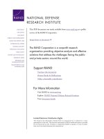
Based on the statistics
published in 2006 by Nanotechnology consumer products inventory, around 600 nano
based products are currently marketed by approximately 322 companies (Nel et al.,
2006). The most recent statistics published in 2009 revealed an increase in the number
of commercialised nano products to 1015, with 605 products in health and fitness, 152 in
household products, 98 in food and beverages, 57 in sporting goods and 19 products in
baby/child products (Analysis, 2009). The analysis revealed silver nanoparticles as the
most commercialised materials. The detailed inventory charts are shown in Figure 1.1.
4
C
o
sm
eti
cs
C
lothin
g
Perso
nal
Car
e
Sporti
ng
Goo
ds
Su
n
scree
n
Filtratio
n
0
50
100
150
200
8 March 2006
25 August 2009
Number of Products
Health and Fitness Subcategory
Health and Fit
n
ess
H
om
e
a
nd G
a
rde
n
Electronics
a
nd Computer
s
Food a
nd
B
e
v
e
rage
C
r
os
s
Cut
t
i
ng
Automot
i
ve
Appl
ia
nc
e
s
G
oods
f
or
Chil
dre
n
0
100
200
300
400
500
600
8 March 2006
25 August 2009
Number of Products
Product Category
Si
l
ver
Carbon
Z
i
n
c
Si
l
ica
/
Si
l
icon
Ti
ta
niu
m
G
o
l
d
0
75
150
225
300
8 March 2006
25 August 2009
Number of Products
Major Materials
0
200
400
600
800
1000
1200
1400
1600
2005 2006 2007 2008 2009 2010 2011
Number of Products
Total Products Listed
Figure 1.1: Updated nano products inventory from 24 countries. The
graphs show increase in the commercialisation of nanoparticles every
year in different areas (Analysis, 2009).
Chapter 1 Toxicity of nanomaterials
5
Human engineered nanoparticles with surfaces designed and functionalised to
perform specific functions are created in large scale these days. It is estimated that the
production of nanoparticles for commercial use will increase from 2300 tons produced
today to 58000 tons by 2020 (Xia et al., 2009). Based on the current sales figures, the
market is expected to exceed one trillion US dollars by 2015 (Xia et al., 2009). Nano
based commercial products are expected to revolutionize areas such as therapeutic
medicine and information technology. Engineered nanomaterials are currently used in
textiles, sporting equipments, medical applications and cosmetics (Bawarski et al.,
2008; Sgobba and Guldi, 2009; Staggers et al., 2008). The wide array of applications
has paved the way for the emergence of multiple branches of nanotechnology,
depending on the applications for which they are designed. Nanobiotechnology,
nanoelectronics, nanomagentics, nanophotonics, nanomechanics, nanolithography,
nanomedicine and nanotoxicology are among a few.
1. 2. Classification of nanomaterials
Nanoparticles can be produced by either
i) ‘Top down’ approach, where bulk materials are broken down to nano size
by milling or etching.
ii) ‘Bottom up’approach that makes nano sized objects by combining atomic
scale materials (Hallock et al., 2008).
Even though many different classifications exist, nanomaterials are mainly
classified based on their composition and shape. Based on composition nanoparticles
are classified as:
Chapter 1 Toxicity of nanomaterials
6
(a) Organic nanoparticles (eg. Polymeric nanoparticles)
(b) Inorganic nanoparticles such as metal nanoparticles (eg. gold, silver)
(c) Organic–inorganic hybrids (eg. nanocomposites)
(d) Carbonaceous nanostructures (eg. Buckyballs)
(e) Liposomes that can be filled with specific materials and
(f) Biological nanoparticles such as proteins.
Based on their shape, nanomaterials are classified as nanotubes, nanoparticles,
nanoprisms, nanocubes, nanosheets and nanorods. Different morphological variants of
nanomaterials are represented in Figure 1.2.