Molecular signatures of gastric cancer an integrated approach to molecular cytogenetics, whole genome copy number and transcriptome profiles
Bạn đang xem bản rút gọn của tài liệu. Xem và tải ngay bản đầy đủ của tài liệu tại đây (13.74 MB, 372 trang )
MOLECULAR SIGNATURES OF GASTRIC
CANCER:
AN INTEGRATED APPROACH TO MOLECULAR
CYTOGENETICS, WHOLE GENOME COPY
NUMBER AND TRANSCRIPTOME PROFILES
LEONG SIEW HONG
(B.Sc., NUS)
A THESIS SUBMITTED
FOR THE DEGREE OF DOCTOR OF PHILOSOPHY
DEPARTMENT OF BIOCHEMISTRY
NATIONAL UNIVERSITY OF SINGAPORE
2009
Acknowledgements
ii
Acknowledgements
It is my greatest honor for being given the golden opportunity to be guided and
mentored by my most dedicated supervisor Dr. Kon Oi Lian. Without her
continual support and encouragements, this thesis will not come to fruition. I
thank her deeply for believing in me and to be that guiding star during my
darkest days in my scientific pursues. I am very grateful to her for always
being open to new ideas and allowing me to chart my own course while
supporting me in every step of the way. She is always there to lend a listening
ear and offering me sound advice. Her passion and dedication for scientific
research had been awe-inspiring and she will always be my role model.
This project, although conceived and performed mostly at the National Cancer
Center, will not be successful without the support and assistance from many
important people. I would especially like to acknowledge Dr. Tan Soo Yong,
Consultant pathologist at SGH. As a member of my TAC committee, his
feedbacks and advice are very much valued. His generosity in sharing his
technical expertise and resources had gone a long way in making this study a
success. I would also like to thank my other advisers and collaborators who
had one way on another been tremendously helpful during the course of this
work. They are: Dr. Patrick Tan (my TAC committee chairman, collaborator
and adviser), Dr. Lai Siang Hui (for histo-pathological assessment of primary
GCs), Dr. Mac Ho and Dr. Tony Lim (in the provision of pre-malignant
gastric tissue samples), Dr. Nallasivam Palanisamy (who spent many hours
relating his molecular cytogenetic experiences).
I am very grateful to Magdalene Koh of the Pathology Department whose
tissue sectioning skills are probably the best in Singapore. Her technical
competency had made a dramatic turnaround in the success of this study. I am
also very blessed to be a member of a wonderful team at NCC. I want to thank
the members of my lab: Louise Lee, (for the tremendous bioinformatic
support), Ng Wai Har (xenoimplantation of tumor samples), Nelson Chen (for
giving me critical feedback in all aspects of my work), Jaichandran (providing
his expert advice in some very challenging experiments) and Frank (my new
Acknowledgements
iii
and enthusiastic member of the lab). To my former colleagues: Mark Tan (my
teacher in the initial stages of microarray data analysis), Cheryl Lee (who
assisted in cell culture), Serene Lok (always there to hear out my frustrations),
Long Yun Chau (now at Harvard), Jaya Visvanathan (Baylor college of
Medicine) and Wee Choon Wei (Temasek Life Sciences). You’ve been great
fun to be with and have certainly made great impact to my research life.
You’ve been the best people I have met in all my life and I cherish every
moment in the lab because you have made working in the lab very enjoyable.
I’m also deeply appreciative to Nelson Chen and Louise Lee who had
dedicated much time and effort to review this thesis and gave very
constructive feedbacks and suggestions. Also not forgetting Ng Kia Min who
had been a great help in the earlier establishment of the TMA-FISH protocols.
I would also like to acknowledge past and current members from Dr. Patrick
Tan’s lab (Wong Kee Yew, Amit Aggarval, Yu Kun and Tao Jiong). Special
thanks to Doris Ma who is always a good friend and forever approachable
whenever administrative affairs need to be tackled. To the folks at The
Wellcome Trust Sanger Institute at Hinxton, Cambridge: Dr Nigel Carter, Ng
Bee Ling, Fu Beiyuan, Yang Fengtang, Susan Gribble and Elena Elenor. The
month-long training had been an eye-opener. I treasure the friendship we’ve
established and I thank you in everyway I could.
I would also specially want to thank Dr. Carol Tang (NNI, Singapore) who
spurred me on in times of frustration and for being my beacon in the undying
passion for scientific discoveries.
I would like to thank the Singapore Millennium Foundation for their
scholarship support in the program and also their generous sponsorship for the
chromosome flow-sorting training at the Sanger Institute.
And finally to the most important people in my life: my family. I will not have
been able to complete this thesis without the continual support of my lovely
wife, who stood by me in this arduous journey. To my very adorable kids, this
thesis is for you too. For Dad and Mum, I have fulfilled your dreams and mine
too! I love you!!
Table of contents
iv
Contents
TITLE PAGE i
ACKNOWLEDGEMENTS ii
TABLE OF CONTENT iv
SUMMARY vii
LIST OF TABLES xi
LIST OF FIGURES xiii
LIST OF ABBREVIATIONS xx
REVIEW OF THE LITERATURE
1. Gastric cancer epidemiology and clinical features 1
2. Gastric cancer etiology 21
3. The African and Asian enigma 50
4. Possible molecular pathogenetic attributes of gastric cancer 66
5. Translocations in epithelial tumors and their possible roles 102
in oncogenesis
MATERIALS AND METHODS
6. Materials and Methods 121
RESULTS AND DISCUSSION
7. Spectral karyotyping of seventeen gastric cancer cell lines 148
7.1 Gastric cancer spectral karyotypes
7.2 Recurrent patterns of translocations
8. Multimodality whole genome characterization of gastric cancer 178
8.1 Whole genome copy number analysis
8.2 Global mRNA expression profiling
8.3 Global miRNA expression profiling
Table of contents
v
9. Dissecting simple rearrangements in gastric cancer cell lines 210
9.1 SNU-1: Cytogenetics
9.2 SNU-1: FISH-walking
9.3 SNU-1: Translocation mechanism
9.4 SNU-1: Molecular consequences
9.5 A balanced translocation in IM95
10. Dissecting complex rearrangements: integrating spectral 235
karyotypes and whole genome copy number profiles
10.1 Global integration of SKY and copy number
analysis
10.2 Copy number transitions and translocation
breakpoints
11. 18q2 translocation in primary gastric cancer 250
11.1 Break-apart FISH probe design and strategy
11.2 TMA construction and FISH assay optimization
11.3 18q2 break-apart FISH assay in primary gastric
adenocarcinomas
11.4 18q2 break-apart in non-gastric cancers
12. Chromosome 18 aberrations and GC histopathology 273
12.1 Clinico-pathological features and 18q21q22
breakpoint
12.2 Chromosome 18 aberrations in metaplasia,
dysplasia, early and late stage gastric cancers
13. Molecular characterization of 18q21q22 breakpoint genes 284
13.1 mRNA expression profiling
13.2 Expression of breakpoint genes by
immunohistochemical staining
Table of contents
vi
14. Precise determination of translocation breakpoints by 295
arraypainting
14.1 Chromosome sorting
14.2 Reverse FISH analysis
14.3 Array painting
CONCLUSION and FUTURE PROSPECTS 311
APPENDIX TABLES 314
APPENDIX FIGURES 334
Summary
vii
Summary
Gastric cancer (GC) is second only to lung cancer as a leading cause of cancer
deaths globally. Of the estimated 1 million newly diagnosed patients
annually
1
, up to 80% have advanced incurable disease. Even after surgery with
curative intent, 60% progress to locoregional and/or distant metastatic
disease
2
. As only surgery is potentially curative for GC, other treatments,
including molecularly targeted agents, are urgently needed for the majority
who are beyond surgical cure. One strategy for developing new curative
treatment is to identify genetic and genomic aberrations that are causal in the
initiation and/or progression of specific cancer types. Discoveries of druggable
cytogenetic signatures
have advanced prognostication and extended the
survival of several hematologic malignancies
3
The notable lack of reliable karyotypic data in most solid cancers has
encouraged the view that translocations are rarely associated with epithelial
cancer development. Fortunately, molecular cytogenetic techniques (e.g.
comparative genomic hybridization, spectral karyotyping, multicolor
fluorescence in situ hybridization (FISH), high resolution copy number and
tiling arrays, genome annotations and bioinformatics) have made solid tumor
cytogenetics more tractable, as demonstrated by recent discoveries of recurrent
translocations in human prostate adenocarcinomas
.
4
and non-small cell lung
cancer
5
. These signature translocations impute shared oncogenic mechanisms
among solid cancers and hematopoetic malignancies
6
Our exploration of this dreaded disease begins with a broad survey of the
current state of knowledge in five chapters. Chapter 1 reviews the current
epidemiological status of GC globally and in Singapore, including a short
.
Summary
viii
discussion of current management and treatment options. Chapter 2 explores
the interplay of various environmental as well as host and/or bacterial
susceptibility factors that may be associated with GC initiation and
progression. In Chapter 3, we examine the African and Asian enigmas –
apparent paradoxes in which high H. pylori infectivity coexists with low GC
prevalence. As cancer is caused and accompanied by structural and functional
genomic alterations, Chapter 4 summarizes and evaluates current
understanding of the molecular pathogenesis of GC. In Chapter 5, we review
signature chromosomal rearrangements in malignant disorders and particularly
note recent discoveries of such translocations in prostate and lung cancers.
Chapter 6 details all methods, materials and resources employed in this
project.
Data generated in this project appear in Chapters 7 – 14. Our work was
directed at five primary goals:
1) To characterize the chromosomal rearrangements, copy number
alterations, differentially expressed mRNAs and miRNAs in a survey
of 17 GC cell lines as a means of documenting the genomic
complexities of GC. These datasets enabled identification of unique
GC signatures based on copy number aberrations and mRNA
expression profiles compared to a well known panel of non-GC cell
lines, the NCI-60 panel. These results are presented in Chapters 7 and
8.
Summary
ix
2) To explore the utility of integrating different molecular cytogenetic
techniques to fully characterize the fusion breakpoints in a simple
rearranged GC cell line, SNU-1, and to propose a translocation
mechanism (Chapter 9).
3) To distinguish “driver” (pathogenic) from “passenger” (collateral)
rearrangements. Data in Chapters 10 and 11 showed that the novel and
recurrent 18q translocation breakpoint in cell lines was also mirrored in
primary GC tumors (but not in non-GC tumors) via a custom-designed
break-apart FISH assay.
4) To ascertain clinico-pathological features of 18q breakpoint-positive
GCs and to determine chromosome 18 status in pre-malignant lesions
(Chapter 12). These include analysis of the expression of two
candidate proteins, Serpin B8 and CD226 antigen by
immunohistochemistry (Chapter 13).
5) To molecularly define junctional DNA of candidate chromosomal
fusions, pure fractions of translocated chromosomes were flow-sorted
and their breakpoints accurately determined using oligo-based array
painting techniques (Chapter 14).
In short, this project highlights the value of integrating a suite of molecular
cytogenetic techniques with other genome-wide analyses in discovering
genomic signatures of GC. This integrated approach makes solid tumor
cytogenetics more tractable, is generalizable and could help to discover
oncogenic chromosomal rearrangements in other common cancers.
Summary
x
References:
1. Garcia, M., et al. Global Cancer Facts & Figures 2007.
( />Facts and_Figures_2007.asp) (2007).
2. Field, K., Michael, M. & Leong, T. Locally advanced and metastatic
gastric cancer: current management and new treatment developments.
Drugs 68, 299-317 (2008).
3. Fröhling, S. & Döhner, H. Chromosomal abnormalities in cancer. N.
Engl. J. Med. 359, 722-734 (2008).
4. Tomlins, S.A., et al. Recurrent fusion of TMPRSS2 and ETS
transcription factor genes in prostate cancer. Science 310, 644-648
(2005).
5. Soda, M., et al. Identification of the transforming EML4-ALK fusion
gene in non-small-cell lung cancer. Nature 448, 561-566 (2007).
6. Mitelman, F., Johansson, B. & Mertens, F. The impact of
translocations and gene fusions on cancer causation. Nat. Rev. Cancer
7, 233-245 (2007).
List of Tables
xi
List of Tables
Chapter 1
Table 1-1 ASR incidence and mortality rates of GC by ethnicity
Table 1-2 ASR incidence of GC in major ethnic groups in
Singapore
Table 1-3 TNM classification and stage grouping (AJCC/UICC)
Table 1-4 Treatment strategies with reference to pathological
stage
Chapter 2
Table 2-1 Mortality rates of Japanese men from Japan compared
to Japanese migrants
Chapter 3
Table 3-1 Cummulative GC incidence rates and H. pylori
seropositivity in major ethnic populations in Singapore
Table 3-2 Univariate analysis of ethnicity as an independent risk
factor
Chapter 4
Table 4-1 Cytogenetic analysis of GC tumors and cell lines (to
date)
Chapter 6
Table 6-1 General information of 17 GC cell lines
Table 6-2 List of BAC clones acquired for FISH assays
Table 6-3 List of GC cell lines and NCI-60 panel of non-GC
cell lines used in copy number analysis
Table 6-4 List of GC cell lines and NCI-60 panel of non-GC
cell lines used to determine mRNA expression
signatures in GCs
List of Tables
xii
Chapter 7
Table 7-1 Summary of composite spectral kayotypes and copy
number analysis of 17 GC cell lines
Table 7-2 List of forty-five recurrent breakpoints in 17 GC cell
lines
Chapter 8
Table 8-1 List of differentially expressed miRNAs and their
experimentally validated targets (to date)
Chapter 9
Table 9-1 Gene expression analysis of down-regulated genes
at the deleted cytoband 4q32.3q35.1
Chapter 10
Table 10-1 List of copy number altered clusters co-localized
with recurrent breakpoint cytobands
Chapter 11
Table 11-1 Scoring thresholds against false positive
classification of break-apart status
Table 11-2 Summary of 18q21q22 break-aparts in GCs and
non-GCs
Chapter 12
Table 12-1 Association analysis of primary GCs by their
18q21q22 break-apart status
Chapter 13
Table 13-1 Serpin B8 expression correlated to 18q12q22
break-apart status of primary GC tumors cores
Table 13-2 CD226 antigen expression in relation to
break-apart status in primary GC tumors cores
Table 13-3 Co-expression of Serpin-B8 and CD226 antigen
expression correlated with break-apart status
List of Figures
xiii
List of Figures
Chapter 1
Figure 1-1 Global incidence and mortality of common cancers
Figure 1-2 Global geographic variation in ASR of gastric cancer
(GC) among males and females
Figure 1-3 GC incidence rates (1973-1997) obtained from 18
cancer registries in Asia, Europe and the USA
Figure 1-4 Anatomic divisions of the human stomach
Figure 1-5 ASR incidence and mortality of GC by ethnicity in
Singapore
Figure 1-6 ASR incidence and mortality of GC in males and
females in Singapore
Figure 1-7 Survival statistics by TNM status
Chapter 2
Figure 2-1 Estimated number of new GC cases and deaths in
developing and developed countries
Chapter 4
Figure 4-1 Partial karyotype of a highly complex GC cell line,
Hs 746T
Figure 4-2 Genome-wide DNA copy number alterations of GC
in publicly available data using CGH and array CGH
Figure 4-3 The TGF-β signaling pathway
Chapter 5
Figure 5-1 Chronology of discoveries of fusion genes in epithelial
tumors
List of Figures
xiv
Figure 5-2 Schematic representation of possible outcomes of
chromosomal translocations
Chapter 6
Figure 6-1 Representative illustration of a H&E stained GC-TMA
section (4 µm)
Figure 6-2 Binary scoring criteria for immunohistochemical
analysis using anti-human Serpin B8
Figure 6-3 Scoring criteria for immunohistochemistry using
antibodies against human CD226 antigen, Serpin B2
and SOCS-6
Chapter 7
Figure 7-1 Schematic diagram of spectral karyotyping performed
on metaphase spreads
Figure 7-2 SKYgram illustrating AGS
Figure 7-3 SKYgram illustrating SNU-1
Figure 7-4 SKYgram illustrating NCI-N87
Figure 7-5 SKYgram illustrating SNU-5
Figure 7-6 SKYgram illustrating SNU-16
Figure 7-7 SKYgram illustrating KATO III
Figure 7-8 SKYgram illustrating Hs 746T
Figure 7-9 SKYgram illustrating IM95
Figure 7-10 SKYgram illustrating MKN7
Figure 7-11 SKYgram illustrating FU97
Figure 7-12 SKYgram illustrating YCC1
Figure 7-13 SKYgram illustrating YCC2
Figure 7-14 SKYgram illustrating YCC3
Figure 7-15 SKYgram illustrating YCC6
List of Figures
xv
Figure 7-16 SKYgram illustrating YCC9
Figure 7-17 SKYgram illustrating YCC11
Figure 7-18 SKYgram illustrating YCC16
Chapter 8
Figure 8-1 Whole genome copy number analysis of 17 GC cell
lines using 100K SNP mapping arrays
Figure 8-2 Classical chromosomal CGH profiles showing copy
number gains and losses in 17 GC cell lines
Figure 8-3 Summary of CGH profiles in 17 GC cell lines and
44/60 primary tumors
Figure 8-4 SOTA analysis showed similar clustering patterns
between 203 primary GCs (training set) versus 17 GC
lines and 44 primary GCs (test set)
Figure 8-5 Hierarchical clustering of copy number alterations
segregating GC from non-GC cell lines
Figure 8-6 Hierarchical clustering of differentially expressed
mRNAs segregating GC from non-GC cell lines
Figure 8-7 63-gene expression signature distinguishing GC from
non-GC cell lines
Figure 8-8 Principal component analysis of the 63-gene
expression signature
Figure 8-9 Representative image of a miRNA microarray
hybridization
Figure 8-10 Hierarchical clustering of differentially expressed
miRNAs of 17 GC cell lines and 40 primary gastric
adenocarcinomas
Figure 8-11 Differentially expressed miRNAs validated using
solution hybridization
Chapter 9
Figure 9-1 G-banded metaphase of SNU-1
List of Figures
xvi
Figure 9-2 Copy number profile of SNU-1 by chromosomal
CGH
Figure 9-3 SKY analysis of SNU-1
Figure 9-4 High resolution 100K SNP mapping array copy
number analysis of Chromosome 4
Figure 9-5 Schematic representation of dual labeled
FISH-walking probes at 4q31.1q32.3
Figure 9-6 FISH-walking analysis defined breakpoint at 4q32.3
Figure 9-7 High resolution 100K SNP mapping array copy
number analysis of Chromosome 1q
Figure 9-8 Schematic representation of dual labeled
FISH-walking probes at 1q24.2q25.3
Figure 9-9 FISH-walking analysis showed a spilt-apart signal
at 1q25.3
Figure 9-10 Schematic representation of dual labeled
FISH-walking probes at 4qter
Figure 9-11 FISH analysis revealed translocation of 4qter to der(1)
Figure 9-12 Schematic representation of FISH-walking probes at
4q26
Figure 9-13 FISH analysis revealed hidden paracentric inversion
at 4q26q32.3
Figure 9-14 A proposed translocation mechanism of SNU-1
Figure 9-15 Classification of the biological processes of genes
mapping to deleted cytoband 4q32.3q35.1
Figure 9-16 SKY analysis of IM95
Figure 9-17 Copy number profile of IM95 by chromosomal
CGH
Figure 9-18 100K SNP mapping array copy number analysis of
Chromosome 1 and 2 in IM95
List of Figures
xvii
Chapter 10
Figure 10-1 Global summary of whole genome copy number
analysis and recurrent translocations in 17 GC
cell lines
Figure 10-2 Copy number transition boundary co-localized to
4q32.3 breakpoint in SNU-1
Figure 10-3 SKY karyotypes of chromosome 18q translocations in
six GC cell lines
Figure 10-4 High resolution copy number analysis to identify
probable copy number transition boundaries
Chapter 11
Figure 11-1 100K SNP mapping array copy number analysis of six
GC cell lines with Chromosome 18 translocation
Figure 11-2 Schematic representation of the physical location of
break-apart probes at chromosome 18
Figure 11-3 Schematic representation of an 18q2 break-apart FISH
assay
Figure 11-4 The "Brute-force" break-apart probe strategy
Figure 11-5 Schematic representation of predicted signal patterns
in the "Brute-force" break-apart FISH assay
Figure 11-6 Optimization and validation of FISH assay on FFPE
tissues
Figure 11-7 Representative images showing 18q21q22
translocation and other 18q aberrations in primary
GCs
Figure 11-8 Immunostaining and immunofluorescence evaluation
of a prostate adenocarcinoma
Figure 11-9 Absence of 18q21q22 break-apart in non-GCs
Chapter 12
Figure 12-1 Correa's multi-step progression model in intestinal type
of GC
List of Figures
xviii
Figure 12-2 Chromosome 18 aberrations in pre-malignant gastric
lesions
Chapter 13
Figure 13-1 Sub-classification of 10 GC cell lines based on their
18q aberrant status
Figure 13-2 Hierarchical clustering of 18q transcripts identified
by SAM segregated GC cell lines by 18q aberrant
status
Figure 13-3 Genes within the break-apart locus selected for
immunohistochemistry
Figure 13-4 Immunohistochemical staining of GC tumor cores
using an anti-human Serpin B8 monoclonal antibody
Figure 13-5 Immunostaining for human pan-cytokeratin AE1/ AE3
of GC tumor cores
Figure 13-6 Immunohistochemical staining of GC tumor cores
using an anti-human antibody for CD226 antigen
expression
Chapter 14
Figure 14-1 Copy number analysis of chromosome 8 in
FU97
Figure 14-2 Flow karyogram of a normal human cell line
GM11321B
Figure 14-3 Flow karyogram of YCC2
Figure 14-4 Flow karyogram of YCC3
Figure 14-5 Flow karyogram of MKN7
Figure 14-6 Flow karyogram of FU97
Figure 14-7 Reverse FISH analysis of flow-sorted chromosome
with translocations in 18q
Figure 14-8 Complete characterization of flow-sorted
chromosomes of YCC2
Figure 14-9 Array painting copy number plots using Agilent
List of Figures
xix
244K DNA array platform on flow-sorted chromosome
spot 3A
Figure 14-10 Array painting copy number plots using Affymetrix
500K SNP mapping array platform on flow-sorted
chromosome spot 3A
Figure 14-11 Array painting of chr 18 copy number plots using
Affymetrix 500K SNP mapping array platform on
whole genome amplified with Genomiphi
Figure 14-12 Dual-labeled fusion FISH assay validated t(6;18)
fusion in YCC2 metaphase
List of Abbreviations
xx
List of Abbreviations
AML Acute Myelogenous Leukemia
APIs Asians and Pacific Islanders
array CGH array Comparative Genomic Hybridization
ASR Age-Standardized Rates
BAC Bacterial Artificial Chromosome
CA Adenocarcinoma
CA3 Chromomycin A3
CGH Comparative Genomic Hybridization
Chr Chromosome
CI Confidence Interval
CML Chronic Myeloid Leukemia
del Deletion
der Derivative
DOP-PCR Degenerate Oligo Primer- Polymerase Chain
Reaction
DSB Double-Strand Breaks
dup Duplication
Dys Dysplasia
FACS Fluorescence-Activated Cell Sorting
FFPE Formalin-Fixed Paraffin-Embedded
FISH Fluorescence in situ Hybridization
GC Gastric Cancer
H&E Hematoxylin and Eosin
List of Abbreviations
xxi
H. pylori Helicobacter pylori
HDGC Hereditary Diffuse Gastric Cancer
Ho Hoechst 33258
hsr Homogeneous staining regions
i Isochromosome
IHC Immunohistochemistry
IM Intestinal Metaplasia
ins Insertion
inv Inversion
ISCN International System for Human Cytogenetic
Nomenclature
Kb Kilobase
LOH Loss of Heterozygosity
mAB monoclonal Antibody
MALT Mucosa-Associated Lymphoid Tissue
mar Marker
Mb Megabase
M-FISH Multi-color FISH
miRNA microRNA
NHEJ Non-Homologous End Joining
NOD-SCID Non Combine Diabetic-Severe Combine
Immune Deficiency
PCA Principal Component Anlaysis
OR Odds Ratio
RR Relative Risk
s.d. Standard deviations
List of Abbreviations
xxii
SKY Spectral Karyotyping
SNP Single Nucleotide Polymorphism
SOTA Self-Organizing Tree Algorithm
t Translocation
TMA Tissue Micro-Array
UV Ultraviolet
Review of the Literature - Chapter 1
1
Review of the Literature
1. Gastric cancer epidemiology and clinical features
1.1 Epidemiology
1.1.1 Global incidence and mortality
Adenocarcinomas account for close to 95% of all malignant gastric tumors,
the remaining being stromal cell tumors, lymphomas, leiomyosarcomas, other
rare sarcomas and carcinoids. Being by far the most common type of
malignant stomach tumor, gastric adenocarcinomas are generally referred to as
gastric cancer (GC) in the research literature, a usage that will be adopted in
this thesis. GC is a significant global health burden. It is the fourth most
common cancer after breast, lung and prostate but the second leading cause of
cancer mortality (after lung cancer). Globally, an estimated 1 million new
cases are diagnosed each year (Figure 1-1), and more than 800,000 deaths
from GC are recorded
annually
1-2
.
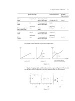
Figure 1-1 Global incidence and mortality of the most common cancers. The
bar chart shows estimates of age-specific cancer rates (Adapted from ref. 2).
Review of the Literature - Chapter 1
2
There is great geographical variation in GC incidence. The highest rates (> 40
per 100,000) are in East Asia (Japan, China and Korea), South and Central
America (Colombia, Costa Rica and El Salvador). North America and most of
Western Europe, in contrast, have low incidence rates (< 10 per 100,000)
3
(Figure 1-2).
Figure 1-2 Global geographic variation in age-standardized incidence rates of
GC among males (upper panel) and females (lower panel), (Adapted from ref.
2).
Review of the Literature - Chapter 1
3
GC incidence rates are 1.8 to 2.0 times higher in males than in females
2
. Apart
from gender, ethnicity appears to be associated with differences in GC
incidence. For example, in the United States, Asians and Pacific Islanders
(APIs) and black men have high age-standardized rates (ASR) of 18.6 and
17.4 cases per 100,000, respectively, compared to 10.0 per 100,000 in
Caucasian men (Table 1-1). Similarly, ASR of API and black women are
almost twice as high at 10.5 and 8.9 per 100,000, respectively versus 4.7 per
100,000 in Caucasian women. Native and Hispanic Americans are also twice
as likely to develop GC compared to the white population
3-5
Table 1-1 Age-adjusted incidence and mortality rates of GC by ethnicity
(2001-2005) based on cancer registries in 17 designated SEER (Surveillance,
Epidemiology and End Results Program) geographic areas in the USA
(Adapted from ref. 3).
. The higher ASR
in African-Americans, Hispanics and Asians may be partly due to their
relatively lower socio-economic status, poor access to healthcare and possibly,
an increased exposure to predisposing infections such as Helicobacter pylori.
In the early 20
1.1.2 Chronological trends
th
Race/Ethnicity Male Female Male Female
All Races 11.3 5.5 5.7 2.9
White 10.0 4.7 5.0 2.5
Black 17.4 8.9 11.5 5.5
Asian/Pacific Islander 18.6 10.5 10.1 5.9
American Indian/Alaska Native 16.8 7.7 9.9 5.2
Hispanic 15.5 9.5 8.7 4.9
Age-adjusted Incidence rates
(2001-2005)
Age-adjusted Mortality rates
(2001-2005)
century, GC was the leading cause of cancer mortality among
men and second after uterine cancer among women in the USA. Since the
1930’s, Western nations have experienced a steady and substantial decline in
GC incidence rates. This striking reduction in GC incidence, especially
among the white population in the US, has been characterized as an