Applications of electrospinning and supercritical carbon dioxide foaming techniques in controlled release and bone regeneration 1
Bạn đang xem bản rút gọn của tài liệu. Xem và tải ngay bản đầy đủ của tài liệu tại đây (189.99 KB, 31 trang )
Chapter 1 1
CHAPTER 1
L
ITERATURE REVIEW AND INTRODUCTION
Tissue engineering is the combination of cell, engineering and material methods, and
application of suitable biochemical and physio-chemical factors to improve or replace
biological functions. While most definitions of tissue engineering cover a broad range of
applications, in practice the term is closely associated with applications that repair or
replace portions of or whole tissues (i.e., bone, cartilage, blood vessels, bladder, etc ).
As growth factors are usually expensive and easily damaged, their delivery to specific
tissue positions efficiently without losing their bioactivity is a major challenge.
Drug delivery refers to the delivery of a pharmaceutical compound to humans or animals.
Apart from drug delivery for anticancer agents, drug delivery for tissue regeneration is
gaining more attention over past decades. For the case of anticancer drug delivery, the
anticancer agents, such as paclitaxel, are employed. As for drug delivery for tissue
engineering, proteins, peptides or DNAs are always utilized. Considering that protein,
peptide and DNA are very easily digested or damaged in organic solvents and cell plasma,
the delivery strategies have been highly emphasized over past years. Most commonly and
traditionally used methods of delivery include non-invasive oral (through the mouth),
nasal, inhalation, and rectal routes. Protein, peptide and DNA, however, could not be
efficiently delivered via these routes as they might be susceptible to degradation and the
Chapter 1 2
most serious weakness is that their concentration in specific tissue might be insufficient
to trigger expected biological responses after going through the dilution of circulation
system. For this reason, local drug delivery devices have been developed. In this way,
high drug concentration can be guaranteed and at the same time systemic toxicity from
drug is minimized.
In order to achieve the aim of tissue growth and remodeling, correct construction of drug
delivery devices for specific tissues is the most crucial challenge, as the interactions
between material, drug and cell are extraordinarily complicated. In general, basic
requirements for drug delivery devices include negligible toxicity, high concentration of
biological factors in tissue, high transfection efficiency of DNA to cells (for the case of
gene delivery), suitable degradation rate of devices with adequate sustained drug release
(Langer and Vacanti, 1993). Over past decades, many release dosage forms have been
developed for drug delivery, such as nanoparticle, microparticle, polymeric disc and film
(Freitas et al., 2005). Unfortunately one common problem with such dosage forms is the
undesirable release profile of drugs. For nanoparticle and microparticle, a burst release at
an early stage together with a very short release course is the major weakness. Xie and
Wang reported that paclitaxel loaded nanoparticles caused serious cytotoxicity but the
effective stage can not sustain (Xie and Wang, 2005). For disc and film, their release
profiles are difficult to tailor and in most cases their release rate is too low (Jackson et al.,
2004). As a result, the drug concentration in target tissue may be insufficient to trigger
expected biological responses. Recently, some researchers have developed several DNA
delivery devices with high gene transcription (Conwell and Huang, 2005; Schreier, 1994;
Chapter 1 3
Tomlinson and Rolland, 1996). There have been two major approaches proposed: the
viral mediated and non-viral mediated gene transfection (Ledley and Ledley, 1998).
Considering the immunological and safety issues of viral vectors, necessity of the
development of non-viral vector systems has been increasingly magnified. Although with
several advantages, namely the lower toxicity and immune responses or no integration
into the genome, non-viral vectors are always unable to transfect cells efficiently due to
the non-optimal device design, in the aspect of interactions between material and gene,
material and cells as well as gene and cells.
In order to tackle the disadvantages and shortcomings with current drug delivery devices,
novel drug delivery devices should be developed. The aim is to obtain and keep high
enough concentration of protein delivered or expressed (for the case of gene delivery) in
tissue, and pose minimal toxicity to environmental cells.
In the following sections, literature reviews on “tissue engineering”, “drug delivery
dosage forms”, and “polymeric material for drug delivery” will be interpreted separately
in more details.
1.1 Tissue engineering
In 2003, the National Science Foundation (NSF)
*
published a report entitled "The
Emergence of Tissue Engineering as a Research Field" (Langer and Vacanti, 1993),
which gives a thorough description of the history of this field. A commonly applied
definition of tissue engineering, as stated by Langer and Vacanti (Langer and Vacanti,
*
Please refer to and
Chapter 1 4
1993), is "an interdisciplinary field that applies the principles of engineering and life
sciences toward the development of biological substitutes that restore, maintain, or
improve tissue function or a whole organ"(Langer and Vacanti, 1993). Tissue engineering
has also been defined as "understanding the principles of tissue growth, and applying this
to produce functional replacement tissue for clinical use" (MacArthur, 2005). A further
description goes on to say that an "underlying supposition of tissue engineering is that the
employment of natural biology of the system will allow for greater success in developing
therapeutic strategies aimed at the replacement, repair, maintenance, and/or enhancement
of tissue function" (Murray et al., 2007).
Powerful developments in the multidisciplinary field of tissue engineering have yielded a
novel set of tissue replacement parts and implementation strategies. Scientific advances
in biomaterials, stem cells, growth and differentiation factors, and biomimetic
environments have created unique opportunities to fabricate tissues in the laboratory from
combinations of engineered extracellular matrices ("scaffolds"), cells, and biologically
active molecules (Langer and Vacanti, 1993). Among the major challenges now facing
tissue engineering is the need for more complex functionality, as well as both functional
and biomechanical stability in laboratory-grown tissues destined for transplantation. The
continued success of tissue engineering, and the eventual development of true human
replacement parts, will grow from the convergence of engineering and basic research
advances in tissue, matrix, growth factor, stem cell, and developmental biology, as well
as materials science and bioinformatics (Donald and Mohammad, 2001).
Chapter 1 5
1.1.1 Cells
Tissue engineering utilizes living cells as engineering materials. Examples include using
living fibroblasts in skin replacement or repair, cartilage repaired with living
chondrocytes, or other types of cells used in other ways (Weng and Wang, 2001). Cells
became available as engineering materials when scientists at Geron Corp.
*
discovered
how to extend telomeres in 1998, producing immortalized cell lines. Before this,
laboratory cultures of healthy, noncancerous mammalian cells would only divide a fixed
number of times, up to the Hayflick limit (Hayflick, 1965).
Cells are often categorized by their source in the following forms:
Autologous cells are obtained from the same individual as that to which they will be
reimplanted. Autologous cells have the fewest problems with rejection and pathogen
transmission, however in some cases might not be available. For example in genetic
disease suitable autologous cells are not available. Also very ill or elderly persons, as
well as patients suffering from severe burns, may not have sufficient quantities of
autologous cells to establish useful cell lines.
Allogeneic cells come from the body of a donor of the same species. While there are
some ethical constraints to the use of human cells for in vitro studies, the employment of
dermal fibroblasts from human foreskin has been demonstrated to be immunologically
safe and thus a viable choice for tissue engineering of skin.
*
Please refer to
Chapter 1 6
Xenogenic cells are those isolated from individuals of another species. In particular
animal cells have been used quite extensively in experiments aimed at the construction of
cardiovascular implants.
Syngeneic or isogenic cells are isolated from genetically identical organisms, such as
twins, clones, or highly inbred research animal models.
Primary cells are from an organism.
Secondary cells are from a cell bank.
Stem cells are undifferentiated cells with the ability to divide in culture and give rise to
different forms of specialized cells. According to their source stem cells are divided into
"adult" and "embryonic" stem cells, the first class being multipotent and the latter mostly
pluripotent; some cells are totipotent, in the earliest stages of the embryo. While there is
still a large ethical debate related with the use of embryonic stem cells, it is thought that
stem cells may be useful for the repair of diseased or damaged tissues, or may be used to
grow new organs.
1.1.2 Growth factors
Growth factor refers to a naturally occurring protein capable of stimulating cellular
proliferation and cellular differentiation. Growth factors are important for regulating a
variety of cellular processes. Typically they act as signaling molecules between cells.
They often promote cell differentiation and maturation, which varies between growth
factors. For example, bone morphogenic proteins stimulate bone cell differentiation,
Chapter 1 7
while fibroblast growth factors and vascular endothelial growth factors stimulate blood
vessel differentiation (angiogenesis).
1.1.2.1 Definition of growth factors
Growth factor is sometimes used interchangeably among scientists with the term cytokine.
Historically, cytokines were associated with hematopoietic (blood forming) cells and
immune system cells (e.g., lymphocytes and tissue cells from spleen, thymus, and lymph
nodes). For the circulatory system and bone marrow in which cells can occur in a liquid
suspension and not bound up in solid tissue, it makes sense for them to communicate by
soluble, circulating protein molecules. However, as different lines of research converged,
it became clear that some of the same signaling proteins the hematopoietic and immune
systems used were also being used by all sorts of other cells and tissues, during
development and in the mature organism.
While growth factor implies a positive effect on cell division, cytokine is a neutral term
with respect to whether a molecule affects proliferation. In this sense, some cytokines can
be growth factors, such as granulocyte colony-stimulating factor (G-CSF) and
granulocyte-macrophage colony-stimulating factor (GM-CSF). However, some cytokines
have an inhibitory effect on cell growth or proliferation. Yet others, such as Fas ligand
(FasL) are used as "death" signals; they cause target cells to undergo programmed cell
death or apoptosis.
Chapter 1 8
1.1.2.2 Examples of growth factors
*
Individual growth factor proteins tend to occur as members of larger families of
structurally and evolutionarily related proteins. There are dozens of growth factor
families such as TGF-beta (transforming growth factor-beta), BMP (bone morphogenic
protein), neurotrophins (NGF, BDNF, and NT3), fibroblast growth factor (FGF), and so
on. Several well known growth factors are:
• Transforming growth factor beta (TGF-β)
• Granulocyte-colony stimulating factor (G-CSF)
• Granulocyte-macrophage colony stimulating factor (GM-CSF)
• Nerve growth factor (NGF)
• Neurotrophins
• Platelet-derived growth factor (PDGF)
• Erythropoietin (EPO)
• Thrombopoietin (TPO)
• Myostatin (GDF-8)
• Growth differentiation factor-9 (GDF9)
• Acidic fibroblast growth factor (aFGF or FGF-1)
• Basic fibroblast growth factor (bFGF or FGF-2)
• Epidermal growth factor (EGF)
• Hepatocyte growth factor (HGF)
*
Please refer to
Chapter 1 9
1.1.2.3 Bone morphogenetic proteins (BMPs)
BMPs are a group of growth factors and cytokines known for their ability to induce the
formation of bone and cartilage (Chen et al., 2004). Originally, seven such proteins were
discovered. Of these, six of them (BMP-2 through BMP-7) belong to the Transforming
growth factor beta superfamily of proteins. Since then, thirteen more BMPs have been
discovered, bringing the total to twenty. BMPs interact with specific receptors on the cell
surface, referred to as bone morphogenetic protein receptors (BMPRs). Signal
transduction through BMPRs results in mobilization of members of the SMAD family of
proteins. The signaling pathways involving BMPs, BMPRs and Smads are important in
the development of the heart, central nervous system, and cartilage, as well as post-natal
bone development. They have an important role during embryonic development on the
embryonic patterning and early skeletal formation. As such, disruption of BMP signaling
can affect the body plan of the developing embryo. For example, BMP-4 and its
inhibitors noggin and chordin help regulate polarity of the embryo (i.e. back to front
patterning). Mutations in BMPs and their inhibitors (such as sclerostin) are associated
with a number of human disorders which affect the skeleton. Several BMPs are also
named “cartilage-derived morphogenetic proteins” (CDMPs), while others are referred to
as “growth differentiation factors” (GDFs).
Chapter 1 10
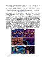
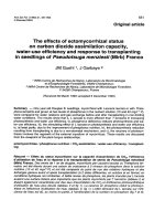
Figure 1.1 Model of human bone morphogenetic protein 2 created using Cn3D
*
Bone morphogenetic protein 2 or BMP-2 (Nickel et al., 2001; Kawamura et al., 2003;
Marie et al., 2003) is a protein that belongs to the TGF-β superfamily of proteins. It, like
other bone morphogenetic proteins, plays an important role in the development of bone
and cartilage. It is involved in the Hedgehog pathway, TGF-beta signaling pathway, and
the Cytokine-cytokine receptor interaction.
1.2 Polymeric materials for drug delivery
1.2.1 Non-degradable materials
The production of drug-loaded (drug in a broad sense including chemical drugs, protein,
DNA, et al.) polymeric pellets and microspheres introduced a new concept in drug
delivery: Drugs can be delivered to tissues in a sustained, continuous and predictable
fashion using polymers as delivery devices. Since the discovery of the first controlled-
release polymer systems in 1960s, new drug delivery devices have become available for
clinical use, including steroid-releasing reservoirs for contraception (Norplant® and
*
Please refer to www.cytok.com/structure.php?start=120
Chapter 1 11
Progestasert®), pilocarpine-releasing devices for glaucoma therapy (Ocusert®) and a
host of new delivery systems for the treatment of cancer. Drug delivery devices are based
on biocompatible polymers, a subset of polymer materials with sufficient
biocompatibility and appropriate physical properties to provide controlled delivery (Fung
and Saltzman, 1997).
The first polymeric controlled-release devices were based on non-degradable polymers,
principally silicone elastomers. In 1964, researchers recognized that certain dye
molecules could penetrate through the walls of silicone tubing (Folkman and Long, 1964;
Folkman and Long, 1966), an observation that leads to the development of reservoir drug
delivery devices, which are hollow polymer tubes filled with a drug suspension. The drug
is released by dissolution into the polymer and then diffusion through the polymer wall, a
mechanism that works for any agent that can dissolve and diffuse through either silicone
or poly(ethylene-co-vinylacetate) (EVAc), the two most commonly used nondegradable
polymers. The Norplant® 5-year contraceptive delivery system, approved by use by
women in the United States since 1990, is based on this technology.
Solid matrices of non-degradable polymers can also be used for long-time drug release.
In comparison to reservoir systems, these devices are simpler (since they are
homogenously and, hence, easier to produce) and potentially safer (since a mechanical
defect in a reservoir device, but not a matrix, can lead to dose dumping). On the other
hand, it is more difficult to achieve constant rates of drug release with non-degradable
matrix; for example, the rate of release of carmustine from an EVAc matrix device drops
Chapter 1 12
continuously during incubation in buffer water. Constant release can sometimes be
achieved by adding rate limiting membranes to homogenous matrices, yielding devices in
which a core of polymer/drug matrix serves as the reservoir. In other cases, water-soluble
crosslinked polymers can be used as matrices (Kim et al., 1992); release is then activated
by swelling of the polymer matrix after exposure to water.
Although non-degradable materials are very stable and can make specific structures
easily, their applications in medical field are strongly hindered for several reasons. First,
when the field expanded from research to application, it was recognized that surgical
removal of drug-depleted delivery systems was difficult, yet leaving non-degradable
foreign materials in the body for an indefinite time period constituted an undesirable
toxicological hazard. Therefore, the physician’s simple desire to have a device that can be
used as an implant and will not require a second surgical intervention for removal
constitutes the most basic reason for the interests in biodegradable polymers. Secondly,
although diffusion-controlled release is an excellent means of achieving predefined rates
of drug delivery, it is limited by the polymer permeability and the characteristics
(molecular weight, solubility) of the drug because diffusion follows Fick’s first law,
which states that the rate of diffusion is proportional to the concentration gradient of the
drug and the diffusion coefficient. Therefore, drugs that have either high molecular
weight (>500 daltons) or poor solubility in the polymer are not amenable to diffusion-
controlled release. Besides these reasons, biodegradation may offer other advantages, For
example, a fractured bone that has been fixated with a rigid, non-biodegradable stainless
implant has a tendency to re-fracture upon removal of the implant. Because the stress is
Chapter 1 13
borne by the rigid stainless steel, the bone has not been able to carry sufficient load
during the healing process. However, an implant prepared from biodegradable polymer
can be engineered to degrade at a rate that will slowly transfer a load to the healing bone.
1.2.2 Biodegradable materials
Biodegradation is defined as the conversion of materials into less complex intermediates
or end products by solubilization, simple hydrolysis, or the action of biologically formed
entities that may be enzymes and other products of the organism. Polymer molecules may,
but not necessarily, break down to produce fragments in this process, and the integrity of
the material decreases as a result of this process (Albertsson and Karlsson, 1990; Vert,
1989; Anderson, 1989). The formed fragments can move away from their site of action
but not necessarily from the body.
In the first half of 20
th
century, research on materials synthesized from glycolic acid and
other hydroxyl acids was abandoned for further development because the resulting
polymers were too unstable for long-term industrial uses. Consequently, the pioneering
studies in the field of controlled drug delivery, begun in the 1960s, used bio-stable
commercial polymers such as polyethylene and silicone rubbers. In these systems, the
release rate of the drug from the polymeric matrix or reservoir device was determined
solely by diffusion; biodegradation of the polymer was not considered at that time.
However, this very instability of early materials-leading to biodegradation-has been
proven to be immensely important in medical applications over the last five decades.
Therefore, much research has been conducted on biodegradable polymer thereafter.
Chapter 1 14
With the development of biodegradable polymers, great progress has been achieved. For
example, polymers prepared from glycolic acid and lactic acid have found a multitude of
uses in the medical industry, beginning with the biodegradable sutures first approved in
the 1960s. Since that time, diverse products based on lactic and glycolic acid-and on
other materials, including poly(dioxanone), poly(trimethylene carbonate) copolymers,
and poly(caprolactone) homo-polymers and copolymers have been accepted for use as
medical devices. In addition to these approved devices, a great deal of research continues
on polyanhydrides, polyorthoesters, polyphosphazenes, and other biodegradable
polymers.
Biodegradable polymers can be either natural or synthetic. In general, synthetic polymers
offer greater advantages than natural materials in that they can be tailored to give a wider
range of properties and more predictable uniformity than materials from natural sources.
However, there are several points to be considered in choosing biodegradable polymers
for medical applications. The general criterion for selecting polymeric biomaterial is to
match the mechanical properties and the time of degradation according to the needs of the
application. An ideal polymer for a particular application would be configured so that it:
• has mechanical properties that match the application, remaining sufficiently
strong until the surrounding tissue has get healed and remodeled;
• does not invoke an inflammatory or toxic response;
• is metabolized in the body after fulfilling its purpose, leaving no trace;
• is easily processable into the final product form;
Chapter 1 15
• demonstrates acceptable shelf life;
• is easily sterilized.
A number of typical properties of biodegradable polymers have been studied (see Table
1.1). The factors affecting the mechanical performance of biodegradable polymers are
those that are well known to the polymer scientists, and include monomer selection,
initiator selection, process conditions, and the presence of additives. These factors in turn
influence the polymer’s hydrophilicity, crystallinity, melting and glass-transition
temperatures, molecular weight, molecular-weight distribution, end groups, sequence
distribution (random versus blocky), and presence of residual monomer or additives. In
addition, the polymer scientist working with biodegradable materials must evaluate each
variable for its effect on biodegradation (Daniels et al., 1990).
Biodegradation has been accompanied by synthesizing polymers that have hydrolytically
unstable linkages in the backbone. The most common chemical functional groups with
this characteristic are esters, anhydrides, orthoesters, and amides. The synthetic
biodegradable polymers are currently being used or investigated for use in wound closure
(sutures, staples), orthopedic fixation devices (pin, rods, screws, tacks, ligaments), dental
applications (guided tissue regeneration), cardiovascular applications (stents, grafts), and
intestinal applications (anastomosis rings). Most of the commercially available
biodegradable devices are polyesters composed of homo-polymers or copolymers of
glycolide and lactide.
Chapter 1 16
Table 1.1 Properties of some biodegradable polymers (adapted from Daniels et al., 1990)
Polymer Melting Point
(ºC)
Glass-transition
Temperature (ºC)
Modulus
(Gpa)
a
Degradation
Time (month)
b
PGA 225-230 35 ~ 40 7.0 6 ~ 12
L-PLA 173-178 60 ~ 65 2.7 > 24
DL-PLA Amorphous 55 ~ 60 1.9 12 ~ 16
PCL 58-63 -65 ~ -60 0.4 > 24
PDO N/A -10 ~ 0 1.5 6 ~ 12
PGA-TMC N/A N/A 2.4 6 ~ 12
85/15 DL-PLGA Amorphous 50 ~ 55 2.0 5 ~ 6
75/25 DL-PLGA Amorphous 50 ~ 55 2.0 4 ~ 5
65/35 DL-PLGA Amorphous 45 ~ 50 2.0 3 ~ 4
50/50 DL-PLGA Amorphous 45 ~ 50 2.0 1 ~ 2
a: tensile strength or flexural modulus
b: time to complete mass loss. Rate also depends on part geometry
1.2.3 Poly(
DL
-lactide-co-glycolide)
Using the polyglycolide and poly(
L
-lactide) properties as a starting point, it is possible to
copolymerize the two monomers to extend the range of homopolymer properties.
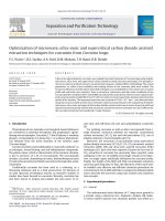
Copolymers of glycolide with both L-lactide and DL-lactide (Figure 1.2) have been
developed for both drug delivery and tissue engineering applications. It is important to
note that there is not a linear relationship between the copolymer composition and the
mechanical and degradation properties of the materials. For example, a copolymer of
50% glycolide and 50% D,L-lactide degrades faster than either homopolymer.
Copolymers of L-lactide with 25-70% glycolide are amorphous due to the disruption of
the regularity of the polymer chain by the other monomer. A copolymer of 90% glycolide
Chapter 1 17
and 10% L-lactide was developed by Ethicon as an absorbable suture material under the
trade name Vicry1. It is absorbed within 3-4 months but has a slightly longer strength-
retention time. It is proved that PLGA 50:50 (50/50 DL-PLG) is the best for drug
delivery due to its favorable degradation rate.
Figure 1.2 Molecular formula of poly(
DL
-lactide-co-glycolide)
Although degradation and mechanical strength of polymeric materials can be tailored to
some extent according to customer’s requirements, no optimal material has been
developed for bone tissue engineering. PLGA 50:50 (50/50 DL-PLG, Table 1.1) is
widely used in drug delivery applications due to its favorable degradation rate, but its
mechanical properties are far from meeting requirements for bone tissue engineering.
Synthetic bone grafts based on metals and some ceramics, like hydroxylapatite and
tricalcium phosphate, have different mechanical properties when compared to human
bone tissue, and this incompatibility often results in implant failure and consequently,
revision surgery (Lane and Bostrom, 1998; Lane et al., 1999). However, the mechanical
strength of HAp may be a good supplement to PLGA-based delivery devices. One of the
purposes of this thesis is to develop PLGA-based novel drug delivery devices, which can
achieve and maintain sufficiently high concentrations of protein or DNA in tissue and
have acceptable mechanical properties. These devices developed in this thesis may be of
Chapter 1 18
importance in combining tissue engineering and drug delivery nicely, with potential in
clinical applications.
1.3 Drug delivery dosage forms
In addition to the attachment of growth factors to polymeric scaffolds, growth factors can
be directly incorporated within scaffolds during the fabrication process. These methods
generally involve the mixing of the polymers with the growth factor before processing to
form the scaffold. The main challenge of this set of methods is to ensure that the
processing conditions do not significantly denature the incorporated growth factors while
allowing the secretion of the growth factor in a sustained manner (Sokolsky-Papkov et al.,
2007).
1.3.1 Hydrogel scaffolds
Hydrogel matrices are physically or chemically crosslinked water-soluble polymers,
which swell to form a gel like substance on exposure to water (Drury and Mooney, 2003).
Hydrogels are formed by the crosslinking of polymer chains to form a scaffold made up
of connected polymer chains. Crosslinking can be done either through physical (UV
irradiation, freeze drying and heating) and chemical means such as ionic crosslinking in
presence of divalent ions or utilization of chemical crosslinkers such as glutaraldehyde
and carbodiimide. Hydrogels can be made from synthetic polymers such as poly(ethylene
glycolide), poly vinyl alcohol, or naturally occurring polymers such as collagen, chitosan
and gelatin. Release of growth factors from hydrogels is believed to be either through
diffusion of the growth factor, mechanical stimulation, or hydrolytic degradation of the
Chapter 1 19
scaffold (Drury and Mooney, 2003). Some examples of hydrogels are reported by
Chenite and coworkers (Chenite et al., 2000). In this particular work hydrogel formation
was induced by temperature, with bone morphogenic protein (rhBMP) introduced into the
hydrogel material (temperature sensitive chitosan-polyol salt combination) during
fabrication and shown to be effective in promoting de-novo bone and cartilage formation
in vivo.
However due to the release kinetics of growth factor through hydrogels being mainly
diffusion controlled through the numerous aqueous channels within the hydrogels,
extended release of the proteins is not easily achieved. Immobilization of the growth
factor within the biodegradable hydrogel seems to improve the release kinetics, with
release of the immobilized factor being controlled by the degradation of the hydrogel.
Several chemical and physical methods exist for the immobilization of growth factors in
hydrogels. Chemical methods involving attachment of reactive groups to the growth
factor for crosslinking with hydrogel polymers have seen increasing use within cartilage
tissue engineering (Holland et al., 2007). Physical methods of immobilization of growth
factors within scaffolds exploits electrostatic interactions that might occur between
charges on growth factors and that on the polymer chains of the hydrogel (Tabata, 2005).
1.3.2 Microspheres based scaffolds
Polymer scaffolds can also be manufactured from growth factor loaded microspheres.
Scaffold fabrication from microspheres could range from fusing of microspheres to form
scaffolds, to formation of microsphere containing composite scaffolds. Growth factor
Chapter 1 20
loaded microsphere can be formed either through chemical cross-linking of aqueous
solutions of natural polymers in the presence of growth factors or by solvent extraction.
The solvent extraction method is mainly used for formation of microspheres from
synthetic polymers. It involves the evaporation of an organic solvent such as
dichloromethane (chloroform and ethyl acetate have also been used), from dispersed oil
droplets containing both the polymer and the growth factor (Freiberg and Zhu, 2004).
Two popular variations of this technique are currently used and are described below. The
double emulsion method involves the dispersion of an aqueous solution of the growth
factor in an organic solution containing the polymer - leading to the formation of the
primary water in oil (W/O) emulsion. A secondary oil in water (O/W) emulsion is formed
by dispersing under continuous mechanical agitation of the primary W/O emulsion in an
aqueous medium containing stabilizers such as poly(vinyl alcohol) (PVA) or
poly(ethylene glycol) (PEG). Microspheres are produced by the evaporation of the
organic solvent from the emulsion droplets. The microspheres are then collected either by
centrifuging or filtration, washed and lyophilised to obtain the free flowing and dried
microspheres. Microspheres prepared from poly(
DL
-lactic-co-glycolic acid) (PLGA) using
the double emulsion method have been used for incorporation of BMP-2 (Hiraoka et. al.,
2006), TGF-β1 (Tabata et al., 1999) and PDGF-BB (Nakahara et al., 2003). The single
emulsion method involves bypassing the primary W/O emulsion stage, by dispersing or
dissolving the growth factor directly into the organic solution of the polymer.
Microspheres are then formed by the subsequent formation of the O/W emulsion,
evaporation of the organic solution from the emulsion droplets. The growth factor could
be micronized by lyophilisation with PEG, CDs etc before dispersing it into the organic
Chapter 1 21
polymer solution to improve its entrapment efficiency (Morita et al., 2000). In the work
carried out investigating the stimulation of angiogenesis within engineered tissues, King
and Patrick (King and Patrick, 2000) were able to produce using the single emulsion
method vascular endothelial growth factor (VEGF) loaded microspheres with a sustained
release of the active growth factor over a 28 day period. Growth factor released from
these microspheres was shown to increase the proliferation of human umbilical vein
endothelial cells in culture. Growth factor loaded microspheres can be used for tissue
engineering applications either by fusing of microspheres directly to form scaffolds, or
combination with other scaffold forming materials to construct composite scaffolds.
In work described by Wei and coworkers (Wei et al., 2006), PDGF-BB (platelet derived
growth factor) loaded microspheres were incorporated in poly(lactic acid) (PLLA) nano-
fibrous scaffold. Due to its structural similarity to collagen (which is a major extracellular
component of bone, cementum, and periodontal ligament (PDL)), the nano-fibrous
structure has been demonstrated to improve cell attachment (Woo et al., 2003), and
possibly to stimulate cell proliferation and differentiation as well (Xu et al., 2004).
Sustained release for days to months of bioactive PDGF-BB was achieved by the
microspheres in scaffold. The authors of this work postulated that composite scaffold
based on 3D nano fibrous material can be used for complex tissue regeneration. Kempen
and Hedberg and their coworkers (Kempen et al., 2003; Hedberg et al., 2002) showed the
application of PLGA and poly(propylene fumarate) (PPF) microspheres in
microsphere/scaffold composite for controlled release of growth factors for bone tissue
engineering.
Chapter 1 22
1.3.3 Porous solid scaffolds
Formation of growth factor loaded porous solid scaffolds can be achieved through several
techniques. The main techniques involve either one-step direct formation of growth factor
loaded scaffolds or formation of growth factor loaded microspheres which can then be
fabricated to form scaffolds.
1.3.3.1. Solvent casting/particulate leaching
Solvent casting-particulate leaching is one of the first techniques that have been utilized
in formation of porous scaffolds for tissue engineering applications, although it is being
replaced by more sophisticated techniques. This technique involves the pouring of the
polymer solution into a bed of salt particles with defined size. Precipitation of the
polymer by evaporation of the polymer solvent under vacuum, followed by leaching of
the salt particles in distilled water leads to the formation of a highly porous scaffold with
well defined pores (Hile et al., 2000). This process of scaffold production is a popular
technique and has been used in the production of angiogenic factors loaded scaffolds for
the induction of angiogenesis (Hile et al., 2000). These factors are emulsified within the
polymer solution prior to the solvent casting-salt leaching process. As the organic solvent
is removed, the polymer precipitates and encapsulates the angiogenic factors.
1.3.3.2. Supercritical processed foam
Denaturation of growth factors during scaffold fabrication has been indicated as the main
cause of activity loss of protein released from scaffold. The need to improve on the
current fabrication methods has led to the creation of scaffolds using supercritical fluids.
Chapter 1 23
The benefits of using these process methodologies are due to the ability to form growth
factor loaded scaffolds in conditions that are favourable to growth factors, avoiding the
need for using organic solvents and harsh process conditions (Kanczler et al., 2007).
Supercritical fluids (SCF) are substances that exist at temperatures and pressures that
exceed its critical temperature and pressure. SCFs combine the properties of the two
phases from which they are formed; they have densities and solvating properties similar
to those of liquids, alongside diffusion and viscosity properties of a gas. CO
2
is the most
common candidate for use as a SCF due to its low toxicity, ease of use and low cost.
Scaffold fabrication using SCF technology (RESS technique) normally involves the
dissolution of the polymer/growth factor complex in supercritical CO
2
(scCO
2
). This is
followed by a rapid expansion of the mixture into a low temperature and pressure
environment which leads to scaffold formation (Vasita and Katti, 2006). Another
technique utilizing the scCO
2
is the PGSS technique, which involves using the scCO
2
to
plasticize the polymer by lowering the glass transition temperatures (Tg) of the polymer.
This technique involves the application of the SCF under pressure into the mixture of
polymer and growth factor until the mixture is saturated. This is followed by
depressurization through a nozzle, leading to the formation of highly porous scaffolds as
the gas comes out of liquefied polymer. This process has the advantages that the starting
material does not have to be soluble in the SCF, and that no organic solvents are required
during processing (Quirk et al., 2004). Mooney and coworkers first showed the
possibility of forming scaffolds using CO
2
, by producing porous polymeric matrices with
carbon dioxide (Mooney et al., 1996; Harris et al., 1998). In further experiments they
incorporated vascular endothelial growth factor (VEGF) during the scaffold fabrication,
Chapter 1 24
and were able to show the release of the active growth factor over a 12 day period
(Murphy et al., 2000). VEGF encapsulated PLA scaffolds have also been shown to
stimulate the formation of blood vessel network in an ex vivo chick chorioallantoic
membrane (CAM) angiogenesis model (Kanczler et al., 2007). In a study carried out by
Yang and colleagues, the effects of recombinant human pleotrophin (PTN) loaded PLGA
scaffolds prepared using the supercritical fluid technology, was compared to a PLGA
scaffold without the loaded growth factor on primary human bone marrow cells.
Negligible cellular growth was observed on PLGA scaffold alone while the PTN loaded
scaffold was shown to induce proliferation of the cell line with total colony formation,
alkaline phosphatase-positive colony formation, and alkaline phosphatase-specific
activity observed with increasing growth factor concentrations (Yang et al., 2003). In
another study carried out by the same group, porous BMP-2-encapsulated poly(
DL
-lactic
acid) (PLA) scaffolds generated by the supercritical fluid process was shown to promote
adhesion, migration, proliferation, and differentiation of human osteoprogenitor cells on
three-dimensional scaffolds. The BMP-2-encapsulated polymer scaffolds showed
evidence of new bone matrix and cartilage formation after subcutaneous implantation
into athymic mice (Yang et al., 2004).
1.3.4 Electrospun fiber
Recently, natural and synthetic polymers have been processed into porous fibrous
scaffolds for tissue engineering applications using the electrospinning process. Scaffolds
generated by electrospinning contain nanoscale fibers with microscale-interconnected
pores, resembling the extracellular matrix (ECM) (Li et al., 2006) and the three-
Chapter 1 25
dimensional nature of the scaffold allows for cells to infiltrate the matrix and proliferate.
Collagen, fibrinogen, chitosan, poly(lactic acid), poly(
L
-lactide-co-caprolactone), and
poly(
D,L
-lactide-co-glycolide) are just a few of the polymers being investigated for use in
electrospun tissue engineering constructs. The electrospinning process produces fibers
with nanoscale diameters through the action of a high electric field (Li et al., 2006). This
process involves the subjecting of a polymer solution in a capillary to an electric field
generated by high voltage differences. When the generated electric field exceeds the
surface tension of the polymer solution, ejection of a polymer jet occurs. This polymer jet
is targeted towards a grounded collector. Enroute to the collector plate, the polymer jet
looses stability, leading to the stretching of the jet, with solid fibers deposited on the
collector in the form of a nonwoven fabric. By the adjustment of solution and operating
parameters, fiber diameter and porosity of fiber mats can be controlled during
electrospinning (Zong et al., 2002; Zong et al., 2003), which makes this a promising
technique for fabrication of tissue-engineered scaffolds. Li and coworkers produced silk
fibroin fiber scaffolds containing bone morphogenetic protein 2 (BMP-2) and/or
nanoparticles of hydroxyapatite (nHAp) using electrospinning, and showed its ability in
inducing in vitro bone formation from human bone marrow-derived mesenchymal stem
cells (hMSCs) (Li et al., 2006). The electrospun scaffolds containing BMP-2 supported
higher calcium deposition and enhanced transcript levels of bone-specific markers than
the electrospun scaffolds without BMP-2, indicating that the scaffolds were an efficient
delivery system for BMP-2.