Applications of electrospinning and supercritical carbon dioxide foaming techniques in controlled release and bone regeneration 3
Bạn đang xem bản rút gọn của tài liệu. Xem và tải ngay bản đầy đủ của tài liệu tại đây (3 MB, 22 trang )
Chapter 4 60
CHAPTER 4
Optimized Bone Regeneration Based on Sustained Release of
Bone Morphogenetic Protein-2 from Three-Dimensional
Fibrous PLGA/HAp Composite Scaffolds
†
4.1 Introduction
Bone is one of the most commonly repaired organs of the body (Langer and Vacanti,
1993; Laurencin et al., 2000; Laurencin et al., 2001). Currently, treatment of fracture and
bone loss associated with trauma, cancer and revision total joint arthroplasty remains a
significant challenge in the field of orthopaedic surgery (Calvert et al., 2003; Crane et al.,
1995; Goldstein et al., 1999; Huang et al., 2005; Constantz et al., 1995; Petite et al., 2000;
Vacanti and Upton, 1994). Due to limitations associated with bone grafts, engineered
biomaterials combined with growth factors have emerged as a new treatment alternative
in bone repair and regeneration. Treatment of fractured bone with cells, growth factors
and biomaterials are the three commonly used ways over the past decade, but it has been
proved that none of the ways can achieve the clinical requirement. Recent research focus
has moved to the application of cells, growth factors and biomaterials in combination.
Using biodegradable, polymeric materials with known biocompatibility, several
researchers have fabricated porous, bioresorbable scaffolds for bone regeneration
†
This chapter highlights the work published in Y.C. Fu, H. Nie, M.L. Ho, C.K. Wang and C.H. Wang.
Optimized Bone Regeneration Based on Sustained Release from Three-Dimensional Fibrous PLGA/HAp
Composite Scaffolds Loaded with Bone Morphogenetic Protein-2. Biotechnol. Bioeng. 99 (4), 996-1006.
2008.
Chapter 4 61
(Coombes and Heckman, 1992; Devin et al., 1996; Thomson et al., 1995).
Over past years, many release dosage forms have been developed for drug or protein
delivery, like nanoparticle and microsphere. However, one common problem is the
existence of a large burst over a narrow time period during the early stage of release.
Fibre has much lower release rate of drug or protein than microsphere because of its
smaller surface/volume ratio (Wei et al., 2006). Biodegradable poly(lactide-co-
glycolide)/hydroxyapatite (PLAGA/HAp) composites have been shown to support the
attachment, growth, and low cytotoxicity in vitro (Nie et al., 2008b). In addition to the
use of biodegradable scaffolds, cells, growth factors and other biological moieties can be
added to the matrix to promote and expedite bone formation. A number of different
growth factors, including bone morphogenetic proteins (BMPs), transforming growth
factor β, platelet-derived growth factor, fibroblast growth factor and insulin growth factor
have been shown to stimulate bone growth, collagen synthesis, and fracture repair both in
vitro and in vivo (Jingushi et al., 1995; Nixon et al., 1998; Pfeilschifter et al., 1993;
Scherping et al., 1997; Thaller et al., 1993). In particular, BMPs are osteoinductive
proteins originally identified in demineralized bone (Urist and Nogami, 1970). They are
known to facilitate bone healing without transferring bone tissues. Among this group of
proteins, BMP-2 has been shown to induce healing in segmental bone defects. Aebli and
colleagues (Aebli et al., 2005) and Saito and colleagues (Saito et al., 2005) reported that
BMPs improve bone regeneration in vivo, and BMP-2 has been found to induce healing
of segmental bone defects.
Chapter 4 62
In a previous study, BMP-2 loaded PLGA/HAp composite scaffolds were successfully
fabricated, and these scaffolds were found to be able to obtain integrity of BMP-2
encapsulated, enhance cell attachment and cause negligible cytotoxicity (Nie et al.,
2008b). The main objective of this study is to examine whether the PLGA/HAp composite
fibrous scaffolds loaded with BMP-2 through electrospinning can improve bone
regeneration. Our hypothesis is that different loading methods of BMP-2 and different
HAp contents in scaffolds can alternate the release profiles of BMP-2 in vivo, therefore
modify the performance of scaffolds in bone regeneration. The current study will first
check the mechanical strength of scaffolds and HAp distribution in scaffolds. Next, the
bioactivity of the produced BMP-2 will be evaluated in vivo using a tibia bone defect
model (see Appendix A1).
4.2 Materials and methods
4.2.1 Materials
Recombinant human bone morphogenetic protein-2 (rhBMP-2) (E. coli expressed, Cat.
No. 355-BEC/CF) and its enzyme-linked immunosorbent assay (ELISA) kit were
purchased from R&D Systems, Inc. (MN, US). Poly(
D,L
-lactide-co-glycolide) (PLGA)
(L/G ratio 50:50, MW 40,000-75,000) and chitosan (medium molecular weight and 75-
85% deacetylated), were procured from Sigma Aldrich (St. Louis, MO, US). HAp
nanocrystals with average diameter of 100nm, dichloromethane (DCM) (Cat. No. DR-
0440), Ketamine Ketalar® and Xylocain® were purchased from Berkeley Advanced
biomaterials Inc. (Berkeley, CA, US), Tedia Company Inc. (Fairfield, OH, US.), Parke-
Davis Taiwan, and AstraZeneca PLC Taiwan, respectively.
Chapter 4 63
4.2.2 Preparation of fibrous scaffolds
In all the experiments of the present work, the fibres were essentially fabricated from
homogeneous emulsions formed from the sonication of organic and aqueous mixture. The
compositons of 4 kinds of scaffold samples and their fabrication can be found in Section
3.2.2 (page 35).
4.3 Characterization of scaffolds
4.3.1 Physical characterization of fibrous scaffolds
Mechanical Property of Fibrous Scaffolds
Field emission scanning electron microscopy (FESEM, JSM-6700F, JEOL Technics Co.
Ltd, Tokyo, Japan) was employed to study the surface morphology of the fibres produced
in each experiment, while the quality of the fibres was determined by tensile strength
testing. The mechanical properties of all fibrous scaffolds (F1, F2, F3, and F4) prepared
in a sheet form (15mm wide x 20mm long x ~150µm thick) were evaluated by applying a
tensile load. Tensile tests of all fibrous scaffolds were conducted by Instron 5848
Microtester with 10 mm/min cross-head speed with a 30mm gauge length. Tensile stress
of each sheet was calculated on the nominal cross-sectional area of the tensile specimens.
Residual Solvent Content in Scaffolds
One of the concerns of pharmaceutical application is the residual solvent content in the
scaffolds fabricated although this factor has seldom been addressed by other fibre
fabrication groups. Before performing further characterizations in vivo, gas
chromatography was used in the present study to determine the residual amount of
Chapter 4 64
Dichloromethane (DCM) remaining in the scaffolds. To quantify the amount of DCM in
the HAp or/and BMP-2 loaded scaffolds, standard solutions with range of DCM
concentrations in N, N Dimethyl Formamide (DMF) from 0.5 to 10 x 10
-6
mL DCM per
mL DMF were prepared and placed in the refrigerator before analysis to prevent
evaporation of the volatile organic solvents. The calibration samples were run using gas
chromatography with mass spectrometry detector (GC-MSD) in order to determine the
peak areas and retention time for DCM. A calibration curve was obtained for peak area of
different concentrations of DCM in DMF. To obtain the residual amount of DCM in the
PLGA/HAp scaffolds obtained from the electrospinning method, 15mg of each sample of
F1-F4 were weighed and dissolved in 5 mL of DMF solution to extract DCM after freeze
drying for 3 days inside a Martin Christ freeze dryer (Martin Christ
Gefriertrocknungsanlagen GmbH, Germany). Next, about 1 mL of the solution was
filtered into standard GC bottles and analyzed using the GC-MSD with an auto-sampler
together with the calibration samples.
4.3.2 In vivo experiments
All procedures were performed in accordance to specifications in the Guidelines for
Animal Experiments of Kaohsiung Medical University and approved by the Institutional
Animal Care and Use Committee (IACUC). As explained in Appendix A1, nude mice
were anesthetized by intraperitoneal injection (3.5 mg/20 g body weight) of Ketamine
(Ketalar®, Parke-Davis, Taiwan) combined with local anesthesia (Xylocain®,
AstraZeneca PLC in Taiwan). One-mm-long tibia bone on the right side of a mouse was
cut out with saw. The part of bone cut was frozen using liquid nitrogen for 5 min. Next,
Chapter 4 65
the fragment was reversed and put back to its original site in the tibia and fixed on both
ends with the other parts of the tibia using an intramedullary needle perfectly (similar to
the application of intramedullary nail in human patients). A scaffold was embedded
around the bone fracture. The wounds were then closed with 4-0 silk sutures. A bone
fragment treated with liquid nitrogen but without seeding any scaffold was used as a
control. Each experiment was performed for 3 nude mice independently unless mentioned
otherwise.
Soft X-ray Observation
After 1, 2, 4, and 6 weeks, the tibia bone fractures were radiographically examined by
soft X-rays (SOFTEX, Model M-100, Japan) at 43 KVP and 2mA for 1.5s. Appropriate
magnification was applied throughout the observation and the resultant micrographs were
compared among all scaffolds together with control.
Semi-Quantification of Fragment’s Contact with Tibia
A semi-quantification method is utilized to compare the performances of all scaffolds at
specific intervals. The tibia bone is hollow and each bone fragment has two ends.
Henceforth, each bone fragment is checked whether all of the 4 corners of the fragment
are connected with tibia bone, or just 1, 2, or 3 of them is (are) connected with it. For
quantification, a score 0, 1, 2, 3, or 4 is assigned to 0, 1, 2, 3, or 4 contact respectively. As
each scaffold is tested in triplicate, the final score for each scaffold at each time point is
recorded with the arithmetic mean of the scores.
Chapter 4 66
Serum BMP-2 Concentration and ALP Activity Measurements
Serum was collected through cardiac puncture and taken out for biochemical assays 1, 2,
4, and 6 weeks after implantation of all scaffolds. The serum BMP-2 concentration was
determined by BMP-2 ELISA kit. To analyze the osteogenic differentiation of bone, the
placental alkaline phosphatase (ALP) activity was determined by Phospha-Light
TM
System (Cat. No. BP300, Applied Biosystems, MA, US), which incorporates Tropix
CSPD
®
chemiluminescent substrate and Emerald
TM
luminescence enhancer for high
sensitivity and wide dynamic range. Serum sample was diluted 2 times with dilution
buffer before 50μL of the resultant sample was transferred into microplate well.
Subsequently 50μL of assay buffer and 50μL of CSPD
®
substrate were added into sample
in well one by one. The incubation times for assay buffer and CSPD
®
are 5 min and 20
min respectively. Finally the ALP activity was detected by microplate luminometer.
Histological Analysis and Immunostain of Bone Tissue
Concurrently, histochemical and immunohistochemical analysis was employed to check
the micro-changes of bone tissue, as a supplement to the X-ray observation. Prior to H&E
and IHC staining, all samples of bone tissue were decalcificated [0.5M EDTA-2H
2
O in
DDW (186.1g/L)], followed by fixation with 4% paraformaldehyde. The resultant
samples were embedded into paraffin wax and 5-μm sections were prepared. Sections
were routinely stained with hematoxylin-eosin. Under the magnification of 400X, all
lacunae within the range of bone fractures were counted and the percentages of lacunae
with cells encapsulated were calculated and compared among all samples and control.
Immunohistochemical staining for Von Willebrand factor (vWF) was performed as
Chapter 4 67
follows. Sections were treated for 9 min with 0.15 mg/L of trypsin in phosphate buffer at
a pH of 7.8 and then incubated overnight at 4 ºC with 1:300 dilution of polyclonal rabbit
antihuman vWF antibody (CHEMICON International, Inc.) Goat antirabbit biotinylated
immunoglobulin (DakoCytomation, Denmark) was used at 1:300 dilution as the
secondary antibody for 60 min at 37 ºC. An avidin-biotin-peroxidase complex (Vector
Laboratories, Burlingame, California) was applied at 1:300 dilution for 60 min at 37 ºC.
Peroxidase activity was detected by 0.4 mg/L of 3, 3
’
-diaminobenzidine in phosphate
buffer at a pH of 7.3, in the presence of 0.12 percent of H
2
O
2
. Then sections were
counterstained with hematoxylin.
4.3.3 Statistical analysis
All the data were statistically analyzed to express the mean ± standard deviation (S.D.).
Student’s t-test was performed and p<0.05 was accepted to be significant.
4.4 Results and discussion
Scaffolds F1-F4 are all densely packed in a three-dimensional manner (Nie et al., 2007).
Fabrication of such densely packed of thin micro- and nano-structured fibres creates
potentially scaffold with a large surface area for the release of BMP-2 as well as
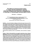
promoting cell interaction and growth (Lazzeri et al., 2005). Mechanical strength testing
was carried out to examine the effect of the addition of HAp on the mechanical property.
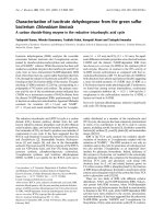
The stress-strain (S-S) curve of the samples was monitored, and representative examples
were shown in Figure 4.1. All different types of fibrous scaffolds showed a similar S-S
pattern, with an initial linear elastic regime, followed by subsequent failure. Compared to
Chapter 4 68
pure PLGA scaffold (F1), PLGA/HAp fibrous scaffolds exhibited a higher initial slope
and lower strain at failure, which was similar to the results obtained by Velayudhan and
colleagues (Velayudhan et al., 2004). It was noted that, among F1-F3, F2 showed the
highest tensile strength, suggesting that the encapsulation of a suitable amount (5%) of
HAp in PLGA contributed to the enhancement of mechanical strength. This was likely
due to the fact that HAp nanoparticles integrated well with PLGA and formed a compact
inorganic-organic composite structure commonly seen in natural bone system. Under the
same loading of HAp (5%), F2 showed much higher maximum tensile strength and lower
strain than F4. This means that the addition of protein solution before (for the case of F2)
and after (for the case of F4) scaffold fabrication affects the mechanical properties of
scaffolds.
Figure 4.1 Comparison of the typical stress-strain curves of fibrous scaffolds.
Chapter 4 69
Table 4.1 DCM residual contents in the four groups of fibrous scaffolds (F1-F4)
F1 F2 F3 F4
DCM residual contents
(ppm)
408 ± 30
342 ± 28
332 ± 31
349 ± 25
A peak area for DCM (=Absorbance x time) was obtained for each sample. Subsequently,
the peak areas for all samples were compared with a calibration curve and the
corresponding DCM concentrations could be calculated and listed in Table 4.1. The
results show that the residual organic solvent content of the scaffolds fabricated using the
electrospinning method was below the safety standards of 500ppm (“Organic Volatile
Impurities”, in United States Pharmacopeia, 1999) after drying in the freeze dryer for 3
days. However, the residual content observed is not very ideal compared with other
dosage forms like nanoparticles or microparticles. This is because the fibrous scaffold has
a much more compact structure, and this could have hindered the evaporation of DCM
from the scaffolds during the fabrication and drying process.
From Table 4.1, it was also observed that the DCM residual content decreases with
increasing HAp content. From 408ppm in scaffold F1, the DCM content dropped to 340-
350ppm in scaffolds F2 and F4, and dropped even further to almost 332ppm in scaffold
F3. The hydrophilicity of HAp could be the contributing factor because the DCM is
hydrophobic and could not exist together well with HAp in scaffolds, and DCM inside
scaffolds with HAp nanoparticles is easier to evaporate because the scaffolds with HAp
Chapter 4 70
nanoparticles (F2, F3 and F4) are more rigid and the strong three dimensional
frameworks give much less resistance to DCM evaporation than F1. As expected,
scaffolds F2 and F4 have the same HAp content and their DCM residual contents are
fairly equal.
Figure 4.2 Time-course of serum BMP-2 concentrations over six weeks after
implantation of fibrous scaffolds F1-F4 (
+
p<0.05 and *p<0.05 by t-test comparison
between the samples).
Figure 4.2 shows the serum BMP-2 concentrations 1, 2, 4, and 6 week(s) after
implantation of scaffolds. F2, F3 and control experienced similar time profiles of serum
BMP-2 concentration and showed their maximal concentrations after 4 weeks, while F4
got the highest serum BMP-2 concentration after 2 weeks and dropped dramatically in
the following weeks. F1 showed significant difference from control, F2, F3 and F4 over 6
weeks, and F4 demonstrated significant difference to all other groups over the initial 2
weeks. As bone healing is a spontaneous process, it is not a surprise to detect BMP-2 in
Chapter 4 71
the serum of the control group. An interesting phenomenon is that F1 shows even lower
serum BMP-2 concentrations than control over the observation period of 6 weeks. This
may be explained by the cytotoxicity of F1. For pure PLGA scaffold F1, its degradation
and resultant acidic environment, combined with its much higher residual solvent
compared with other scaffolds as shown in Table 4.1, may destroy peripheral cells and
hinder the spontaneous secretion of BMP-2. The in-vitro release profiles of BMP-2 from
the 4 scaffolds in PBS have been reported in our previous work (Nie et al., 2008b). As
shown in Figure 3.6 (Chapter 3), the percentage release rate of BMP-2 is the highest for
scaffold F4 at the early stage, with more than 96% of the protein being released within
the first 15 days of the in-vitro release study. Since protein was loaded after the fibrous
scaffolds were fabricated, the protein molecules were essentially located outside the
fibres and remained in the interstitial spaces within the 3D network. Hence, it is easier for
the protein molecules to diffuse into the release medium without requiring the fibres to
undergo biodegradation before they can be released. For F1-F3, the scaffolds with higher
HAp contents release BMP-2 faster, but they share very similar release profiles over a 2-
month period, close to a linear mode.
Concurrently, ALP activity in serum 1, 2, 4, 6 week(s) after implantation was
investigated. As shown in Figure 4.3, different time profiles were observed for different
samples. Control showed a gradual increase of ALP activity over 6 weeks, which was a
sign of spontaneous repair of bone defect. Similar to the control group, bone defect in F1
group experienced spontaneous healing and the ALP activity increased with time, with
the highest ALP activity being observed at week 6. The general trend of ALP for F4 is
Chapter 4 72
significantly different from all other groups. It burst over the first 2 weeks and decreased
in the following 4 weeks. ALP activity is a good indicator for analyzing the activity of
osteogenic differentiation of cytoclasts. High ALP activity refers to high differentiation
rate of cytoclast and correspondingly high bone healing rate. For example, F4 showed the
highest ALP activity over the initial 2 weeks, demonstrating that the cytoclast
differentiation rate and bone healing rate were both higher than other groups. After 2
weeks, ALP activity decreased to a very low level. This shows that cytoclast
differentiation rate is quite low and indirectly proves that bone healing has almost
concluded, as evidenced by X-ray photograph.
Figure 4.3 Time-course of serum ALP activity over six weeks after implantation of
fibrous scaffolds F1-F4 (*p<0.05 by t-test comparison between the samples).
Chapter 4 73
Correlation was carried out between the curves of BMP-2 and ALP for each scaffold
together with control using the Kendall tau rank correlation method. The results are
tabulated in Table 4.2. It is noted that BMP-2 concentrations for F4 in weeks 4 and 6 are
the same but the corresponding ALP activities are different. As a result, P and τ have
multiple solutions. From Table 4.2, one can see that τ values for all scaffolds and control
are all between 0 and 1, which means that BMP-2 and ALP curves for all scaffolds
together with control are positively correlated over the period of 6 weeks. The correlation
between BMP-2 and ALP is very obvious for F4. BMP-2 surged to a high level in week 1
and reached the highest in week 2, but it decreased to a low plateau in week 4. Compared
to other groups, the sustained high concentration in F4 over the first two weeks attracted
more stem cells, which experienced migration, proliferation and differentiation. ALP
level is an indirect indicator for the rate of new bone formation. The highest ALP level
for the F4 group (against F1, F2 and F3) in the first 2 weeks verifies the most active new
bone formation during this time period. F3 releases BMP-2 in a sustained mode till week
6 because of HAp effect, but its effect on bone growth in vivo is not obvious at all. It is
postulated that the corresponding level of BMP-2 concentration did not reach the
triggering level for effective bone regeneration. Therefore, our conclusion is that
maintaining a high enough BMP-2 concentration over the first 2 weeks can facilitate
efficient bone formation.
Table 4.2 Statistical analysis of the correlation between serum BMP-2 and ALP activity
Ctrl F1 F2 F3 F4
P
5 4 4 6 6 (5)
τ
0.667 0.333 0.333 1 1 (0.666)
Chapter 4 74
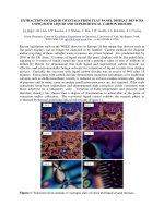
Figure 4.4 Radiographs of nude mice tibias after 1, 2, 4, and 6 week(s) of implantation of
scaffolds F1-F4. Bone fragment without implantation of any scaffold is taken as the
control. White arrows identify bone defects.
Chapter 4 75
Table 4.3 Comparison of performance scores for all samples at specific intervals*
Control F1 F2 F3 F4
Week 1
0.00 0.00 0.33 0.67 1.33
Week 2
1.67 2.00 1.00 2.00 2.00
Week 4
2.67 3.33 2.67 3.33 3.33
Week 6
3.67 4.00 4.00 3.67 4.00
* The number shown is the arithmetic average of triplicate samples.
Soft X-ray photographs can clearly demonstrate the outcome of treatment with different
scaffolds. Figure 4.4 shows soft X-ray photographs of mice tibia fractures 1, 2, 4 and 6
week(s) after implantation of scaffolds together with control. Apparently, one can find
that the bone ends from control and F1 group were as sharp as the post-operation case
and there was no significant bone regeneration after 4 weeks, especially the delayed
union of bone fractures were clearly shown by white arrows in micrographs. In contrast,
those from F2- F4 groups showed wide and dull bone ends, indicating the formation of
new bone after 4 weeks. This was especially evident for the bone ends where the two
disconnected sections on tibia were found to form new bridges after 4 weeks. The above
result demonstrates clearly that BMP-2 released from F2-F4 within the first 4 weeks took
effect and helped in bone regeneration. As confirmed by serum BMP-2 measurements,
the average BMP-2 concentration for F4 is the highest as compared with all other groups
over observational range. Table 4.3 shows the results for performances of all scaffolds at
1, 2, 4, and 6 week(s) by a semi-quantification method. From this table, one can see that
within the first week, F2-F4 demonstrate better performances than F1 and control,
showing that scaffolds F2-F4 take effects at a very early stage. Over the following 3
Chapter 4 76
weeks, F2 slows down and F1 catches up to the same score as F3 and F4. At the week 6,
all bone defects have healed perfectly for scaffolds F1, F2 and F4, while control and F3
are only partially repaired then.
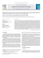
Figure 4.5 Histological specimens from nude mice tibias after 1, 2, 4, and 6 week(s) of
implantation of scaffolds F1-F4. Bone fragment without implantation of any scaffold is
taken as the control. Blue circles identify bone fragments. Original magnification is 40X
for all.
Chapter 4 77
One can examine the callus formation by the observation of bone fragments from H&E
staining micrographs shown in Figure 4.5. In the figure, the fragmental defects 1 week
after implantation are circled in blue for easier reading. From the micrographs, obvious
callus formation around bone defects can be seen in all samples. After 6 weeks, bone
fractures for F2, F3 and F4 have perfectly healed, especially for F2 and F4, while F1 and
control have not yet fully healed. Comparing the results of all samples after 4 weeks, F4
demonstrates the best performances. In Figure 4.6, numerous osteoclast-like cells
(identified by blue arrows) were observed to reabsorb the trabecular bone throughout the
defect at this time-point.
Figure 4.6 Regeneration of the marrow space and callus near fragmental defects at 4
weeks after treatment with F4 (magnification: 100X). Osteoclast diffusion (red arrow) to
defect makes necessary preparation for bone regeneration, while increased osteoclast
resorption (blue arrows) of the callus initiates the process of bone remodeling at four
weeks.
IHC staining can show the formation of blood vessels in bone fractures. After treatment
of bone fragments in liquid nitrogen for 5 minutes, cells and vessels in fragments should
Chapter 4 78
have both been destroyed, which just represents the osteoconductivity of bone graft. On
the other hand, it can be seen as a good sign of bone regeneration if newly formed bone
cells and blood vessels can be detected in bone fragments after some treatment. Von
Willebrand factor is a large multimeric glycoprotein and produced constitutively in
endothelium during blood vessel formation. After treatment of IHC staining, vWF can be
stained as brown color. Therefore brown color staining detected in bone fragments can be
seen as the formation and growth of new blood vessels. H&E staining analysis above has
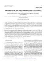
shown bone healing after treatment of all scaffolds. Since the largest difference is seen at
4 weeks, IHC staining analysis at 4 weeks of treatment is employed to confirm the
finding that F2-F4 are better than F1 and control. Blue circles are applied to show
fragmental defects in 40X micrographs and blue arrows are employed to show the
lacunae with or without newly formed cells (Figure 4.7). From this figure, one can see
that less brown color staining is observed for control and F1, but F2-F4 show more of
brown staining. The observations demonstrate that the cells in bone fragments treated by
F2-F4 are refreshing and growing, accompanied by neovascularization. The
neovasculization represents the new vessels ingrowth and this process can bring more
bone marrow stromal cells to facilitate bone healing and substitute the dead bone more
quickly. The brown color zones detected in F3 and F4 (Figure 4.7) demonstrates the
advantage of F3 and F4 over other groups on bringing bone marrow stromal cells to bone
defects and making preparation for stromal cell differentiation.
Chapter 4 79
Figure 4.7 Immunohistochemical specimens from nude mice tibias after 1, 2, 4, and 6
week(s) of implantation of scaffolds F1-F4. Bone fragment without implantation of any
scaffold is taken as control. Blue circles and arrows identify bone fragments and lacunae,
respectively.
Osteoconduction refers to the ability of some materials to serve as a scaffold on which
bone cells can attach, migrate, grow and divide. In this way, the bone healing response is
"conducted" through the graft site. In contrast, induction of bone formation refers to the
Chapter 4 80
capacity of many normal chemicals in the body to stimulate primitive "stem cells" or
immature bone cells to grow and mature, forming healthy bone tissue. In this study, the
responsibility of fixation and osteoconduction was taken by the dead bone segment and
intra-medullary wire (see Appendix A1). While the scaffolds were just placed near the
fractures and took the role as a BMP-2 source. The osteoconductive enhancement of HAp
nanoparticles in some scaffolds was not significant as they were not exactly placed in the
defects. In this study, we focused on osteoinductive role of BMP-2 and HAp
nanoparticles were acting as a regulator of BMP-2 release rate. Higher amount of HAp
can enhance BMP-2 release from scaffolds, as shown in the in vitro release profiles
(Figure 3.6).
Our previous study shows that BMP-2 loaded pure PLGA fibrous scaffold (F1) can
release BMP-2 in a sustained mode and the corresponding BMP-2 integrity is well
maintained (Nie et al., 2008b). However, its conformational structure is partially
damaged, with significant difference to native BMP-2 on α helix percentage. One of the
possible reasons for structural changes is the long-time contact between DCM and BMP-
2 during the fabrication process. This is in direct contrast to other cases where BMP-2
molecules can attach themselves to HAp nanoparticles and avoid the direct contact with
DCM over a long period of time. Another important reason is that HAp can compensate
the pH change caused by the degradation by-products of PLGA. For the case of F1, no
HAp is agglomerated into that scaffold and the acidic environment following PLGA
degradation tends to damage the secondary structures of BMP-2. For scaffolds F2, F3 and
F4, BMP-2 integrity and secondary structures are well maintained in vitro, and their in
vivo performances on defect healing are also better than F1. This observation proves that
Chapter 4 81
F2, F3 and F4 can keep BMP-2 activity better than F1.
4.5 Conclusions
BMP-2 is easy to be digested by enzyme once it is exposed to serum in vivo. Sustained
release provides the best strategy to maintain high level of BMP-2 in the local area and
that is the main motivation to design the release profile of scaffolds. This study
investigated two methods to load BMP-2 into three dimensional fibrous scaffolds using
an electrospinning method, including encapsulating into fibres or coating on fibre surface,
and the results revealed that BMP-2 encapsulated into fibres retained its biological
activity in vitro and in vivo. The addition of suitable amount of HAp nanoparticles can
enhance scaffold tensile strength and decrease residual solvent content. Animal
experiments demonstrated that BMP-2 loaded pure PLGA scaffold (F1) can’t keep the
bioactivity of BMP-2 in vivo and has no effect on bone healing. The bioactivity of BMP-
2 released from F2-F4, where BMP-2 was encapsulated inside fibres (F2 and F3) or just
coated on the surface of fibres (F4), was well maintained in vivo and better performance
of bone healing was observed. Hence, it can be concluded that the HAp and BMP-2
encapsulated fibrous scaffolds (F2-F4) are promising as BMP-2 delivery devices for bone
regeneration.