The role of GRIM 19 in xenopus embryo development
Bạn đang xem bản rút gọn của tài liệu. Xem và tải ngay bản đầy đủ của tài liệu tại đây (3.75 MB, 159 trang )
THE ROLE OF GRIM-19 IN XENOPUS EMBRYO
DEVELOPMENT
CHEN YONG
(M.Med. Wuhan Univ.)
A THESIS SUBMITTED
FOR THE DEGREE OF DOCTOR OF PHILOSOPHY
INSTITUTE OF MOLECULAR AND CELL BIOLOGY
NATIONAL UNIVERSITY OF SINGAPORE
2006
Acknowledgements
i
Acknowledgments
I would like to express my sincere gratitude to my supervisor, Dr. Xinmin Cao, for
providing me with the opportunity to pursue my Ph.D. research work in her laboratory. I
am grateful to Dr. Xinmin Cao for her guidance and support throughout my graduate
studies.
I am thankful to my graduate supervisory committee, Drs. Alan G Porter, Walter
Hunziker and Yun-jin Jiang for their constructive suggestions and critical comments.
I would especially like to thank Dr. Jianlin Fu, Wai Hong Yuen and all the other
staff in the transgenic frog facility for providing excellent technical support and an ideal
working environment for animal model generation and phenotype analysis. I also thank
Ke Guo, Jie Li and Zeng Qi for histological analysis, and Chee Peng Ng for EM.
I am grateful to Drs. Alirio J. Melendez and Farazeela Bte Mohod Ibrahim in the
Department of Physiology, National University of Singapore, and Dr. Andrew L. Miller in
the Department of Biology, Hong Kong University of Science and Techlology, for helpful
discussion, technical assistance and collaboration in the area of calcium signaling. I also
thank Dr. Katherine E. Yutzey in the Children’s Hospital Research Foundation, Cincinnati,
OH, for providing Nkx 2.5 promoter constructs.
Thanks also go to all past and present members of the CXM laboratory for their
discussion, good suggestions, technical assistance and friendship.
I am deeply grateful to Xing Chen and John Tng for their critical comments on my
thesis writing.
Finally, my deepest appreciation goes to my parents and my wife for their
consistent love, support and encouragement through out the years.
List of publications
ii
List of Publications
Chen Y
, Yuen W., Fu J., Huang G., Melendez A. J., Ibrahim F.B., Lu H., and Cao X.
Mitochondrial respiratory chain controls intracellular calcium signaling and NFAT
activity essential for heart formation in Xenopus. Mol. Cell. Biol. (under revision)
Emerald B.S.*, Chen Y.*, Zhu T., Zhu Z., Lee K.O., Gluckman P.D. and Lobie P.E. (2007)
alpha CP1 mediates stabilization of hTERT mRNA by autocrine human growth hormone.
J. Bio. Chem. (Published online on 2006 Nov 3)
* Authors contributed equally to this work.
Huang G., Chen Y., Lu H., and Cao X. (2006) Coupling mitochondrial respiratory chain
to cell death: an essential role of mitochondrial complex I in the interferon-beta and
retinoic acid-induced cancer cell death. Cell Death Differ. (Published online on 2006 Jul 7)
Zhang X., Zhu T., Chen Y
., Mertani H.C., Lee K.O., and Lobie P.E. (2003) Human
growth hormone-regulated HOXA1 is a human mammary epithelial oncogene. J Biol
Chem 278, 7580-7590.
Table of Contents
iii
Table of Contents
Acknowledgements……………………………………………………………………….i
List of Publications……………………………………………………………………….ii
Table of Contents………………………………………………………………… …….iii
Summary……………………………………………………………………………… viii
Abbreviation……………………………………………………………………….…… x
List of Figures and Tables………………………………………………………… … xiv
Chapter 1 General introduction………………………………….…………………… …1
1.1.Mitochondria respiratory chain…………………………………… ………………2
1.1.1. Oxidative phosphorylation……………………………………………………2
1.1.2. Components of MRC……………………… ……………………………….4
1.1.2.1. NADH:ubiquinone oxidoreductase (Complex I)…………………………5
1.1.2.2. Succinate:ubiquinone oxidoreductase ( complex II)………………….… 6
1.1.2.3. Ubiquinol:cytochrome c oxidoreductase (Complex III)……………….…7
1.1.2.4. Cytochrome c oxidase (Complex IV)……………………………….……9
1.1.2.5. ATP synthase (Complex V)………………………………………………9
1.1.3. MRC diseases………………………………………………… ……………10
1.1.4. GRIM19 - a subunit of MRC complex 1……………………………………13
1.2. Intracellular calcium signaling ……………………………… …………………15
1.2.1. Regulation of calcium mobilization. ……………………………………… 16
1.2.1.1. Calcium ON mechanism……………… ………………………………17
1.2.1.2. Calcium OFF mechanism……………… …………… ………………20
Table of Contents
iv
1.2.2. Calcium-calcineurin-NFAT signalling pathway ……………………………23
1.2.2.1. Structure and function of calcineurin……………………………………24
1.2.2.2. Structure and function of NFAT…………………………………… …25
1.2.3. Role of NFAT in cardiogenesis…………………………… ………………28
1.3. Cardiogenesis………… …………………………………………………………31
1.3.1. Molecular pattern in cardiaogenesis…………… ………………………….32
1.3.2. The role of Nkx2.5 in cardiogenesis ……………………………………… 37
1.3.3. Transcriptional regulation of Nkx2.5 ……………………………………….39
1.4. Rationale of this thesis ………………………………………… ……………….41
Chapter 2 Material and Methods…………………………………………………………43
2.1. Materials …………………………………………………………………… …44
2.2. Constructtion of plasmids…………………………………… …………… …44
2.3. Cell culture ……………………………………………………………… …… 45
2.4. Preparation of DH5α Escherichia coli competent cells…………………… … 45
2.5. DNA transformation ……………………………………………………………46
2.6. LIPOFECTAMINE™ DNA transfection………………………………… …46
2.7. Xenopus embryo manipulation …………………………………………… …47
2.8. Isolation of cDNA clones of Xenopus laevis GRIM-19………… ……… …48
2.8.1. Prepare Xenopus tropicalis GRIM-19 cDNA probe………… ……… 48
2.8.2. Screening of Xenopus laevis oocyte cDNA library………… ……… …48
2.9. QuikChange™ Site-Directed Mutagenesis………… …………………… …49
2.10. Prepare RNA probe or caped mRNA by in vitro transcription……………… 50
2.11. Whole-mount in situ hybridization………… ……… …51
Table of Contents
v
2.12. Histological analysis ………… ……… …52
2.13. Transmission electron microscopy… …53
2.14. In vitro transcription and translation… …53
2.15. Si RNA… …54
2.16. Western blotting… …54
2.17. Intracellular calcium measurement… …55
2.18. Luciferase reporter assay… …56
2.19. RT-PCR… …56
2.20. Electrophoretic mobility shift assay (EMSA) … …57
2.21. Mitochondrial complex I oxidative phosphorylation assay……………………58
2.22. Whole-mount in situ TUNEL staining…………………………………………59
2.23. Statistical Analysis…………………………………………………………… 59
Chapter 3 Mitochondrial respiratory chain complex I is essential for heart formation in
Xenopus……………………………………………………………………….60
3.1. Introduction……………………………………………………………………….61
3.2. Results…………………………………………………………………………….64
3.2.1 Cloning and expression pattern of XGRIM-19 in Xenopus laevis……………64
3.2.2 Knockdown of XGRIM-19 impairs MRC complex I activity in Xenopus
embryos.………………………………………………………………….….66
3.2.3 Knockdown of XGRIM-19 causes heart defect in Xenopus embryos……… 69
3.2.4 Knockdown of XGRIM-19 down-regulates cardiac gene expression and NFAT
activity……………………………………………………………………….74
Table of Contents
vi
3.2.5 Constitutively activated NFATc4 rescues the heart defect in XGRIM-19 KD
embryos …………………………………………………………………….78
3.2.6 NFATc4 rescues the defects of sarcomere formation in the heart muscles… 80
3.2.7 Knockdown of XGRIM-19 or NDUFS3 impairs calcium mobilization and
calcium-induced NFAT activity…………………………………………… 82
3.3 Discussion………………………………………………………………………….87
Chapter 4 NFAT regulated Nkx2.5 expression in transcriptional level…………………91
4.1. Introduction………………………………………………………………….……92
4.2. Results…………………………………………………………………………….95
4.2.1. Constitutively active NFATc4 rescued Nkx2.5 expression in GRIM-19 KD
Xenopus embryos………………………………………………………………95
4.2.2. Nkx2.5 gene expression is NFAT dependent during RA-induced cardiac
differentiation of P19 cells…………………………………………………….96
4.2.3. Predicted conserved NFAT and its cofactor binding elements are localized in the
promoter region of Nkx2.5 genes.………………………………… …… 100
4.2.4. NFATc4 interacted with NFAT binding elements in Nkx2.5 gene promoter.103
4.2.5. NFATc4 up-regulates Nkx2.5 expression on transcriptional level……… …106
4.3. Discussion…………………………… ……………………………………….110
Chapter 5 General discussion………………………………… ………………………114
5.1. GRM-19 knocking-down Xenopus as a model for studying the MRC functions in
early embryonic development……………………………………………… ….115
5.2. MRC activity is crucial for triggering intracellular calcium mobilization and NFAT
activity……………………………………………………………………………116
Table of Contents
vii
5.3. NFAT is a transcriptional regulator of Nkx2.5…………………… ……………117
5.4. A model of regulation of heart development by MRC……………………… …118
References………………………………………………………………………………121
Summary
viii
Summary
The mitochondrial respiratory chain (MRC) plays a crucial role in cellular energy
production, which is needed for cell division, movement, secretion, and activation of
signaling pathways. MRC mutations cause diseases with multi-system disorders
including encephalopathies, myopathies and cardiomyopathies, which occur in 1 per
10,000 live births in humans (Triepels et al., 2001). Depletion of MRC activity results in
severe abnormalities in embryo development and leads to embryonic lethality (Huang et
al., 2004; Larsson et al., 1998). The lack of an adequate animal model imposes limits on
our current understanding of molecular processes in MRC-dependent embryonic
development and the pathogenesis of these MRC diseases. To address this issue, GRIM-
19, a newly identified MRC complex I subunit, was knocked down in Xenopus embryos.
The embryos exhibited typical phenotypes associated with mitochondrial diseases
including retarded growth, mitochondrial proliferation, and moderately serious levels of
neural, eye, and muscle tissue disorders. However, the most striking phenotype exhibited
is that of defective heart formation. This can be rescued by reintroduction of human
GRIM-19 mRNA. The heart tube failed to loop in most of GRIM19 knocked-down
embryos, and the expression of several cardiac markers such as Nkx2.5 and its
downstream gene, MLC2, and cardiac actin, were also reduced. Upon further
investigation, we found that the activity of NFAT, a family of transcription factors that
contributes to early organ development, was down-regulated in GRIM-19 knockdown
embryos. Furthermore, expression of a constitutively active form of mouse NFATc4 in
these embryos could restore normal heart development. NFAT activity is controlled by
Summary
ix
the calcium-dependent phosphatase protein, calcineurin, which suggests that calcium
signaling may be disrupted by GRIM-19 knockdown. Indeed, both the calcium response
and calcium-induced NFAT activity were impaired in cell lines of knocked-down GRIM-
19, and NDUFS3, another complex I subunit. We also showed that NFAT can rescue
expression of Nkx2.5 in GRIM-19 knocked-down embryos; NFAT binds on directly
Nkx2.5 promoter and up-regulates Nkx2.5 transcription. Our data demonstrates the
essential role of the MRC in heart formation and sheds light on the signal transduction
and gene expression cascades involved in this process.
Abbreviation
x
Abbreviation
ANT 1 adenine nucleotide translocator 1
AR activation region
AP1 activator protein 1
ATP adenosine triphosphate
BMP bone morphogenetic protein
BNP b type natriuretic peptide
BN-PAGE blue native polyacrylamide gel electrophoresis
CamK calcium/calmodulin-dependent protein kinase
CA cardiac actin
CA-NFATc4 constitutively active NFATc4
CNS central nervous system
CICR Ca
2+
induced Ca
2+
release
CR conserved region
CRACs Ca
2+
release-activated Ca
2+
channels
CREB CRE binding protein
CsA cyclosporin A
CSMDHs Ca
2+
-sensitive mitochondrial dehydrogenases
cTnl cardiac troponin-l
cTnT cardiac troponin-T
Cyt cytochrome
DMEM Dulbecco’s modified Eagle’s medium
DMSO Dimethyl sulfoxide
Abbreviation
xi
EMSA electrophoretic mobility shift assays
EMT endocardial-mesenchymal transformation
ER endoplasmic reticulum
FAD
flavin adenine nucleotide; oxidized state
FADH
2
flavin adenine nucleotide; reduced state
FBS fetal bovine serum
FGF fibroblast growth factors
GATAs GATA binding transcription factors
GSH superoxide dismutase and glutathione peroxidase
GRIM-19 genes associated with retinoid-IFN-induced mortality-19
Hand1/2 heart and neural crest derivatives expressed transcript 1 or 2
Hsp60 heat shock protein 60kd
ICRAC Ca
2+
release-activated Ca
2+
current
IFN-β interferon-β
ΙL interleukin
InsP
3
1, 4, 5-trisphosphate
InsP
3
Rs InsP
3
receptors
JNK Jun N-terminal kinase
KD knocked down
LD lipid droplet
MEF MADS-box transcription factor
MHC myosin heavy chain
MIB mitochondrial isolation buffer
Abbreviation
xii
MLC myosin light chain
MO morpholino oligonucleotides
MRC mitochondrial respiratory chain
mtDNA mitochondrial DNA
nDNA nuclear DNA
NAD Nicotinamide adenine dinucleotide; oxidized state
NADH Nicotinamide adenine dinucleotide; reduced state
NCX Na
+
/Ca
2+
exchanger
NFAT nuclear factor of activated T cells
Nkx2.5 NK2 transcription factor related, locus 5
FMN Flavin MonoNucleotide
O
2
superoxide radicals
OCT3 octamer binding protein 3
OH· hydroxyl radicals
ORF open reading frame
OXPHOS oxidative phosphorylation
PBS phosphate-buffered saline
PLC phospholipase C
PM plasma membrane
PMA phorbol 12 myristate 13-acetate
PMCA Plasma membrane Ca
2+
ATPase
Q ubiquinone
QH
2
reduced ubiquinol
QH· ubisemiquinone radicals
Abbreviation
xiii
RA retinoic acid
ROCCs receptor-operated Ca
2+
channels
RyRs Ryanodine receptors
SERCA sarco-endoplasmic reticulum ATPase
siRNA small interfering RNA
SR sarcoplasmic reticulum
Tbx T-box transcription factor
TGFβ transformation growth factor-β
TCR T cell receptor
TFAM mitochondrial transcription factor A
TUNEL terminal deoxynucleotidyl transferase biotin-dUTP nick nnd labeling
VEGF Vascular endothelial growth factor
VDAC voltage-dependent anion channel
VOCCs voltage-opened Ca
2+
channels
List of Figures and Tables
xiv
List of Figures and Tables
Figure 1.1. Schematic of morphology and function of MRC…………………………… 5
Figure 1.2. Regulation of calcium dynamics and homeostasis. …………………………17
Figure 1.3. Schematic of role of mitonchondria in calcium dynamics………………… 22
Figure 1.4. Calcium-Calcineurin-NFAT pathway………………………………… … 23
Figure 1.5. Schematic of NFAT domain (based on mouse NFATc2).………………….26
Figure 1.6. Schematic of transcriptional network involved in cardiogenesis………… 35
Figure 3.1 Comparison of the amino acid sequence of GRIM-19 between Xenopus laevis,
Xenopus tropicalis, mouse, and human……………………… …………….65
Figure 3.2A In situ hybridization of XGRIM-19 in Xenopus embryos…………………65
Figure 3.2(B and C) XGRIM-19 mRNA and protein expression pattern during embryo
development………….……………………………………… ……………66
Figure 3.3 Knockdown efficiency of XGRIM-19 and its effect on the complex I
activity………………………………………………………………………67
Figure 3.4 Knockdown of GRIM-19 impairs complex I activity……………………….68
Figure 3.5 General morphology and cardiovascular formation in XGRIM-19 KD
embryos……………………………………………………………………….69
Figure 3.6 Knockdown of XGRIM-19 causes multi-system disorder in Xenopus
embryos……………………………………………………………………….70
Figure 3.7 Knockdown of XGRIM-19 causes heart defect in Xenopus embryos……….72
Figure 3.8 Inhibition of complex I activity causes heart defect in Xenopus embryos… 73
Figure 3.9Depletion of XGRIM-19 down-regulates expression of several cardiac genes75
Figure 3.10 Depletion of XGRIM-19 affects expression of specific cardiovascular genes
List of Figures and Tables
xv
during different embryonic stage……………………………………… ….76
Figure 3.11 Depletion of XGRIM-19 compromises NFAT activity…………………… 77
Figure 3.12 Comparison of NFATc4 and CA-NFATc4 activity in MCF-7 cells……… 78
Figure 3.13 CA-NFATc4 partially rescues the heart defect in XGRIM-19 KD embryos.79
Figure 3.14 Ultrastructure of heart and skeletal muscle from Xenopus embryos at
stage 45…………………………………………………………………… 81
Figure 3.15 GRIM-19 knockdown compromises intracellular calcium mobilization… 84
Figure 3.16 GRIM-19 knockdown compromises NFAT activity……………………… 85
Figure 3.17 Knockdown efficiency of GRIM-19 and NDUFS3 in HeLa and Jurkat
Cells…………………………………………………………………………86
Figure 4.1 CA-NFATc4 rescues Nkx2.5 expression in XGRIM-19 KD embryos………96
Figure 4.2 Low concentration of RA induces Nkx2.5 gene expression and cardiac
Differentiation……………………………………………………………….98
Figure 4.3 Nkx2.5 gene expression is NFAT dependent…………………………… …99
Figure 4.4 Schematic diagram of 10.7 kb 5’-flanking sequence of mouse Nkx2.5
promoter…………………………………………………………………….100
Figure 4.5 Comparison of the mouse Nkx2.5 CR1 with rat, dog and human
sequences………………………………………………………………… 102
Figure 4.6 Comparison of the mouse Nkx2.5 CR2 (partial) with rat, dog and human
sequences………………………………………………………………… 103
Figure 4.7(A-B) NFATc4 interacted with NFAT binding elements in Nkx2.5 gene
promoter…………………………………………………………………….………… 104
Figure 4.7(C-D) NFATc4 interacted with NFAT binding elements in Nkx2.5 gene
promoter……………………………………… ………………………………………105
List of Figures and Tables
xvi
Figure 4.8 NFATc4 up-regulates transcriptional activity of Nkx2.5 enhancer……… 108
Figure 4.9 NFATc4 up-regulates transcriptional activity of Nkx2.5 3 kb promoter… 109
Figure 5.1 A model of regulation of heart development by MRC…………………… 119
Table 1 Primer sequences for RT-PCR…………………………………………………57
Table 2 Specific gene expression in control and XGRIM-19 KD embryos ……………74
Chapter 1
1
Chapter 1
General introduction
Chapter 1
2
1.1. Mitochondrion respiratory chain (MRC)
Cells need energy to move, contract, divide and produce secretary products to
communicate with other cells. The primary energy currency inside cells is adenosine
triphosphate (ATP), a high energy phosphate nucleotide. Hydrolysis of ATP releases
energy, which meets the need of various biological reactions in cells. ATP is
manufactured by several cellular process including glycolysis, photosynthesis and
oxidative phosphorylation. The majority of ATP production in eukaryotic cells is fulfilled
by oxidative phosphorylation in mitochondria. Mitochondria are believed to have evolved
from aerobic bacteria which colonized primordial eukaryotic cells that lacked aerobic
metabolism (Wallace, 2005). Mitochondria endowed eukaryotic cells with the ability to
produce ATP by oxidative phosphorylation, a much more efficient way to generate energy
than through anaerobic glycolysis. The Mitochondrion is a double membrane bound
organelle in eukaryotic cells. It contains four compartments: the outer membrane which
encloses the organelle, the inner membrane which folds inside forming shelve-like
structures called “cristae”, the inner membrane space, and the matrix which is localized
inside the inner membrane. Oxidative phosphorylation and ATP synthesis are performed
by the mitochondrial respiratory chain (MRC) located on the inner membrane of the
mitochondria.
1.1.1. Oxidative phosphorylation.
Oxidative phosphorylation is the main source of generating ATP in cells. The
energy of cells comes from oxidation of fuel molecules such as lipids and carbohydrates,
especially glucose. Three biochemical reaction steps are needed to convert energy from
Chapter 1
3
these energy-containing molecules into ATP. In the first step, glucose or fatty acids are
broken-down and converted to acetyl CoA (acetyl coenzyme A) and carbon dioxide. The
energy released from these processes is used to generate ATP as well as NADH and
FADH
2
. The breakdown of glucose in this step is termed glycolysis, in which glucose is
broken down into two three-carbon molecules known as pyruvate. Glycolysis yields two
pyruvate molecules, and a net gain of 2 ATP and two NADH per glucose. The overall
reaction is:
1 Glucose + 2 NAD
+
+ 2 P
i
+ 2 ADP → 2 pyruvate + 2 NADH + 2 ATP + 2 H
2
O
In the absence of oxygen, pyruvate is not metabolized via aerobic respiration but
converted to waste products such as lactate in the cytoplasm. However, in the presence of
oxygen, pyruvate translocates to the matrix of mitochondria where it is converted to acetyl
CoA and proceeds to the next step, the citric-acid cycle (also named Krebs cycle). During
this process, the acetyl CoA is further broken down into carbon dioxide and releases the
energy to generate ATP, NADH and FADH
2
. The net energy gain in the citric acid is
1ATP, 3 NADH, and 1 FADH
2
per acetyl CoA. The overall reaction is:
acetyl CoA + 3 NAD
+
+ FAD + ADP + 2 P
i
2 CO
2
+3 NADH + 3 H
+
+ FADH
2
+ ATP
Thus, only limited energy from the breakdown of glucose is used for generation of
ATP during glycolysis and citric-acid cycle. The majority of energy is transfered to
NADH and FADH
2
which are used to produce ATP by the third process termed oxidative
phosphorylation. Oxidative phosphorylation is the process in which ATP is formed as a
result of the transfer of electrons from NADH and FADH
2
to O2 by protein complexes of
the mitochondria respiratory chain within mitochondria inner membrane. During this
process, protons are pumped from the mitochondrial matrix into the intermembrane space
to generate a transmembrane proton potential as a result of electron flow. The protons then
Chapter 1
4
flow back to the matrix via ATP synthase on the inner membrane where the proton
potential energy is used to produce ATP. A total of 10 protons are ejected to the
intermembrane space for every 2 electrons which are transferred from NADH to oxygen.
Oxidation of FADH
2
also transfers 6 protons from the matrix to the intermembrane space.
Production of 1 ATP requires 4 protons flowing back to the matrix. Thus each NADH
molecule contributes enough proton motive force to generate 2.5 ATP. Each FADH
2
molecule generates 1.5 ATP. Altogether, through oxidation of one glucose molecule, the 8
NADH and 2 FADH
2
molecules account for production of more than 23 ATP by oxidative
phosphorylation. The total ATP production from aerobically metabolized glucose is
around 30 ATP in comparison with to the 2 from anaerobic glycolysis. Therefore, aerobic
respiration is approximately 15 times more efficient than anaerobic.
1.1.2. Components of mitochondrial respiratory chain MRC
The mitochondrial respiratory chain (MRC) catalyzes oxidative phosphorylation
which plays a crucial role in aerobic respiration of cells. The MRC consists of five multi-
subunit complexes (complexes I-V) and two additional electron carriers: coenzyme Q10
and cytochrome c. MRC complexes I-IV, coenzyme Q10 and cytochrome c act as electron
carriers which transfer two electrons from reducing substrates (NADH and FADH
2
) to
molecular oxygen. Thus the electron carriers are said to form an electron-transport chain.
MRC complexes I, III and IV are also proton pumps which simultaneously pump protons
from the matrix to the intermembrane space to generate a proton gradient across the inner
mitochondrial membrane. The electrochemical energy of this gradient is then used to drive
ATP synthesis by complex V. An overview of morphology and function of MRC is
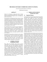
illustrated in Figure 1.1.
Chapter 1
5
Adopted from electron transport chain lecture by Antony Crofts
Figure 1.1. Schematic of morphology and function of MRC.
1.1.2.1. NADH:ubiquinone oxidoreductase (Complex I)
NADH:ubiquinone oxidoreductase (complex I) of the MRC catalyzes the first step
of electron transfer. It catalyzes the oxidation of NADH, the reduction of ubiquinone, and
the transfer of 4H
+
across the mitochondrial inner membrane. Complex1 is the largest
complex composed of at least 46 structural subunits in humans. Among them, 7 subunits
are encoded by mitochondrial DNA (mtDNA), while others are encoded by nuclear DNA
(nDNA). The 46 subunits of complex1 form a boot shape, which contains two sub-
Complex I
NADH:ubiquinone
oxidoreductase
Complex II
Succinate:ubiquinone
oxidoreductase
Complex III
ubiquinol:cytochrome c
oxidoreductase
Complex IV
cytochrome c oxidase
Complex V
ATP synthase
NADH
FMN
FeS
red
Centers
UQ
NAD
+
FMNH
2
FeS
ox
Centers
UQH
2
succinate
FAD
FeS
ox
Centers
UQ
fumarate
FADH
2
FeS
ox
Centers
UQH
2
UQH
2
2FeS
ox
2cyt c
1red
2cyt c
ox
UQ
2FeS
red
2cyt c
1ox
2cyt c
red
2cyt c
red
2Cu
Aox
2cyt a
red
2cyt a
3ox
Cu
B
2cyt c
ox
2Cu
Ared
2cyt a
ox
2cyt a
3red
Cu
B
H
2
O
½O
2
+H
2
Chapter 1
6
complex domains. The peripheral domain corresponding to the “ankle” of the boot
protrudes from the mitochondrial inner membrane to the matrix. The inner membrane
domain (the “foot” of boot) contains hydrophobic proteins and is bounded in the inner
membrane. Electron transfer starts from the peripheral domain of complex 1 where NADH
is oxidized and 2 electrons are transferred to Flavin MonoNucleotide (FMN). The
electrons are then passed to the iron-sulfur centers which are also located in the
hydrophilic peripheral domain. Through the iron-sulfur centers, the electrons are finally
transferred to ubiquinone (also named coenzyme Q, CoQ or Q) which is close to the
interface between the peripheral and intra-membrane domains. Simultaneously,
ubiquinone (Q) takes up two protons from the matrix side, to form fully reduced ubiquinol
(QH
2
). The hydrophobic ubiquinol feeds into a ubiquinone pool inside the inner
membrane and diffuses to complex III. Complex I produce 1 QH
2
, per NADH oxidized.
During the process of electron transfer from NADH to ubiqinone, complex I pumps 4
protons across the coupling membrane to generate an inner membrane proton potential.
The total reaction of complex1 can be described as:
NADH + H
+
+ Q + 4H
+
N
<==> NAD
+
+ QH
2
+ 4H
+
P
In this chapter,
N
means N-side (the protochemically negative matrix side).
P
means P-side
(the protochemically positive inter-membrane-space side of mitochondrial inner
membrane).
1.1.2.2. Succinate:ubiquinone oxidoreductase ( Complex II)
FADH
2
is oxidized by succinate:ubiquinone oxidoreductase (complex II).
Complex II is the smallest complex, containing only 4 nuclear coded proteins. The
complex II is an important enzyme complex in both the citric-acid cycle and the
Chapter 1
7
mitochondrial respiratory chain. During the citric-acid cycle, complex II oxidizes
succinate to fumarate. The electrons from succinate are accepted by FAD which is
subsequently reduced to FADH
2
during oxidation of succinate to fumarate. FADH
2
is then
reoxidized by electron transfer through a series of three iron-sulfur centrers of complex II
to ubiquinone, yielding QH
2
. The energy released from oxidation of succinate and FADH
2
is inadequate to pump H
+
. Therefore, this complex only generates one QH
2
per succinate
oxidized and pumps no protons across the inner membrane. The total reaction of complex
II can be described as:
succinate + Q <==> fumarate + QH
2
Both complex I and complex II transfer electrons to ubiquinone which then ferries
the electrons to complex III. Ubiquinone is the only non-protein electron carrier of the
mitochondrial respiratory chain. It is very hydrophobic and dissolves within the lipid core
of the inner membrane. The quinine ring of ubiquinone accepts 2 electrons and is reduced
to ubiquinol (QH
2
). Ubiquinone can also accept a single electron to generate
ubisemiquinone radicals (QH·). QH· can be very dangerous as they generate superoxide
radicals (O
2
). O
2
has limited reactivity with lipids. However, O
2
can be dismutated to
H
2
O
2
. The H
2
O
2
will undergo the fenton reaction to form hydroxyl radicals( OH·) which
is far more reactive and lethally destructive than O
2
. In order to cope with these free
oxygen radicals, mitochondria contain superoxide dismutase and glutathione peroxidase
(GSH) to reduce free oxygen radicals to H
2
O.
1.1.2.3. Ubiquinol:cytochrome c oxidoreductase (Complex III)
Complex III accepts the electrons from ubiquinol (QH
2
), passes the electrons to
cytochrome c (cyt c) and transports protons across the inner membrane from the matrix
Chapter 1
8
site to the inter membrane space. The oxidation of every QH
2
produces 2 cyt c
red
(reduced
cytochrome c) and pumps 4 proton. Human complex III contains 11 subunits. Among the
subunits, only cytochome b (cyt b) is encoded by mtDNA. The process in which complex
III transfers electron from the two-electron-carrying QH
2
to the single-electron carrying
cyt c is catalyzed by three subunits: cyt b, cyt c
1
and an iron sulfur protein through a two-
step Q cycle. In the first step, one QH
2
gives up its two electrons to complex III. One
electron passes through the iron sulfur protein and cyt c
1
to the oxidized cyt c (cyt c
ox
) to
form cyt-c
red
. The other electron is carried through two cyto-b centres and delivered to
ubiqinone to form a semiquinone (QH·). Once QH
2
is oxidized, it releases its two H
+
to
the inter membrane space. The same process happens again with another QH
2
. The second
QH
2
contributes its two electrons to produce one more cyt c
red
and fully reduces the QH·
to QH
2
. Again, it pumps two H
+
into the intermembrane space. Thus the Q cycle
consumes two QH
2
, but generates only one in return. In the whole process, there is a net
oxidation of just one QH
2
and reduction of two cyt c, and 4 H
+
ions are pumped across the
inner membrane. The total reaction of complex III can be described as:
QH
2
+ 2 cyt-c
ox
+ 2H
+
N
<==> Q + 2 cyt-c
red
+ 4H
+
P
Cytochrome c is a small, water soluble protein which transfers electrons from
complex III to complex IV. It is among the three types (a,b,c) of cytochromes containing
a heme group. Cytochrome c contains a heme-c prosthetic group. The Fe ion in the heme
group can either be in the oxidized (Fe
3+
) or the reduced (Fe
2+
) form. This makes the Fe
ion of cyt c severe as an electron carrier for transfer of electrons between complex III and
complex IV. Besides being an essential component of the electron transfer chain, cyt c is
also an intermediary in apoptosis. Pro-apoptotic stimuli can trigger the release of cyt c
from mitochondria into cytosol where it activates a caspase cascade. The caspases are