Elucidating the role of dok 3 in b cell receptor signaling using gene knockout mice 2
Bạn đang xem bản rút gọn của tài liệu. Xem và tải ngay bản đầy đủ của tài liệu tại đây (1.54 MB, 132 trang )
1
1.1 Innate and adaptive immunity
The human body encounters numerous foreign substances, of which, some are
microbes that can elicit an immune response. The physiological function of the human
immune system is to recognize and eliminate invading microbes. Defense against
microbes is mediated by the early reactions of innate immunity and later responses of
adaptive immunity. Innate immunity consists of mechanisms that exist prior to an
encounter with microbes, are rapidly activated, and react similarly to repeated infection.
In contrast to innate immunity, more highly evolved defense mechanisms are stimulated
by exposure to infectious agents and increase in magnitude and defensive capabilities
with each successive exposure to a particular microbe. Because this form of immunity
develops as a response to infection and adapts to the infection, it is called adaptive
immunity.
Innate immunity provides the early lines of defense against microbes. This system
uses pattern recognition receptors to recognize invariant structures that are shared by
microbes. Most of them are often essential for survival of the microbes, such as
lipopolysaccharides (LPS) that constitute the cell wall in gram-negative bacteria or
unmethylated microbial DNA, thus limiting the capacity of microbes to evade detection
(Akira, 2006). The principle components of innate immunity includes physical and
chemical barriers such as epithelia and antimicrobial substances produced at epithelial
surfaces; phagocytic cells and natural killer cells; and blood proteins such as
complements factors and cytokines.
In contrast to innate immunity, there are two types of adaptive immune responses,
called the humoral immunity and cell-mediated immunity, which are mediated by
2
different components of the immune system and function to eliminate different types of
microbes. Neutralization of foreign extracellular microbes and soluble microbial toxins is
mediated by humoral immunity by means of secretory molecules such as antibodies
produced by B lymphocytes. Eradication of intracellular bacteria and viruses requires
cell-mediated immunity, also called cellular immunity. Intracellular microbes could
evade circulating antibodies, survive and proliferate inside host cells. Cell-mediated
immunity is mediated by circulating lymphocytes, mainly T lymphocytes and
macrophages, which promotes the lysis of infected host cells or destruction of microbes
by phagocytosis, respectively.
1.2 B lymphocytes
The human body contains approximately 2 X 10
12
lymphocytes, of which 5-15%
are B cells. B cells are small (6-10 µm) and have a dense nucleus and little cytoplasm.
They were discovered together with T cells in the early 1960s and can be distinguish
phenotypically only by analysis of cell surface markers. B lymphocytes were so called
because in birds they were found to mature in an organ called the bursa of Fabricius
(Glick, 1991). In mammals, no anatomic equivalent of the bursa exists, and the early
stages of B cell maturation occur in the bone marrow and in the fetus liver. Thus, “B”
lymphocytes refer to bursa-derived lymphocytes or bone marrow-derived lymphocytes.
The humoral immune response is the aspect of immunity that is mediated by
antibodies produced by B cells and is so called because it involves substances found in
the humours, or body fluids. The concept of humoral immunity developed based on
analysizing antibacterial components in the serum. Hans Buchner is credited with the
development of the humoral theory. In 1890 he described alexins, or “protective
3
substances”, which exist in the serum and other bodily fluids and are capable of killing
microorganisms. Alexins, later redefined as "complement" by Paul Ehrlich, were shown
to be the soluble components of the innate response. In the same year, Emil von Behring
and Shibasabo Kitasato in Koch’s laboratory discovered that injecting diphtheria toxin
into animals produces a serum containing an antitoxin that provided passive anti-
diphtheria immunity to people (Behring and Kitasato, 1965). This component in serum is
later termed antibodies or immunoglobulins (Igs).
B lymphocytes constitute an unique and vital element of both innate and adaptive
immune system. They are the sole antibody production factories of our immune system.
Naturally occurring or secreting antibodies are distributed throughout our body, in the
blood streams, in the mucosal tissues, and to a lesser extent, the interstitial fluid of the
tissues. These Y-shaped secreted antibodies, of which there are four classes (IgM, IgG,
IgA and IgE), are composed of two heavy and two light chains. Each of the millions of
different antibody molecules synthesized is capable of recognizing a different antigen. B
cells achieve this very large repertoire of antibodies by complex mechanisms such as
V(D)J recombination Ig genes as well as other processes including somatic
hypermutation and gene conversion.
Apart from its pivotal role in antibody production, B lymphocytes also participate
in adaptive immune responses by functioning as antigen presenting cells. The ability of B
lymphocytes to capture, process and present antigens to T cells is requisite for normal
humoral immune responses. B lymphocytes are remarkably efficient at presenting protein
antigens that bind to their Ig receptors to class II MHC-restricted CD4
+
helper T cells. Ig
molecules function as high-affinity binding sites for capturing antigens at limiting
4
concentrations. In addition, Ig receptor-mediated endocytosis leads to an intracellular
pathway of protein traffic that favors recycling of antigens and optimizes the processing
of these antigens. As a result, small amounts of endocytosed antigens are capable of
associating with MHC molecules and being presented in a form that can be specifically
recognized by T lymphocytes. The antigen presenting function of B cells is particularly
important in secondary antibody responses, which require low concentrations of antigens
and MHC-restricted T-B cell cooperation.
1.2.1 Subpopulations of B lymphocytes
B cells are divided into different types according to the expression of surface
molecules and anatomical location. Two major B cell subsets, designated B-1 and B-2
(B-0 or follicular B), exist in human and mice. B-1 cells are located in the peritoneal and
pleural cavities (Hardy, 2006). They expressed high levels of surface IgM, low levels of
B220 and IgD and moderate levels of CD5, and absence of CD23. In contrast,
conventional, or B-2, cells are the predominant B cells found in secondary lymphoid
organs, such as spleen and lymph node, and in the circulation. B-2 cells express high
levels of B220, IgD and CD23 and moderate levels of IgM and lack surface CD5
expression. The B-1 population can be further subdivided into B-1a and B-1b
subpopulations. The B-1b “sister population” lacks surface CD5 but shares the other
attributes of B-1a cells, such as natural antibody production and low in B220 expression.
Another subset of B cells is the marginal zone (MZ) B cells and is normally found in the
spleen (Martin and Kearney, 2000). Unlike follicular B-2 cells that express high levels of
IgD and CD23, with either high or low levels of IgM, MZ B cells express high levels of
IgM and very low levels of IgD and CD23.
5
1.2.2 B cell development
B cell development is a highly regulated multi-step process that proceeds through
the ordered maturation of a B cell from a committed precursor and ends with the
generation of an immunocompetent mature B cell (Hardy and Hayakawa, 2001).
Mammalian B cells develop from lymphoid progenitors in the bone marrow (Osmond,
1990). In the bone marrow, hematopoietic stem cells differentiate into common
lymphocyte progenitors (CLP) that can develop into mature B cells. The main goal of this
developmental process is to generate a population of cells expressing a diverse repertoire
of B-cell antigen receptors (BCRs), with different specificities that are capable of
recognizing and responding to new and recurring pathogens (Kurosaki, 2000). In contrast
to other cell lineages, developmental progression of B cells relies on a mechanism of
selection that ensures the survival of B cells expressing BCRs with certain characteristics
and specificities (Seagal and Melamed, 2003). This selection process not only serves to
promote the development of B cells with functional antigen receptors (positive selection)
but also provides a mechanism for the elimination of clones that are able to respond to
endogenous or self-antigens (negative selection). Importantly, the ordered assembly and
expression of individual components of the BCR drives the maturation process;
development is arrested unless a specific component of the BCR has been successfully
expressed (Benschop and Cambier, 1999). These points of developmental arrest have
defined checkpoints where the B cell interrogates the functionality of the BCR, and only
those B cells in which proper assembly has occurred are selected for continued
developmental progression.
6
The mature form of the BCR consists of two functional units, the antigen
recognition unit formed by the immunoglobulin (Ig) heavy (H) and light (L) chains, and
the signaling unit formed by the proteins Igα (CD79a) and Igβ (CD79b). The process of
B-cell development is highly dependent on assembly of a competent BCR. The signaling
proteins Igα and Igβ are expressed first, beginning at the earliest defined cell stage
committed to the B lineage, the pro-B cell. The genes encoding the Ig heavy and light
chains are comprised of a series of segments termed variable (V), diversity (D), and
joining (J), which are brought together by a site specific recombination process termed
V(D)J recombination (Chen and Alt, 1993). During the pro-B stage, the Ig heavy-chain
locus is in the process of rearrangement. Despite the absence of heavy-chain protein,
there is some indication that Igα and Igβ may be expressed at the pro-B-cell surface.
Although Igα and Igβ function at the pro-B stage is unclear, it has been proposed that this
complex constitutes a pro-B-cell receptor that may signal continued development to the
pre-B cell stage.
Following successful recombination of the heavy chain, this protein is assembled
into the pre-BCR complex together with the surrogate light chain (SLC) proteins, VpreB,
λ5, Igα and Igβ (Burrows et al., 2002). The pre-BCR then provides the context where the
functionality of the newly synthesized heavy chain is tested, and signals generated by this
receptor allow the transition to the pre-B stage. Pre-BCR signaling is also necessary for
allelic exclusion of the un-rearranged heavy-chain locus, light-chain recombination, and
the proliferative expansion of pre-B cells that occurs prior to light-chain recombination.
Successful light-chain recombination at the pre-B stage is followed by displacement of
the SLC proteins in the receptor complex. Assembly and surface expression of the mature
7
BCR containing light and heavy chains marks the transition to the immature stage. It is at
the immature stage that the BCR is first able to interact with conventional polymorphic
ligands. In contrast to previous selection steps that tested the signaling capacity of the
receptor complex, selection at the immature stage is designed to test the receptor–ligand
interaction (Benschop and Cambier, 1999; Seagal and Melamed, 2003). Intimate contact
between the immature B cell and bone marrow stromal cells allows receptors capable of
recognizing self-antigens to be identified and eliminated through a variety of mechanisms
collectively termed ‘negative selection’. Non-self-reactive B cells exit to the periphery
and reach the spleen, where they may still be tested for reactivity against self-antigens
before transiting to the mature stage.
Immature B cells in the spleen are further divided into transitional 1 (T1) and
transitional 2 (T2) cells by virtue of their cell-surface phenotype (Rudin and Thompson,
1998). The phenotype of T1 cells most closely resembles that of bone marrow immature
B cells. T1 cells inhabit the spleen’s red pulp and give rise to T2 and mature naive B
cells, when entering into the spleen follicles. In contrast to immature B cells that undergo
negative selection following ligand binding, mature B cells initiate pathways that lead to
proliferation and further differentiation into antibody producing B cells (plasma cells)
(Manz et al., 2002) or memory B cells (Tsiagbe et al., 1992).
1.3 B cell receptor signal transduction
Most lymphocytes such as T cells, B cells, natural killer cells and macrophages
express specific cell surface receptors that enable them to recognize foreign microbial
antigens. T cells, via T cell receptors (TCR), recognize foreign protein antigens presented
to them on a major histocompatibility complex (MHC) molecule by our own antigen
8
presenting cells; B cells utilize B cell receptors (BCR) to distinguish protein and non-
protein antigens. On the other hand, Toll-like receptors (TLR), known to be expressed by
many cell types, are capable of identifying diverse classes of non-protein pathogen-
associated molecular patterns (PAMPS) present in microbes such as unmethylated
bacterial DNA, double-stranded and single-stranded viral RNA and bacterial cell
membrane constituents such as lipopolysaccharides (LPS) (Akira and Hemmi, 2003;
Benschop and Cambier, 1999; Seagal and Melamed, 2003). Binding of antigens to these
membrane-bound receptors trigger a wide variety of cellular responses. The distinct
intracellular signaling cascades downstream of each receptor relays messages to the
nucleus, transcribing new gene products that lead to cell proliferation, cell differentiation,
cell migration, cytokine production and even cell death, just to name a few.
The B cell receptor (BCR) is an integral membrane protein complex that is
composed of an antigen binding subunit (two Ig H chains and two Ig L chains) and a
signaling subunit (a disulfide-linked heterodimers of Igα and Igβ). Igα and Igβ each
contain a sequences motif of approximately 26 amino acids residues within their
cytoplasmic regions called immunoreceptor tyrosine activation motif (ITAM). The ITAM
consists of two YXXL motifs separated by 6–8 amino acids, where Y stands for tyrosine,
L is for Leucine and X stands for any amino acid. Both tyrosine residues in the ITAM
become phosphorylated upon receptor aggregation and initiates signal transduction by
providing specific binding site for Src-homology (SH) 2 domain containing effectors,
including several classes of cytoplasmic protein tyrosine kinases (PTKs), which
phosphorylate intracellular enzymes and adaptor molecules. Such phosphorylation events
cause increased levels of intracellular calcium, activation of phosphatidylinositol 3-kinase
9
(PI3-K), cytoskeletal reorganization, transcriptional activation, and, finally, B cell
maturation, proliferation, and antibody secretion.
A temporal analysis of the activity of the PTKs after BCR aggregation has shown
that the Src family kinases such as Lyn or Blk is activated first, mediating the
phosphorylation of the ITAM tyrosines. The doubly phosphorylated ITAM provides a
binding site for the PTK, Spleen-associated tyrosine kinase (Syk), which binds via its
tandem SH2 domains. Once bound, Syk becomes phosphorylated on tyrosine residue
519, and its activity is greatly increased. In addition, Bruton’s tyrosine kinase (Btk) is
activated. These receptor-associated proteins undergo transphosphorylation, which then
initiates SH2-mediated recruitment of other signal transduction molecules.
Among the proteins recruited is B-cell linker protein (BLNK)/SH2 domain-
containing leukocyte-specific phosphoprotein of 65 kDa (SLP-65). BLNK lacks catalytic
or enzymatic activity, but instead, functions as an adapter between the receptor complex
and downstream signaling proteins. BLNK contains several modules that facilitate
protein-protein and protein-lipid interactions between members of the signaling cascade.
Tyrosine phosphorylation of BLNK is mediated by Syk and occurs in the cytoplasm.
Once phosphorylated, BLNK associates with many other cytoplasmic signaling proteins,
such as phospholipase C-γ2 (PLC-γ2), Vav (a guanine nucleotide exchange factor for the
Rho/Rac family of GTPases), and the Shc/Grb2/son of sevenless (SOS) complex and
promotes their translocation to the membrane compartment, thus, promoting the
formation of the BCR signalosome. The four most intensively studied biochemical
signaling pathways associated with the activated BCR are the Ras GTPase,
10
phosphoinositide 3-kinase (PI3-K)/protein kinase B (PKB), phospholipase C-γ2 (PLC-
γ2), and Rho GTPase pathways.
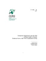
Diagram 1 B cell receptor signaling pathways (obtain from Cell Signaling
Technology website)
11
Receptor aggregation rapidly activates Src family kinases, including Lyn, Blk and Fyn,
Syk and Btk tyrosine kinases, initiating complex signaling cascades involving multiple
adaptors, kinases, phosphatases, G-proteins and transcription factors. The complexity of
BCR signaling permits many distinct outcomes, including proliferation, differentiation,
apoptosis, survival and tolerance. Many other transmembrane proteins, some of which are
receptors, are known to modulate specific elements of BCR signaling. A few of these,
including CD45, CD19, CD22, and FcγRIIB1 (CD32), are indicated above in yellow.
1.3.1 The PLCγ2 pathway
One of the signaling pathways shown to be activated by the BCR is the
phosphoinositide pathway which involves the hydrolysis of the phosphatidylinositol 4,5-
bisphosphate (PI4,5-P
2
) by PLCγ2 to form diacylglycerol (DAG) and inositol 1,4,5-
triphosphate (IP
3
) (Kurosaki et al., 2000). Both molecules act as second messengers, with
the former critical for activating protein kinase C (PKC) family members, and the latter
inducing the release of calcium from intracellular stores by binding to IP
3
receptors on
the endoplasmic reticulum (ER). One important substrate of PKC appears to be the
cAMP response element binding protein (CREB) transcription factor, which is regulated
by cAMP in some cell types but by PKC in B cells (Xie and Rothstein, 1995; Xie et al.,
1996). Calcium elevation in B cells leads to the activation of both calcium-calmodulin-
dependent protein kinase II and the calmodulin-activated protein serine/threonine
phosphatase, calcineurin. Among the events regulated by calcium elevation are the
phosphorylation of the transcription factor Ets-1 and the calcineurin-regulated
dephosphorylation of the cytosolic component of the nuclear factor of activated T cells
12
(NFATc), which causes NFATc translocation from the cytoplasm to the nucleus where it
can participate in transcriptional regulation.
1.3.2 The Ras signaling pathway
BCR stimulation also leads to the activation of Ras, which in turn activates the
classical mitogen-activated protein kinase (MAPK) pathway. The MAPK cascades
constitute a group of signal transduction pathways characterized by successive
phosphorylation of coupled serine/threonine or dual specificity kinases. The conserved
signaling modules consist of a MAPK, a MAPK kinase (MAPKK), normally a dual-
specificity kinase that phosphorylates MAPK on threonine and tyrosine residues, and a
MAPKK kinase (MAPKKK), which phosphorylates MAPKK on serine and activates it.
The best characterized MAPK pathway is the extracellular-signal regulated kinase
(ERK) pathway. The classical MAP kinases, ERK-1 and ERK-2 are important for the
ability of the BCR to stimulate proliferation of resting splenic B cells. BCR ITAM
phosphorylation recruits Shc that forms a complex with Grb2 and SOS resulting in Ras
activation and initiation of the Ras/Raf/MEK cascade. This pathway receives a primary
signal from Ras-GTP, which binds directly to Raf-1, the MAPKKK in this cascade.
Activated Raf-1 activates MEK-1 and MEK-2 (the MAPKK), which both in turn
phosphorylate ERK-1 and ERK-2. Phosphorylated ERKs form dimers, a step required for
nuclear translocation and subsequent phosphorylation of transcriptional regulatory
proteins, including Fos, Jun and members of the Ets family (Cohen, 1997).
1.3.3 The Rho GTPase pathway
Rho GTPases are instrumental in the organization of actin cytoskeleton, but
also for the control of gene expression. Although these proteins have been classically
13
implicated in chemotaxis, there are now clear indications on how differential
signaling toward other, more specific functions, such as phagocytosis or the
production of reactive oxygen species (Van Hennik and Hordijk, 2005).
Unlike the
ERK pathway, the c-Jun N-terminal kinase (JNK)/ stress-activated protein kinase
(SAPK) pathway is delivered by another family of small GTP-binding proteins, the Rho
GTPases Rac1 and Cdc42 (Coso et al., 1995; Teramoto et al., 1996a; Teramoto et al.,
1996b). Rac1 and Cdc42 activate NF-κB (Perona et al., 1997) and the serum response
factor transcription factors
(SRF) (Hill et al., 1995). The JNK cascade elements are
positioned in a conventional signaling cascade involving MKK1-4 (the MAPKKK),
SEK1 (the MAPKK) and JNK (Tibbles et al., 1996). JNK translocates to the nucleus
where it can regulate the activity of multiple transcription factors. Among the targets of
this cascade are the transcriptional factors c-Jun and ATF-2. JNK phosphorylates c-Jun in
its N-terminal region, greatly promoting its transcriptional activating ability (Treisman,
1996).
Regulation of the p38 MAPK is also achieved through a serine kinase cascade and
Rac1 and Cdc42. Unlike the requirement for both PKC and calcium in JNK activation,
maximum p38 activation appears to require only PKC, not calcium. As with other MAPK
cascades, the membrane-proximal component is a MAPKKK, typically a MEKK or a
mixed lineage kinase (MLK). The MAPKKK phosphorylates and activates MKK3/6, the
p38 MAPK kinases. MKK3/6 can also be activated directly by ASK1, which is
stimulated by apoptotic stimuli. p38 MAPK is involved in regulation of HSP27 and MK2
(MAPKAPK-2), MK3 (MAPKAPK-3) and several transcription factors including ATF-2,
14
Stat1, the Max/Myc complex, MEF-2, Elk-1 and indirectly CREB via activation of
MSK1.
1.3.4 The PI3-K pathway
A fourth signaling pathway important for BCR signaling involves the activation
of phosphatidylinositol 3-kinase (PI3-K), which phosphorylates the inositol ring of
phosphatidylinositol-4,5-biphosphate at the 3 position. The product of this reaction,
phosphatidylinositol-3,4,5-triphosphate (PIP
3
), is known to recruit PLCγ (Falasca et al.,
1998), as well as the kinases Btk (Buhl et al., 1999) and Akt (Gold et al., 1999). These
and other effectors bind via pleckstrin homology (PH) domain interactions with PIP
3
to
the plasma membrane where, in the context of additional modifications, they are
activated. While Akt activation generates cell survival signals, PLCγ and Btk activation
leads to phosphoinositide hydrolysis yielding inositotl-1,3,4-triphosphate, which mediates
mobilization of calcium (Fluckiger et al., 1998). PIP
3
is also essential for full activation
of MAP kinases and regulates the activity of NF-κB (Kane et al., 1999). Loss of PIP
3
generation, through genetic ablation of the regulatory subunit of PI-3K p85α, results in
an absence of mature B cells in the periphery (Fruman et al., 1999; Suzuki et al., 1999).
Thus signaling cascades that mediate production of PIP
3
are critical not only for active
BCR-mediated responses but also for B cell development and survival.
1.4 Negative regulators of B cell receptor signaling
Although early studies focused principally on the role of positive signals on
lymphocyte activation, increasing evidence suggests that each mode of cellular activation
is finely regulated by an integrated series of both positive and negative regulators. Upon
the complete elimination of invaders, auto-regulatory and negative feedback mechanisms
15
step in to maintain homeostasis and returning the immune system to basal resting state.
Unchecked or prolonged activation of immune responses are detrimental to the host.
Several mechanisms exist to prevent inappropriate B-cell activation and to avoid
generation of autoreactive antibodies and autoimmune diseases. One type of negative
regulation involves the direct or indirect recruitment of inhibitory signals by a large
group of receptors carrying immunoreceptor tyrosine-based inhibitory motifs (ITIM) in
their cytoplasmic domains. Example of such inhibitory receptors are PD-1, which recruits
Src homology 2 (SH2) domain-containing protein tyrosine phosphatases (SHPs)
(Okazaki et al., 2001), as well as FcγRIIB, which binds the SH2 domain-containing
inositol-5 phosphatase-1 (SHIP-1) (Coggeshall, 1998; Ono et al., 1996). Ligation-induced
phosphorylation of ITIM motifs in the cytoplasmic tails of most of these receptors results
in the recruitment of phosphatases. These two classes of phosphatases prevent B-cell
activation by inhibiting critical steps in the BCR signaling cascade. For example, the
phosphatidylinositol-3 kinase (PI-3K) pathway plays a central role in regulating
numerous biologic processes, including survival, adhesion, migration, metabolic activity,
proliferation, differentiation, and cell activation through the generation of the potent
second messenger PI-3,4,5-trisphosphate (PI-3,4,5-P(3) or PIP
3
). To ensure that
activation of this pathway is appropriately suppressed/terminated, the ubiquitously
expressed tumor suppressor phosphatase and tensin homolog deleted on chromosome 10
(PTEN) hydrolyzes PI-3,4,5-P(3) to PI-4,5-P(2), whereas SHIP-1 and SHIP-2 break it
down to PI-3,4-P(2).
In addition to cell-surface receptors, it is now clear that adaptor molecules are also
involved in inhibiting signal transduction by mediating protein-protein or protein-lipid
16
interactions. Adaptor or docking proteins are non-enzymatic but possess multiple
modular domains responsible for recruiting signaling proteins to activated receptors,
nucleating intermolecular complexes and positively or negatively modulating effector
protein activity by inducing conformational changes or phosphorylation/
dephosphorylation. Adaptor molecules are commonly defined as proteins that possess
protein-protein or protein-lipid interaction domains. However, many enzymes can be
considered to function as adaptors proteins, because they additionally contain protein- or
lipid-binding modules. For example, the Src-family PTK, Lyn has one SH2 domain and
one SH3 domain, as well as an enzymatic domain. The interaction modules in adaptors
and enzymes act to localize proteins to specific subcellular sites, control enzymatic
activities and direct the formation of multiprotein complexes – all of which, in turn,
contribute to the qualitative and quantitative control of B cell signaling.
1.4.1 FcγRIIB receptor
FcγRIIB receptors are single-chain molecules bearing IgG-binding sites in their
extracellular domains and cytoplasmic domains containing an immunoreceptor tyrosine-
based inhibitory motif (ITIM), a 13-amino acid sequence required for inhibitory function
(Muta et al., 1994). The comparison of sequences of several inhibitory receptors revealed
the conservation of a valine or an isoleucine residue at position Tyr-2, resulting in the
characteristic ITIM sequence V/IxYxxL. FcγRIIB receptor is expressed on B cells,
macrophages, neutrophils and mast cells. FcγRIIB receptors suppress cellular activation
by promoting dephosphorylation reactions, resulting from the recruitment of SHIP-1 to
the ITIM. SHIP-1 decreases the cellular levels of phosphatidylinositol PIP
3
, ultimately
preventing the influx of extracellular calcium. FcγRIIB inhibition of cell activation was
17
also proposed to be mediated by the adaptor protein Dok-1 through suppressing of ERK
activation (Yamanashi et al., 2000; Ott et al., 2002)
FcγRIIB1 receptor isoforms are preferentially expressed in B lymphocytes, and
are involved in the negative regulation of antibody production and B cell proliferation
(Van den Herik-Oudijk IE et al., 1994). FcγRIIB1 receptors are important factors in
controlling the amplitude of B cell activation in response to antigen. Specific IgG
antibodies that bind to the BCR through their Fab region can interact through their Fc
portion with inhibitory FcγRIIB1 on B cells and deliver an inhibitory signal.
Coaggregation of FcγRIIB1 with the BCR inhibited independently ligated receptors
whose signaling required PIP
3
. Negative signaling by FcγRIIB1 represents a feedback
suppression system that functions to inhibit B cell activation, proliferation and antibody
production (Daeron et al., 1995).
FcγRIIB2 isoforms are coexpressed with activating FcγR in primary monocytes,
neutrophils and monocyte-derived dendritic cells. In mast cells and basophils,
coclustering of FcγRIIB1 and FcγRIIB2 isoforms with activating FcγR caused inhibition
of degranulation. Follicular dendritic cells (FDC) express high levels of FcγRIIB.
FcγRIIB receptors expressed on FDC in germinal centers are involved in the retention of
immune complexes and in the generation of recall responses. A defect in the maturation
of FDC in the germinal centers was noticed in FcγRIIB-deficient animals. Because of
their ability to inhibit cell activation, FcγRIIB receptors are believed to promote
noninflammatory clearance of immune complexes. The inhibitory capacity of FcγRIIB2
in cells of the mononuclear phagocyte system is exerted upon coaggregation with ITAM-
bearing FcγRs, leading to inhibition of effector functions. In monocytes and macrophages,
18
FcγRIIB2 receptors regulate the production of cytokines and the amplitude of
inflammation in response to immune complexes.
1.4.2 SH2-containing inositol 5’-phosphatase-1 (SHIP-1)
An unknown tyrosine-phosphorylated p145 protein was isolated, together with
Shc, while using a Grb2 C-terminus SH3 domain-based affinity chromatography to
identify potential binding partners of Grb2 upon IL-3 stimulation (Damen et al., 1996).
Its predicted amino acid sequence revealed an amino-terminal SH2 domain, a central 5’-
phosphatase domain, two NPXY sequences and a proline rich C-terminal tail. This
protein was named SH2-containing inositol phosphatase-1 or SHIP-1. Ware et al.
described cloning of human SHIP from a human megakaryocytic cell line cDNA library
using 2 nonoverlapping mouse SHIP cDNA fragments as probes (Ware et al., 1996). This
interesting novel gene was also independently cloned by three groups using different
approaches (Drayer et al., 1996; Kavanaugh et al., 1996; Lioubin et al., 1996). Northern
blot analysis suggested that human SHIP-1 is expressed as a 5.3-kb mRNA in bone
marrow and a wide variety of other tissues. Sequence analysis of the cDNA predicted a
protein of 1188 amino acids exhibiting 87.2% overall sequence identity with mouse
SHIP-1. Liu et al. studied the expression of the ship gene during mouse development (Liu
et al., 1998). They found that the gene is expressed in late primitive-streak stage embryos
(7.5 days postcoitum), when hematopoiesis is thought to begin, and the expression is
restricted to the hematopoietic lineage. In adult mice, SHIP-1 expression continues in
most cells of hematopoietic origin, including granulocytes, monocytes, and lymphocytes,
and is also found in the spermatids of the testis. Furthermore, the level of SHIP-1
expression is developmentally regulated during T-cell maturation. These results
19
suggested a possible role for SHIP-1 in the differentiation and maintenance of the
hematopoietic lineages and in spermatogenesis.
SHIP-1 acts by hydrolyzing inositol metabolites phosphorylated at the 5’ position
of the inositol ring, namely, phosphatidylinositol 3,4,5-triphosphate [PI(3,4,5)P
3
] and
1,3,4,5-tetrakisphosphates [I(1,3,4,5)P
4
]. The membrane-bound PI(3,4,5)P
3
is critical for
binding and membrane recruitment of pleckstrin homology (PH) domain containing
molecules like the PTK Btk, a pivotal effector of B-cell activation (Bolland et al., 1998),
and the serine-threonine-specific protein kinase Akt/PKB, a prosurvival factor (Carver et
al., 2000; Baran et al., 2003). By converting PI(3,4,5)P
3
to PI(3,4)P
2
, SHIP-1 precludes
activation of these PH domain bearing effectors and can prevent B-cell activation
(Helgason et al., 2000; Liu et al., 1998; Brauweiler et al., 2000). In support of this idea, it
has been reported that B cells freshly isolated from SHIP-1
-/-
mice exhibited augmented
BCR-induced proliferation. Moreover, in vivo B-cell maturation is accelerated in SHIP-1
-
/-
mice (Helgason et al., 2000; Liu et al., 1998; Brauweiler et al., 2000).
20
Diagram 2 SHIP-1-mediated inhibition of cellular activation. (Ravetch and Lanier,
2000)
Although viable and fertile, SHIP-1
-/-
mice failed to thrive, and survival was only
40% by 14 weeks of age (Helgason et al., 1998). The mice exhibited a
myeloproliferative-like syndrome with consolidation of the lungs caused by infiltration of
macrophages (Helgason et al., 1998; Oh et al., 2007). They concluded that SHIP-1 plays
a crucial role in modulating cytokine signaling within the hematopoietic system.
The primary mode of recruitment of SHIP-1 in activated B cells is believed to
involve FcγRIIB. Engagement of FcγRIIB by the Fc portion of immunoglobulin G (IgG)
present in immune complexes (which are generated as a consequence of productive B-
cell activation) results in tyrosine phosphorylation of the ITIM of FcγRIIB, thus
triggering binding of the SHIP-1 SH2 domain and membrane translocation of SHIP-1.
Analyses of ex vivo B cells or B-cell lines lacking SHIP-1 have provided evidence that
FcγRIIB-associated SHIP-1 inhibits B-cell activation by preventing BCR-induced
21
PI(3,4,5)P3 accumulation, activation of Btk (Bolland et al., 1998) and Akt/PKB (Aman et
al., 1998), calcium fluxes (Okada et al., 1998; Hashimoto et al., 1999), and ERK
activation (Brauweiler et al., 2001; Ganesan et al., 2006; Ono et al., 1997; Pearse et al.,
1999). There are also FcγRIIB-independent mechanisms for recruiting SHIP-1 in B cells.
In agreement with this, it has been reported that SHIP-1-deficient B cells display
enhanced BCR-elicited PI(3,4,5)P3 generation and Akt activation even in the absence of
FcγRIIB coligation. While the exact mechanism of recruitment of SHIP-1 in this setting
is not known, it likely involves interactions with other molecules. This view is also
consistent with the finding that SHIP-1 can associate with intracellular adaptor molecules
like Shc (Tridandapani et al., 1999; Tridandapani et al., 1997), Grb2 (Poe et al., 2000)
and Dok-1 (Kepley et al., 2004; Abramson and Pecht, 2002), -2 (Dong et al., 2006) and -
3 (Robson et al., 2004; Lemay et al., 2000).
SHIP-1
-/-
mast cells were found to be far more prone to degranulation, after the
crosslinking of IgE preloaded cells (Huber et al., 1998). IgE alone also stimulated
massive degranulation in SHIP-1
-/-
but not wildtype mast cells. This degranulation with
IgE alone, which may be due to low levels of IgE aggregates, correlated with a higher
and more sustained intracellular calcium level than that observed with wildtype cells and
was dependent on the entry of extracellular calcium. The results showed the critical role
that SHIP plays in setting the threshold for degranulation and demonstrated that SHIP
directly modulates a 'positive-acting' receptor.
1.4.3 Lyn tyrosine kinase
The Src family kinase Lyn might support BCR activation by phosphorylating the
first tyrosine of the ITAM sequence and other BCR proximal signaling elements and
22
presumably by stimulating oxidase activity (Hibbs and Dunn, 1997). Apart from its
positive effect on BCR signaling, however, Lyn participates in a negative feedback loop
that terminates BCR signaling (Chan et al., 1998). Upon activation, Lyn phosphorylates
inhibitory receptors like CD22 and CD72, which then bind phosphatases that
dephosphorylate the ITAMs and inhibit signal transduction. Indeed, B cells from Lyn-
deficient mice do not show drastic developmental defects and are hyperreactive rather
than hyporeactive. Furthermore, Lyn-deficient mice show a high susceptibility to the
development of autoimmune diseases, characterized by circulating autoreactive
antibodies and the deposition of IgG immune complexes in the kidney (Silver et al.,
2006; Hibbs et al., 1995b). These results demonstrate the importance of Lyn as an
inhibitory element of BCR signaling, whereas Lyn’s role as a positive signaling element
of BCR signaling is only seen if Lyn deficiency is combined with deficiencies of other
signaling elements, such as Btk. In the chicken B cell line DT40, Lyn deficiency results
in the reduction of both calcium mobilization and tyrosine phosphorylation of substrate
proteins (Takata et al., 1994).
B cells from Lyn
-/-
mice exhibit hyperproliferation to BCR and BCR-FcγRIIB
stimulation, along with enhanced MAP kinase activation and Ca
2+
influx (Wang et al.,
1996; Chan et al. 1997a,b; Nishizumi et al., 1998). This hyper-responsiveness is thought
to be a cause of the autoimmune disease that develops in Lyn
-/-
mice (Hibbs et al., 1995;
Nishizumi et al., 1995). The phosphorylation of Dok-1 under both conditions are Lyn-
dependent as shown using Lyn
-/-
B cells, there is loss of Dok-1 phosphorylation. This
data thus suggest that Dok-1 could act in pathways downstream of Lyn.
23
1.4.4 C-terminal Src tyrosine kinase (Csk)
Csk was originally purified as a kinase which can phosphorylate the negative
regulatory tyrosine residue (Tyr-527) of c-Src, thereby suppressing their activity, and was
subsequently shown to phosphorylate other members of the Src PTKs, such as Lyn, Fyn,
Yes and Lck, at their C-terminal tyrosine residues in vitro (Nada et al., 1991). The amino-
terminus of Csk contains SH3 and SH2 domains and a kinase domain at its carboxyl-
terminus. Csk
-/-
embryos exhibit defects in the neural tube and die between day 9 and
day 10 of gestation (Imamoto and Soriano, 1993; Nada et al., 1993). Cells derived from
these embryos exhibit an increase in activity of Src and the related Fyn kinase. Csk is a
potent inhibitor of immunoreceptor signaling in T cells and macrophages, but not in B
cells and mast cells. In Csk
-/-
DT40 B cells, Lyn and Syk became constitutively
phosphorylated but cells still required additional signals from BCR to elicit other
biochemical events such as calcium mobilization (Hata et al., 1994).
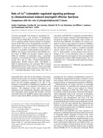
1.4.5 Downstream of tyrosine kinases (Dok)
Dok (downstream of tyrosine kinases) adaptor proteins constitute a family of
seven members, consisting Dok-1 (p62
dok
), Dok-2 (p56
dok-2
, Dok-R, FRIP), Dok-3 (Dok-
L), Dok-4 (IRS-5), Dok-5 (IRS-6), Dok-6 and Dok-7 (Diagram). Dok-1 and Dok-2 are
highly related
in structure and are preferentially expressed, together with
Dok-3, in
hematopoietic cells. Dok-1 and Dok-2
are expressed in the T cell lineage, whereas B cells
express
Dok-1 and Dok-3. Dok-4 is strongly expressed in non-hematopoietic organs,
particularly intestines, kidneys and lungs. Dok-5 is expressed most exclusively in the
central nervous system. Dok-6 is highly expressed in developing central nervous system.
Dok-7 is preferentially expressed in skeletal muscles and heart. These proteins though
24
having diverse expression profiles, are structurally similar,
containing an N-terminal
module composed of tandem pleckstrin homology (PH)-phosphotyrosine binding (PTB)
domains
followed by a region rich in binding motifs to Src homology
(SH)2 and SH3
domains at their C-terminus. These multiple modular domains are responsible for
recruiting signaling proteins, PTB domain is known to bind phosphotyrosine-containing
motif and phosphorylated tyrosines in carboxy-terminal region can act as docking sites
for SH2-containing signaling molecules. PH domain can be involved in membrane
localization by binding phospholipids in the plasma membrane.
25
PH
PTB
PH
PTB
PH
PTB
PH
PTB
PH
PTB
PH
PTB
Y
1
1
1
1
1
482
412
444
325
306
Y Y YY Y Y Y Y Y Y
YY YY
YY YY
Y
YY Y Y Y
Y
Y
YYY
Y
Recruitment to
membrane
phospholipids
Recruitment to
membrane receptors,
bind effector
molecules
Potential tyrosine kinases sites,
association with effector
molecules containing SH2, SH3
or PTB domain
Structure of Dok family
1 331
p62 Dok-1
p56 Dok-2, DokR, FRIP
Dok-3, DokL
Dok-4, IRS5
Dok-5, IRS6
Dok-6
PH
PTB
1 504
Dok-7
YYYY
YY YY
PH
PTB
PH
PTB
PH
PTB
PH
PTB
PH
PTB
PH
PTB
Y
1
1
1
1
1
482
412
444
325
306
Y Y YY Y Y Y Y Y Y
YY YY
YY YY
Y
YY Y Y Y
Y
Y
YYY
Y
Recruitment to
membrane
phospholipids
Recruitment to
membrane receptors,
bind effector
molecules
Potential tyrosine kinases sites,
association with effector
molecules containing SH2, SH3
or PTB domain
Structure of Dok family
1 331
p62 Dok-1
p56 Dok-2, DokR, FRIP
Dok-3, DokL
Dok-4, IRS5
Dok-5, IRS6
Dok-6
PH
PTB
1 504
Dok-7
YYYY
YY YY
Diagram 3 Structure and domains of Dok family members
Dok family members share conserved structure, consisting of a PH domain (green), PTB
domain (yellow) and multiple consensus tyrosine residues that can be phosphorylated
upon activation.