Functional interactions of protein tyrosine phosphatase alpha (PTPa) and src in mouse development and integrin singaling investigation of double PTPa src deficient mice and cells
Bạn đang xem bản rút gọn của tài liệu. Xem và tải ngay bản đầy đủ của tài liệu tại đây (3.26 MB, 215 trang )
Functional Interactions of Protein Tyrosine Phosphatase Alpha
(PTPα) and Src in Mouse Development and Integrin Signaling:
Investigation of Double PTPα/Src-Deficient Mice and Cells
CHEN MIN
NATIONAL UNIVERSITY OF SINGAPORE
2007
Functional Interactions of Protein Tyrosine Phosphatase Alpha
(PTPα) and Src in Mouse Development and Integrin Signaling:
Investigation of Double PTPα/Src-Deficient Mice and Cells
CHEN MIN
(M.Sc., Shanghai Medical University)
(B.Med., Shanghai Medical University)
A THESIS SUBMITTED
FOR THE DEGREE OF DOCTOR OF PHILOSOPHY
INSTITUTE OF MOLECULAR AND CELL BIOLOGY
NATIONAL UNIVERSITY OF SINGAPORE
2007
i
Acknowledgements
I would like to take this opportunity to express my sincere gratitude to my supervisor,
Professor Catherine J PALLEN, although word is not always enough. Thanks to her
great scientific guidance, encouragement, and endless patience, I can go through my
graduate study, and complete my thesis with her kind help and careful proofreading.
I am thankful for my supervisory committee members, Dr. Pauline JOHNSON and
Dr. Michael COX, in Canada; Dr. Xinmin Cao, Dr. Kongpeng LAM, and Dr
Borluen Tang in Singapore. Their thoughtful ideas and broad knowledge helped me a
lot for the progress of my project.
I was so luck to have a chance to work with such nice people either in Singapore or in
Canada. I am thankful for their generosity for sharing the reagents and information,
and their wisdom and broad knowledge made my life in the lab more interesting.
I don’t think I have guts to pursue the degree without the persistent and strong support
from my husband, Yan XU. I want to let him know that his understanding is always
precious, and I really appreciate that. I am forever indebted to my parents. I want to
say thanks to them for their understanding and encouragement. I am so grateful for my
parents-in-law to take care of my adorable son without any complaint.
ii
Table of Contents
Acknowledgements…………………………………………………………………… i
Table of contents……………………………………………………………………….ii
List of tables………………………………………………………………………….viii
List of figures……………………………………………………………………… ix
Abbreviations………………………………………………………………………… xi
Summary…………………………………………………………………………… xiii
CHAPTER1 Introduction ……………………………………………………………1
1.1 Protein phosphorylation……………………………………………… 1
1.2 Protein tyrosine phosphatase (PTP) superfamily 2
1.3 Catalytic mechanism of protein tyrosine phosphatases………… 5
1.4 Receptor-like PTPs and their features ……………………………… 6
1.5 Receptor-like tyrosine phosphatase alpha (PTPα) ……………………8
1.5.1 Overview of PTPα …………………………………………… 8
1.5.2 Biological properties of PTPα ……………………………… 10
1.5.2.1 Substrates of PTPα.………………………………10
1.5.2.2 Biological functions of PTPα …………………… 10
1.5.3 Combinatorial regulation of PTPα catalytic activity and
specificity …………………………………………………….13
1.5.3.1 Dimerization………………………………… 13
1.5.3.2 Phosphorylation…………………………………….14
1.5.3.3 Protein-protein interactions…… ………………… 16
1.5.3.4 Proteolysis …………….………………………… 18
1.5.3.5 Oxidation……………………………………………18
1.6 PTPs involved in regulating Src family kinases (SFKs)………… 18
1.6.1 Structure and regulation of SFKs …………………………….19
1.6.2 Regulation of SFK activity by PTPs ……………………… 23
iii
1.6.2.1 PTPα… …………………………………………….23
1.6.2.2 CD45……………………………………………… 24
1.6.2.3 PTP1B………………………………………………26
1.6.2.4 SHP1 and SHP2…………………………………….27
1.7 Integrin signaling…………………………………………….…… 28
1.7.1 Kinases in integrin-mediated signaling……………………… 31
1.7.1.1 Focal adhesion kinase (FAK)…………………….…31
1.7.1.2 SFKs in integrin signaling……………………….….34
1.7.2 PTPs in integrin signaling………………………… ……… 36
1.7.2.1 PTPα …………………………………………….….36
1.7.2.2 SHP2……………………………………………… 38
1.7.2.3 PTP1B…………………………………………… 39
1.7.2.4 PTP-PEST……………………………………… 41
1.7.2.5 PTEN……………………………………………….42
1.7.3 Summary of integrin-induced signaling events… ………….43
1.8 Research rationale and objectives…………………………………… 44
CHAPTER 2: Materials and Methods…………………………………………… 47
2.1 Mouse genotyping……………………… 47
2.1.1 DNA extraction from mouse tail tips or embryonic yolk
sacs………………………………………………… 47
2.1.2 Genotyping for PTPα and Src ……………… 47
2.2 Generation of PTPα/Src double mutant mice………………….… 48
2.3 Embryonic dissection………………………………………….…… 50
2.4 Mouse growth observation………………………………………… 50
2.5 Histological staining……………………………………….………….50
2.6 Experiments with mouse embryonic fibroblasts…………………… 51
2.6.1 Derivation of mouse embryonic fibroblasts………………… 51
2.6.2 Other mouse embryonic fibroblasts………………………… 52
2.6.3 Cell proliferation assay……………………………………… 52
iv
2.6.4 Cells stimulation with extracellular matrix (ECM)
components………………………………………………………… 53
2.6.5 Cell adhesion, spreading and migration assays……………….53
2.6.5.1 Cell adhesion assay ……………………………… 53
2.6.5.2 Cell spreading assay …………………………… ….54
2.6.5.3 Cell migration assay……………………………….54
2.7 Immunofluorescent staining……………………………….………….55
2.8 Protein analysis……………………………………………………….56
2.8.1 Cell lysis……………………………….………… …………56
2.8.2 Determination of protein concentration …… …….……… 56
2.8.3 Immunoblotting……………………………………… … 57
2.8.4 Immunoprecipitation …………………………… …… 57
2.8.5 Quantification of proteins…………………………… … 58
2.9 Transient transfection…………………………………….………… 58
2.9.1 Plasmids amplification and purification ……… …….………58
2.9.2 Cell culture and transient transfection………………………59
2.10 Treatment of cells with inhibitors……………………………… 60
2.10.1 PP2 and PP3 treatment………………………… ……… 60
2.10.2 Cytochalasin D treatment…………………… … ……… 60
2.11 PTPα adenovirus expression system…………………… 61
2.11.1 Generation of pKS-PTPαY789F (∆ Pac) ………………….61
2.11.2 pAdEasy transfer vector (pShuttleCMV) subcloning …… 62
2.11.3 Generation of pAdEasy recombinant plasmids in bacterial
cells …… 63
2.11.4 Transfection of recombinant pAdEasy plasmid into Qbi-293A
cells……… …………………………………………… 63
2.11.5 Confirmation of adenoviral-mediated PTPα expression… 64
2.11.6 Amplification of viral particles …………… ………… 64
v
2.11.7 Cesium chloride (CsCl) purification of recombinant
adenovirus ……………………….……………… …… 65
2.11.8 Viral particles titration.……….………… ……………….66
2.11.9 Re-introduction of wild type and mutant PTPα into PTPα
-/-
cells by recombinant adenovirus infection …… ……… 67
CHAPTER 3: Characterization of PTPα
-/-
Src
-/-
Double Knockout Mice …… 68
3.1 Overview …………………………………………………………… 68
3.2 Results ……………………………………………………………… 68
3.2.1 Combined ablation of PTPα and Src does not result in
embryonic lethality … ………………………………… 69
3.2.2 Post-natal survival and growth of double mutant PTPα
-/-
Src
-/-
mice ……………………………………… ………… 72
3.2.3 The combined ablation of PTPα and Src does not affect
organogenesis ………………………………………… 76
3.3 Discussion…………………………………………………………….81
CHAPTER 4: Phenotypes of PTPα/Src Double Mutant Mouse Embryonic
fibroblasts (α/s DKO)……… 86
4.1 Overview ……………………………………………………….… 86
4.2 Results …………………………………………………………… 87
4.2.1 Mouse embryonic fibroblasts deficient in both PTPα and
Src display a distinctive morphology after spontaneous
immortalization … 87
4.2.2 α/s DKO cells are defective in fibronectin-induced cell
adhesion and spreading ………………………………… 91
4.2.3 Integrin-induced cytoskeletal organization is altered in α/s
DKO cells …………………………………… ………… 95
4.2.4 Integrin-induced FAK tyr397 phosphorylation is not affected
in α/s DKO cells …… … ……………….101
4.2.5 Constitutive activation of Erk is a consequence of the
combined absence of PTPα and Src……………………….104
4.3 Discussion………………………………… 105
vi
CHAPTER 5: Integrin-induced PTPα Tyrosine Phosphorylation is Required for
cytoskeletal Reorganization and Cell Migration ……………… 112
5.1 Overview ……………………………………………………………112
5.2 Results ………………………………………………………………113
5.2.1 Integrin-induced tyrosine phosphorylation of PTPα … …113
5.2.2 SFKs are essential for FN-induced PTPα tyrosine
phosphorylation ………………………………….………115
5.2.3 Catalytically inactive mutant PTPαDM or Tyr789 mutant
PTPαY789F is not phosphorylated upon integrin
stimulation……………………………………… …… 116
5.2.4 Integrin-induced PTPα phosphorylation is dependent on an
intact actin cytoskeleton and FAK …… ……………… 119
5.2.5 A PTPα adenoviral expression system efficiently reintroduces
wild type and mutant forms of PTPα into PTPα
-/-
fibroblasts………………………………………… … 120
5.2.6 PTPα phosphorylation at Tyr789 is not required for integrin-
induced Src/Fyn activation and FAK or paxillin
phosphorylation ……………………… …………… 121
5.2.7 PTPα Tyr789 phosphorylation is required for integrin induced
cell spreading and cytoskeletal organization…………… 126
5.2.8 PTPα Tyr789 phosphorylation is required for integrin-
stimulated cell migration …… ………………………… 129
5.2.9 The cell detachment-induced dephosphorylation of PTPα is
not due to auto-dephosphorylation ……………………… 130
5.2.10 SHP2 is not the phosphatase responsible for detachment-
induced dephosphorylation of PTPα …………………131
5.3 Discussion ……………………………………………………… 133
CHAPTER 6: GENREAL DISSCUSSION and CONCLUSIONS……………136
6.1 Roles of PTPα and Src in embryonic development ……………… 136
6.1.1 PTPα is not essential for embryonic development, but is
required for normal hippocampal development and proper
function ………………………………………………… 137
vii
6.1.2 SFKs play essential but redundant roles in embryonic
development ………………….………………………… 139
6.1.3 A combined deficiency in PTPα and Src does not affect
mouse embryonic development, but does increase postnatal
mortality 141
6.2 The roles of PTPα and Src in integrin signaling ……………… …143
6.2.1 SFKs are required for integrin signaling ………………….143
6.2.2 Role of PTPα as an activator of SFKs in integrin
signaling………………………………………………… 145
6.2.3 Additional roles of PTPα-mediated SFK activation in integrin
signaling ……………… ………………………….…….146
6.3 Regulation of PTPα by integrin stimulation………………….…… 151
6.3.1 PTPα is tyrosine phosphorylated upon integrin stimulation,
but this is not required for SFK activation … ….……… 151
6.3.2 PTPα Y789 phosphorylation is required for integrin-
stimulated cell spreading and migration.………………… 155
6.3.3 Two roles of PTPα in integrin signaling …………….… 156
6.3.4 The reciprocal link between integrin-induced PTPα
phosphorylation and cytoskeletal organization may underlie
the defects observed in α/s DKO cells ……………….157
6.4 Overall summary ………………………………………………… 159
6.5 Future directions ………………………………………….…………162
Reference ………………………………………………………………………… 164
Publications ……………………………………………………………………… 199
viii
LIST of TABLES
2.1 Primer sequences used in PCR reactions for mouse PTPα and Src
genotyping…………………………………………………………………….49
2.2 Sequences of primers used to generate the PTPαY789F mutant ……….……62
3.1 Embryos obtained from heterozygous PTPα/Src intercrosses (PTPα
+/-
Src
+/-
) x
(PTPα
+/-
Src
+/-
) ………………………………………………………….…… 70
3.2 Embryos obtained from homozygous PTPα and heterozygous Src intercrosses
(PTPα
-/-
Src
+/-
) x (PTPα
-/-
Src
+/-
) …………………………………… …… 71
3.3 Pups obtained from homozygous PTPα and heterozygous Src intercrosses
(PTPα
-/-
Src
+/-
) x (PTPα
-/-
Src
+/-
)……………………………………………….73
3.4 Weights of organs…………………………………………………………… 79
3.5 Organ weight as a percentage of body weight……………………………… 79
4.1 Integrin-induced structural and morphological properties of wild type, PTPα
-/-
,
Src
-/-
, and α/s DKO fibroblasts…………………………… ………………100
6.1 Summary of phenotypes of PTPα
-/-
, Src
-/-
, and PTPα
-/-
Src
-/-
mice ………….142
6.2 Summary of phenotypes of PTPα
-/-
, Src
-/-
, and α/s DKO (PTPα
-/-
Src
-/-
)
fibroblasts in response to FN stimulation ………………………………… 150
ix
LIST of FIGURES
1.1 Schematic diagram of structures of selected members of the PTP
superfamily…………………………………………………………………… 4
1.2 PTPα catalytic activity and specificity are regulated by phosphorylation and
protein-protein interaction .………………………………………………… 17
1.3 Structural organization of SFK proteins …………………………………… 20
1.4 Mechanisms involved in the activation of SFKs …………………………… 23
1.5 Schematic representation of integrin signal transduction and downstream
events emanating from integrin stimulation ………………………………….30
1.6 Organization of the domains of focal adhesion kinase (FAK) ……………….32
1.7 Integrin-stimulated tyrosine phosphorylation and signaling events ………….44
2.1 Schematic diagrams of PTPα and mutants ………………………………… 59
3.1 Body weights of wild type (PTPα
+/+
Src
+/+
), single PTPα/Src mutant (PTPα
-/-
Src
+/+
and PTPα
+/+
Src
-/-
), and PTPα/Src double mutant (PTPα
-/-
Src
-/-
) mice at 7
to 21 days after birth………………………………………………………… 75
3.2 Histological analysis of organs obtained from mice with different genotypes
(PTPα
+/+
Src
+/+
, PTPα
-/-
Src
+/+
PTPα
+/+
Src
-/-
PTPα
-/-
Src
-/-
) at three weeks of
age …………………………………………………………………………….80
4.1 Cell morphology, filamentous actin organization and localization of paxillin
and cortactin in wild type (WT), PTPα
-/-
, Src
-/-
and α/s DKO (PTPα
-/-
Src
-/-
)
fibroblasts …………………………………………………………………… 90
4.2 Cell proliferation assay …………………………………………………… 91
4.3 α/s DKO cells exhibit defective integrin-induced cell adhesion …………… 93
4.4 α/s DKO cells exhibit an integrin-mediated cell spreading defect that is more
severe than that of PTPα
-/-
and Src
-/-
cells ………………………………… 95
4.5 FN-induced actin stress fiber assembly and focal adhesion formation in wild
type, PTPα
-/-
, Src
-/-
and α/s DKO fibroblasts ……………………………… 98
4.6 Integrin-induced phosphorylation of FAK at Tyr397 and Tyr576 in single and
double knockout fibroblasts lacking Src and/or PTPα …………………… 103
4.7 Erk 1/2 activation status in single and double knockout fibroblasts lacking Src
and/or PTPα …………………………………………………………………105
x
5.1 Integrin-induced tyrosine phosphorylation of PTPα ……………………… 114
5.2 SFKs are essential for integrin-induced PTPα phosphorylation ……………116
5.3 Catalytic activity of PTPα is required for integrin-stimulated and SFK-
mediated PTPα phosphorylation at Tyr789 …………………………………118
5.4 Integrin-induced PTPα phosphorylation is dependent on an intact cytoskeletal
organization and FAK is required for integrin-stimulated PTPα
phosphorylation …………………………………………………………… 120
5.5 Re-introduction of wild type and mutant forms of PTPα into PTPα
-/-
fibroblasts
by adenoviral infection …………………………………………………… 121
5.6 PTPα Tyr789 phosphorylation is not required for Src/Fyn activation upon
integrin stimulation…………………………………………………….…….124
5.7 PTPα Tyr789 phosphorylation is not required for integrin-stimulated FAK or
paxillin phosphorylation ………………………………………………… 125
5.8 PTPα catalytic activity and phosphorylation at Y789 are required for integrin-
induced cell spreading, assembly of actin stress fibers and focal adhesion
formation ……………………………………………………………………128
5.9 Haptotactic migration assay towards FN ………………………………… 129
5.10 Catalytically inactive PTPα (PTPαDM) is dephosphorylated upon cell
detachment………………………………………………………………… 131
5.11 PTPα is dephosphorylated upon detachment in SHP2
-/-
cells …………… 132
6.1 A diagram of integrin-induced PTPα phosphorylation ………………… 153
6.2 A proposed model of the two roles of PTPα in integrin signaling ……… 157
6.3 Integrin signaling transduction pathways ………………………………… 161
xi
ABBREVIATION
Ala: Alanine
Asp: Aspartic acid
ATCC: American type culture collection
β-ME: β-mercaptoethanol
BCR: B cell receptor
BHK-IR: Baby hamster kidney cells overexpressing insulin receptor
BRET: Bioluminescence resonance energy transfer
BSA: Bovine serum albumin
CAAX motif: C-terminal prenylation motif
CADTK: Calcium-dependent tyrosine kinase
CAH: Carbonic anhydrase-like domain
CaM: Camodulin
CIP: Calf intestine phosphatase
CNS: Central nervous system
CPE: Cytopathic effect
CsCl: Cesium chloride
Csk: C-terminal Src kinase
Cys: Cysteine
D1: Membrane proximal domain
D2: Membrane distal domain
DMEM: Dulbecco’s modified Eagle medium
DSP: Dual specific phosphatase
ECM: Extracellular matrix
ECL: Enhanced chemiluminescence
EGF: Epidermal growth factor
EGFR: EGF receptor
ER: Endoplasmic reticulum
FAK: Focal adhesion kinase
FAT: Focal adhesion targeting
FBS: Fetal bovine serum
FGF: Fibroblasts growth factors
FN: Fibronectin
FN-III: Fibronectin type III
FRET: Fluorescence resonance energy transfer
FRNK: FAK-related non-kinase
GAP: GTPase activating protein
GRCP: G protein coupled receptor
h: Hour
HRP: Horseradish peroxidase
ICAM: Intercellular adhesion molecule
Ig-like: Immunoglobulin-like
IPTG:
Isopropyl-1-thio-β-D-galactopyranoside
IR: Insulin receptor
IRS-1: Insulin receptor substrate-1
JAK: Janus kinase
Kv1.2 channel: Potassium channel
LAR: Leukocyte common antigen related protein
LMW-PTP: Low molecular weight PTP
xii
LTP: Long term potentiation
mACh: m1 musarinic acetylcholine
mAChR: Receptor for mACh
MAM: Meprin/A5/PTPµ domain
Min: Minute
NCAM: Neural adhesion molecule
NMDARs: N-methyl-D-aspartate receptor
PBS: Phosphate buffered saline
PCR: Polymerase chain reaction
PEST motif: Proline (P), glutamic acid (E), serine (S) and threonine (T)
PDGF: Platelet-derived growth factor
PDGFR: PDGF receptor
PI3-K: Phosphoinositide-3 kinase
PI(4,5)P2: Phosphatidylinositol 4,5-biophosphate
PI(3,4,5)P3: Phosphatidylinositol 3,4,5-trisphosphate
PKC: Protein kinase C
PLL: Poly-L-lysine
PMSF: Phenylmethylsulfonyl fluoride
pNPP: para-nitrophenyl phosphatase
PPs: Protein phosphatases
PP2: 3-(4-chlorophenyl)1-(1,1-dimethylethy)-1H-pyrazolo[3,4-d]
pyrimidin-4-amine
PP3: 4-amino-7-phenylpyrazol[3,4-d]pyrimidine
PRLs: Protein tyrosine phosphatases from regenerating liver
PRNK: Pyk2-related non-kinase
PSD-95: Post synaptic density 95
PTEN: Phosphatase and tensin homolog deleted on chromosome 10
PTP: Protein tyrosine phosphatase
PTK: Protein tyrosine kinase
PTPα: Protein tyrosine phosphatase alpha
PVDF: Polyvinylidene difluoride
Pyk2: Proline-rich tyrosine kinase
RIPA: Radioimmunoprecipitation assay
RPTP: Receptor like PTP
ROS: Reactive oxygen species
SDS: Sodium dodecyl sulfate
SDS-PAGE: SDS-polyacrymide gel electrophoresis
SFKs: Src family kinases
SH2: Src homology 2 domain
SH3: Src homology 3 domain
SHP: SH2 domain-containing PTP
SHPS-1: SHP2 substrate 1
siRNA: Small interfering RNA
SIRPα: Signal regulatory protein alpha
TCID
50
: Tissue culture infectious dose 50
TCR: T cell receptor
Tyr: Tyrosine
Val: Valine
WPD: Trp-Pro-Asp
xiii
Summary
The requirement for the dual expression of PTPα and Src for mouse embryonic
development and integrin signaling was investigated using genetically modified mutant
mice or cells with a deficiency in both PTPα and Src. PTPα/Src homozygous double
mutant mice were generated by intercrossing PTPα
+/-
Src
+/-
or PTPα
-/-
Src
+/-
mice.
Mouse fibroblasts were isolated from embryos of appropriate genotypes, and
spontaneously immortalized.
The combined ablation of PTPα and Src does not result in embryonic lethality, but
appears to increase the rate of postnatal mortality between birth and three weeks of age.
PTPα/Src double mutant mice exhibit toothlessness and growth retardation manifested
in reduced overall body weight and reduced weight of many major organs, similar to
defects in single mutant Src-null mice. These findings suggest that the dual expression
of PTPα and Src is not essential for mouse embryonic development and that the
observed defects are mainly attributable to the ablation of Src. Despite the absence of
additional unique defects in PTPα/Src double mutant mice, the enhanced incidence of
postnatal mortality suggests that the residual activity of Fyn, Yes and/or other SFKs in
PTPα
-/-
Src
-/-
mice can be insufficient for normal maturation to the adulthood.
Mouse embryonic fibroblasts with a combined absence of PTPα and Src display
reduced adhesion to and spreading on fibronectin (FN), accompanied by altered
cytoskeletal organization, that is distinct from or more severe than the defects in single
mutant PTPα
-/-
and Src
-/-
cells. FN-stimulated FAK Tyr397 phosphorylation is
xiv
comparable to that in wild type cells, even though this is reduced in PTPα
-/-
cells and
delayed in Src
-/-
cells. Typically, downregulation of Erk is observed upon cell
detachment from the substratum, however, in the double mutant cells, Erk remains
fully activated when the cells are placed in suspension. These observed defects in cells
dually deficient in PTPα and Src suggest that PTPα-mediated SFK activation is
essential for integrin signaling, and plays a negative feedback role in orchestrating
integrin signaling.
To determine how PTPα is regulated upon integrin stimulation and how a signal
emanating from integrin is transduced to PTPα, thus linking the actions of PTPα to
SFKs, the phosphorylation status of PTPα was investigated. PTPα is phosphorylated at
Tyr789 upon integrin stimulation, and SFKs (either Src or Fyn/Yes) are required for
full integrin-induced PTPα phosphorylation. Further investigations show that integrin-
stimulated phosphorylation of PTPα depends on PTPα catalytic activity, the formation
of SFK-FAK complex, and on an intact cytoskeleton. Unlike mitosis, PTPα
phosphorylation is not required for integrin-induced Src and Fyn activation. However,
it is essential for downstream events that promote cytoskeletal reorganization, focal
adhesion formation, and cell migration. These findings identify and distinguish two
roles of PTPα in integrin signaling, an early role as an SFK activator which is not
phosphorylation dependent, and a later role in cytoskeletal organization that requires
its phosphorylation.
These studies demonstrate that PTPα and Src are two interconnected molecules with
reciprocal interactions in the complex network of integrin signaling. PTPα functions in
integrin signaling both as an SFK activator and an SFK effector.
1
CHAPTER 1
Introduction
1. 1 Protein phosphorylation
Phosphorylation of proteins on serine/threonine or tyrosine residues is a rapid,
reversible post-translational modification that functions as a specific “switching”
mechanism to form or disrupt regulatory connections between proteins. Reversible
protein phosphorylation is crucial for the regulation of numerous cellular events,
including cell growth and tissue differentiation, inter-cellular communication, as well
as immune responses. Protein phosphorylation is a highly regulated process by which
information can be shuttled from the cell surface to the nucleus. The balance of protein
phosphorylation is under the control of kinases and phosphatases.
Compared to protein phosphorylation in general, phosphorylation on tyrosine residues
is extensively utilized only in multicellular eukaryotes. Tyrosine phosphorylation plays
key roles in many biological processes including proliferation, differentiation,
migration, and survival, and is also important in coordinating processes among
neighbouring cells in embryogenesis and organ development, as well as tissue
homeostasis (Hunter 1995). Abnormalities in tyrosine phosphorylation can lead to
numerous inherited or acquired human diseases. Transmembrane and intracellular
protein tyrosine kinases (PTKs) are activated by extracellular signals and generate
phosphotyrosyl proteins, either by auto- or substrate phosphorylation. The first PTK to
be identified was v-Src (a Rous sarcoma virus protein) which can transform cells
through initiating tyrosine phosphorylation-based signaling events (Brugge et al., 1977;
2
Eckhart et al., 1979; Hunter et al., 1980; Sefton et al., 1980). Based on the current
genome sequence information, it is estimated that there are more than 100 PTKs
encoded by the human genome (Alonso et al., 2004). It is generally agreed that
tyrosine phosphorylation is regulated by the equal and balanced actions of PTKs and
protein tyrosine phosphatases (PTPs), but the first PTP was purified and characterized
in 1988 (Tonks et al., 1988), ten years following the first identification of PTKs
(Brugge et al., 1977; Eckhart et al., 1979; Hunter et al., 1980; Sefton et al., 1980).
Subsequently, a large number of PTPs were identified through cDNA cloning using
polymerase chain reaction (PCR) and low-stringency hybridization techniques. Recent
findings have led to the understanding that PTPs play specific and active, even
dominant, roles in setting the levels of tyrosine phosphorylation in cells and in the
regulation of physiological processes (Fischer et al., 1991; Walton et al., 1993; Tonks
et al., 1996; Mustelin et al., 2003). PTP1B was the first PTP to be identified (Tonks et
al., 1988). A surprising finding was that PTP1B did not show any significant overall
sequence similarity to the serine/threonine protein phosphatases (PPs), indicating that
it evolved separately. After nearly twenty years of investigation of PTPs, it is now
clear that they comprise a large superfamily of related enzymes (Fig. 1.1). There are
107 genes in the human genome that encode members of four PTP families (Alonso et
al., 2004). Moreover, PTPs are highly specific, not only for particular phosphorylated
proteins but also for non-protein (i.e. phospholipid) substrates.
1.2 Protein tyrosine phosphatase (PTP) superfamily
Protein phosphatases can be generally divided into two main groups, PPs and PTPs,
based on their substrate specificity. PPs, such as PP1 and PP2A, specifically hydrolyze
serine/threonine phosphoesters (P-Ser/Thr) and comprise a large family of metallo-
3
protein enzymes whose functions within the cells are extremely diverse and highly
regulated. Catalysis by PPs has been proposed to proceed by a direct attack of an
activated water molecule on the phosphorus center of the substrate, without phosphoryl
transfer to the enzyme (Egloff et al., 1995). PPs play in a variety of key roles in
biological processes including embryonic development, cell proliferation, and death.
Unlike PPs, PTPs dephosphorylate phosphotyrosine (P-Tyr) and possess different
characteristics of protein structure and catalytic mechanism. PTPs do not share any
sequence similarity with PPs, and they do not require metal ions for catalysis (Dixon
1995; Denu et al., 1996).
PTPs are characterized by a conserved active sequence motif within the catalytic
domain of ∼240 amino acid residues (PTP domain). The catalytic domain of each PTP
displays 30-40% identity among individual enzymes. Outside the conserved catalytic
domain, the amino acid sequences of PTPs vary greatly. The first PTP crystal structure,
that of PTP1B (Barford et al., 1994; Jia et al., 1995), provided insights into what
structural elements constituted the minimum PTP catalytic domain and suggested
which region defined substrate specificity. Subsequent crystal structures of other PTPs
have confirmed and refined these general characteristics. All PTPs possess at least one
catalytic domain, and each PTP is composed of at least one conserved domain
characterized by a signature motif CX
5
R containing cysteine and arginine residues
known to be essential for PTP catalytic activity (Guan et al., 1991).
Based on overall structure and subcellular localization, PTPs are divided into two
groups, receptor-like (RPTPs) and intracellular PTPs (Fig. 1.1). The ligands for RPTPs
are largely unknown. RPTPs consist of an extracellular domain, one transmembrane
4
spanning region, and two tandem intracellular PTP domains. The exception is a small
subgroup of RPTPs that only contain a single PTP domain, such as PTPβ or PTP-PS.
PTP1B SHP1/2 PTP-
PEST
PTEN
Tyrosine-specific
VH-1
DSPs
PTPβ PTPα
Receptor-like PTPs
PTPε
TM
CD45 LAR
PTPδ
PTPσ
Intracellular PTPs
PTPζ
PTPγ
TCPTP
PTPμ
PTPλ
PTPκ
MKP-1
Heavily glycosylated
PTP domain
FN-III
Ig-like
CAH-like
PEST
CX
5
R motif
SH2 domain
CAAX motif
C2 lipid binding
MAM domain
CH2A/B
CDC25
D1
D2
D1 D1
D2 D2
D1
D2
LMW-
PTP
PRLs
D2
D1
PTP1B SHP1/2 PTP-
PEST
PTEN
Tyrosine-specific
VH-1
DSPs
PTPβ PTPα
Receptor-like PTPs
PTPε
TM
CD45 LAR
PTPδ
PTPσ
Intracellular PTPs
PTPζ
PTPγ
TCPTP
PTPμ
PTPλ
PTPκ
MKP-1
Heavily glycosylated
PTP domain
FN-III
Ig-like
CAH-like
PEST
CX
5
R motif
SH2 domain
CAAX motif
C2 lipid binding
MAM domain
CH2A/B
CDC25
D1
D2
D1 D1
D2 D2
D1
D2
LMW-
PTP
LMW-
PTP
PRLsPRLs
D2
D1
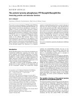
Figure 1.1. Schematic diagram of structures of selected members of the PTP
superfamily. The inset box shows some of the various structural motifs that can be
found in the PTPs (FN-III: Fibronectin type-III; MAM: Meprin/A5/PTPμ domain; Ig:
Immunoglobulin-like domain; CAH: Carbonic anhydrase-like; C(X
5
)R: PTP signature
motif; SH2: Src homology 2 domain; PEST: Pro-Glu-Ser-Thr motif; CH2A/B:
sequence homology found in Cdc25; CAAX: prenylation motif).
5
Intracellular PTPs possess a single catalytic domain with flanking regions often
containing novel protein-protein interaction or targeting domains that direct the
enzymes to specific intracellular locations. DSPs (dual specific PTPs) are an additional
family of intracellular PTPs that can dephosphorylate both P-Ser/Thr and P-Tyr. PTEN
is classified as a DSP, and in addition to its protein tyrosine phosphatase activity, it
plays a major role in cells as a lipid phosphatase. Another member of the intracellular
PTPs is LMW-PTP (low molecular weight PTP), an 18 KDa enzyme that is widely
expressed in many cells. It has been shown that LMW-PTP can dephosphorylate the
PDGF receptor or p190RhoGAP in PDGF signaling (Chiarugi et al., 2000). The PRLs
are 3 closely related intracellular PTPs with a unique (among PTPs) C-terminal
prenylation motif (CAAX motif), and the expression of PRL-3 is upregulated in colon
cancer metastases (Zeng et al., 2000; Saha et al., 2001).
1.3 Catalytic mechanism of protein tyrosine phosphatases
Crystal structures of the catalytic domains of PTP1B (Barford et al., 1994; Jia et al.,
1995), Yop51 PTP (Stuckey et al., 1994; Fauman et al., 1996), VHR (Yuvaniyama et
al., 1996) and LMW-PTPs (Su et al., 1994), in conjunction with the kinetic data from
in vitro assays, have provided bases for understanding the specificity of
phosphotyrosine recognition and the mechanism of catalysis. Enzymological and
mutational studies have elucidated that all PTPs share a common two-step catalytic
mechanism. The first step is the formation of a covalent thiophosphate intermediate
through transfer of the phosphate group on the substrate to the essential cysteine
residue in the PTP active site. This is a rate-limiting step in most PTP-mediated
catalyses. The second step is the hydrolysis of the phosphate group from the
intermediate and recovery of the enzyme (Guan et al., 1991; Zhang et al., 1995).
6
The PTP signature motif C(X
5
)R within each PTP domain forms a continuous
phosphate-binding loop located at the base of the catalytic cleft, with the invariant
cysteine residue (C) at the bottom of the active cleft (Neel et al., 1997). Only the side
chain of a P-Tyr residue in a target substrate is of sufficient length to reach the
catalytic cysteine residues at the bottom of the cleft; while P-Ser and P-Thr are too
short to be dephosphorylated (Dixon 1995). A WPD loop located ~30 amino acids N-
terminal to the active site is important for PTP-mediated catalytic hydrolysis. The
aspartic acid residue (Asp) within the WPD loop serves as a general acid for the
formation of a phospho-enzyme intermediate (Barford et al., 1994; Zhang et al., 1994;
Jia et al., 1995). Upon substrate binding, the phosphorylated tyrosine residue of the
substrate fits into the catalytic cleft and the WPD loop moves closer to the
phosphotyrosine residue. The phenyl ring of the phosphotyrosine residue is protonated
by the Asp of the WPD loop, facilitating a nucleophilic attack by the essential cysteine
residue on the phosphoester bond and resulting in the formation of a thiophosphate
intermediate (Guan et al., 1991; Cho et al., 1992; Wo et al., 1992). The intermediate
then undergoes hydrolysis by a water molecule which is hydrogen bonded to the WPD
Asp, releasing the phosphate moiety and allowing the recovery of the active enzyme
(Lohse et al., 1997; Zhang 1998). The mutation of Asp to Ala in the WPD loop
dramatically reduces PTP catalytic activity, converting the enzyme into a substrate-
trapping mutant (Denu et al., 1996; Wu et al., 1996; Flint et al., 1997; Lohse et al.,
1997).
1.4 Receptor-like PTPs and their features
Generally, RPTPs consist of an intracellular segment containing one or two PTP
domains, a single transmembrane domain and a variable extracellular segment.
7
Interestingly, the second PTP domain of most RPTPs displays little or no catalytic
activity, suggesting that the second PTP domain may have another role (Streuli et al.,
1990; Cho et al., 1992). The diversity of extracellular segments of RPTPs presumably
reflects an equivalent diversity in the ligands to which they may respond, although the
ligands for the RPTPs have generally not been identified. RPTPs can be subdivided
into five types based on common features found in the extracellular domains (Fig. 1.1).
Type I RPTPs are represented by the hematopoietic cell restricted CD45 family, which
has multiple isoforms that vary in size of the extracellular domain, and arise from the
differential splicing of exons 4, 5, and 6 (Thomas et al., 1987). CD45 was first
identified as a major surface protein on nucleated hematopoietic cells, and is critical
for classical antigen receptor signaling by modulating SFK activity (Ostergaard et al.,
1989; Thomas 1989; Guttinger et al., 1992; Cahir McFarland et al., 1993). Type II
RPTPs are LAR (leukocyte common antigen related protein)-like PTPs, including
LAR, PTPδ, and PTPσ. The extracellular segments of this type of PTP consist of three
immunoglobulin-like (Ig-like) repeats and four to eight type-III fibronectin (FN-III)
repeats depending on the alternative splicing. They are expressed as pro-proteins and
undergo a proteolytic process to generate functional LAR-PTPs (Streuli et al., 1988;
Streuli et al., 1992). With the exception of LAR that is widely expressed, most other
LAR-like PTPs are preferentially expressed in neurons, and are implicated in neuronal
development (Tian et al., 1991; Thompson et al., 2003). Type III RPTPs are
characterized by eight fibronectin type-III like repeats within their extracellular
domains, such as PTPβ. PTPα and PTPε are type IV PTPs, and the members of this
small group generally have a short, heavily glycosylated extracellular domain. PTPα
has been shown to play roles in cell proliferation, transformation, and neuronal
8
differentiation (Pallen 2003). Type V molecules include PTPξ and PTPγ, which have
an N-terminal carbonic anhydrase-like domain (CAH-like).
1.5 Receptor-like protein tyrosine phosphatase alpha (PTPα)
1.5.1 Overview of PTPα
PTPα, isolated by many groups using PCR-based PTP identification and cloning, is a
widely expressed transmembrane molecule that is particularly highly expressed in
brain (Kaplan et al., 1990; Krueger et al., 1990; Matthews et al., 1990; Sap et al., 1990).
It is a ∼130 kDa membrane-spanning PTP that has a very short and heavily
glycosylated extracellular domain that is connected to two classic intracellular catalytic
domains (termed D1 and D2) (Fig. 1.2). The ligand for PTPα has not been identified.
Three alternatively spliced variants of PTPα have been reported. The smallest isoform
lacking any insertions is a ubiquitously expressed 123 amino acid form of PTPα,
with/on which most studies have been conducted. A second isoform is expressed in
brain, skeletal muscle and certain differentiated cell types that contains a 9 amino acid
insertion in the juxtamembrane extracellular domain arising from the alternative
splicing of a 27 base pair mini-exon (Kaplan et al., 1990; Krueger et al., 1990). These
two isoforms have similar catalytic activities in vitro, including towards the SFKs Fyn
and Src. However, when expressed in cultured cells, the larger isoform is twice as
effective in promoting the transforming activity of Src (Kapp et al., 2007). A third
splice variant of PTPα contains a 36 amino acid insertion within the first catalytic
domain (Matthews et al., 1990), however no studies have examined the expression of
this isoform or the effects of this insertion on PTPα function. Another unusual
characteristic of PTPα is that the membrane distal domain (PTPα-D2) of PTPα has
detectable catalytic activity, although this is much lower than that of the membrane
9
proximal domain (PTPα-D1) (Wang et al., 1991; Lim et al., 1997). Most first or
membrane proximal domains (D1) of RPTPs such as CD45 or LAR are catalytically
active, whereas the second or membrane distal domains (D2) have either no detectable
or extremely low in vitro activity, usually less than 0.1% of that of D1 (Streuli et al.,
1990; Cho et al., 1992). The D2 domain of PTPα displays about 10% of D1 activity
towards the low molecular weight substrate para-nitrophenyl phosphate (pNPP) (Lim
et al., 1997; Wu et al., 1997). The relatively high activity of PTPα-D2 towards pNPP is
due to two factors, the higher intrinsic activity of PTPα-D2 compared to that of the D2
domains of other RPTPs, and/or the lower activity of PTPα-D1 compared to that of D1
of other RPTPs (Lim et al., 1997; Wu et al., 1997). PTPα-D2 has catalytic activity
towards pNPP, but it does not display the same relative level of activity towards
phosphotyrosyl peptide substrates. Sequence alignment of these two domains has
revealed that the different catalytic activity and substrate recognition result from only
two amino acid differences between D1 and D2, and this has been confirmed by the
point mutation of each residue in PTPα-D2 to its variant counterpart in PTPα-D1 (Val
536 to Tyr or Glu 671 to Asp). The mutation of these two residues within PTPα-D2
positively affects the catalytic efficiency of D2 towards both pNPP and phosphotyrosyl
peptide (Lim et al., 1998; Buist et al., 1999). The differences in PTPα-D2 catalytic
activity and substrate specificity from PTPα-D1 indicate disparate functions of these
two domains. The evolutionary conservation and intrinsically low activity of the PTP-
D2 domain infer a non-enzymatic role of this domain in PTP functions, perhaps
through effecting protein-protein interactions that could regulate specific PTP targeting
and substrate localization.