Synthesis and stress analysis of germanium nanocrystals embedded in dielectric matrices 3
Bạn đang xem bản rút gọn của tài liệu. Xem và tải ngay bản đầy đủ của tài liệu tại đây (770.74 KB, 18 trang )
Chapter 6 Results & Discussions III
- 124 -
Chapter 6
Results & Discussions III:
Analysis of Stress Development of Ge
Nanocrystals in Silicon Oxide Matrix
6.1 Introduction
There has been an intense interest in nanocrystals embedded in a silicon
oxide matrix in the hope that the quantum confinement effect will make the
indirect bandgap semiconductors (e.g. Si or Ge) become more efficient light
emitters. Recently, Takeoka et al. had demonstrated the strong size dependence of
the photoluminescence (PL) spectra from the Ge nanocrystals embedded in a
silicon oxide matrix and suggested it to be linked to electron-hole pairs confined
in the nanocrystals [1]. However, there is a relatively unexplored area in the
research of Ge nanocrystals in silicon oxide matrix, namely, the stress
development of the nanocrystals in such a system.
Wellner et al.
[2] had concluded from their Raman results of Ge
nanocrystals in silicon oxide that it was strongly affected by compressive stress.
The Ge nanocrystals were obtained from annealing silicon oxide layers implanted
with Ge. In our earlier work, it is also noticed the that the Raman peak position
Chapter 6 Results & Discussions III
- 125 -
changed in our co-sputtered samples when rapid thermal annealed under different
conditions [3]. Moreover, in the pervious chapter, we have pointed out that the
changes in the Raman peak position of samples are associated with the
mechanical stress experienced by the nanocrystals.
This stress occurs mainly because the SiO
2
is unable to accommodate the
growing Ge nucleus. Wellner et al. and Borany et al. have argued that the
compressive stress might be explained by the fact that Ge undergoes a volume
expansion of about 6% during the liquid to solid phase transition [2,4]. Yi et al. [5]
agreed with Wellner et al. and suggested that the observed pressure exerted on the
nanocrystals are mainly attributed to a volumetric difference as the nanocrystals
reside in a matrix cavity that is too small.
In this chapter, we report results of a study on the development of
mechanical stress experienced by Ge nanocrystals in a silicon oxide matrix using
a combination of Raman and transmission electron microscopy techniques. We
have followed the procedure outlined by Sharp et al. [6,7] to secure free-standing
Ge nanocrystals from those originally embedded in the silicon oxide matrix. This
enables us to examine the effect of the compressive stress exerted on the
nanocrystals on the Raman spectra that is independent of the size effect and
discuss the origin of such stress. The samples used in this work are the same as
those used in Chapter 4 and consists of Sample A, B and C with Ge concentration
estimated to be around 3, 10 and 15 at.%.
Chapter 6 Results & Discussions III
- 126 -
6.2 Liberating the Ge nanocrystals
Recently, Sharp et al. has shown in his work that a mixture solution of 1:1
HF:H
2
O is able to selectively etch away the oxide while retaining a substantial
amount of the nanocrystals on top of the substrate as free-standing nanocrystals.
Moreover, it was also found this etching process does not significantly affect
nanocrystal size and that the structure of the Ge nanocrystals remains stable even
after prolonged exposure to the environment [5-7]. We followed such procedure
and dipped the samples in the HF:H
2
O solution with occasional shaking of the
sample holder to enhance the etching process. After ensuring that the oxide film
has been successfully removed (which can be judged by a colour change of the
film), the sample was removed from the solution and left to dry in air.
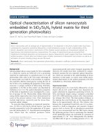
Figure 6.1: (a)-(c) Schematic diagrams of the process of obtaining free
standing Ge nanocrystals from the annealed co-sputtered samples
via our careful etching experiments. (d) XTEM micrograph of
Sample A annealed at 1000°C in forming gas (10% H
2
, 90% N
2
)
for 15 minutes after selective removal of oxide matrix and (e) high
magnification TEM of 6.1(d).
Chapter 6 Results & Discussions III
- 127 -
The above mentioned process is shown in a schematic in Figure 6.1. As
the oxide is being etched away, the embedded nanocrystals will be released from
the oxide film. The nanocrystals are found to accumulate at the surface after a
complete removal of the SiO
2
, as can be seen in Figure 6.1(d). The nanocrystals
retain their crystallinity, showing clear lattice fringes as can be seen from Figure
6.1(e). The attractive van der Waals forces are likely to be responsible for the
build-up of nanocrystals. It has also been shown that these nanocrystals remain
crystalline and un-oxidized up to at least 5 months even when directly exposed to
air [5].
6.3 Calculation of stress experienced by the Ge nanocrystals
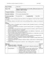
Typical Raman spectra of pre- and post etched annealed samples are
shown in Figure 6.2. The frequency of the phonon Raman band depends on the
masses and positions of the atoms, the inter-atomic forces (i.e. force constants of
the bonds) and the bond length. Therefore, any effects altering these features will
produce a change in the frequency of the band. For instance, a tensile stress will
result in an increase in the lattice spacing and, hence, a decrease in the
wavenumber of the vibrational mode. In the case of compressive stress, the
decrease of the lattice parameter yields a corresponding increase of the vibrational
frequency. From the downward shift towards relatively lower frequency in the
Raman peaks between the pre- and post-etched samples, it suggests that the
nanocrystals experienced compressive stress.
Chapter 6 Results & Discussions III
- 128 -
Figure 6.2: A set of typical Normalized Raman spectra of samples annealed
for 15 minutes at 900°C before and after selective oxide matrix
removal.
Several effects can contribute to the stress state of the nanocrystal: the
surface tension between the matrix and nanocrystal, the different thermal
expansion coefficients of Ge and silicon oxide, and the equilibrium size of the
matrix cavity in which the nanocrystal resides. In order to account for the
temperature changes during processing and characterization, one must consider
the thermal expansion coefficients of silica and Ge, the latter being an order of
magnitude higher than the former. The differential expansion contributes to a
tensile stress that reduces slightly the compressive stress state of the nanocrystal
when quenched to room temperature.
Chapter 6 Results & Discussions III
- 129 -
However, considering all these sources of stress, one may conclude that
the observed pressure is mainly attributable to a volumetric difference, i.e. the
nanocrystal resides in a matrix cavity that is too small. This volumetric difference
may arise from the matrix atoms not being able to move rapidly enough to
accommodate the nanocrystal as it is growing due to the fast growth rate
experienced by the nanocrystals as a result of enhanced diffusivity at elevated
annealing temperatures, or the stress could be caused by the volumetric expansion
of Ge (~6%) [2,4] when it solidifies from the liquid phase to the solid phase.
By comparing the Raman spectra of our pre- and post-etched annealed
samples, the hydrostatic pressure, P, in the nanocrystals can be estimated as [8]
)2(3
12110
SS
P
etchedembedded
+
−
=
γω
ωω
(6.1)
where ω
embedded
and ω
etched
are the Raman peak positions of the nanocrystals
embedded in silicon oxide and free-standing Ge nanocrystals respectively, and ω
0
is the Raman peak position of bulk Ge, γ = 0.89 cm
-2
is the mode-Grüneisen
parameter [8],
S
11
and S
12
are components of the elastic compliance tensor with
S
11
+ 2S
12
= 0.44 ×10
-12
dyn
-1
cm
2
.
6.3.1 Effect of annealing time on the stress state of the nanocrystals
In order to study the influence of annealing time on the formation and the
stress state of the nanocrystals, samples A were subjected to the conventional
furnace annealing between 700 to 1000°C for 15 and 60 minutes, and their Raman
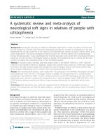
spectra are shown in Figures 6.3 and 6.4, respectively. Figure 6.3 shows that a
small peak at 305 cm
−1
is observed for sample annealed at 700°C, indicating the
Chapter 6 Results & Discussions III
- 130 -
growth of Ge nanocrystal in the oxide matrix. The peak intensity increases and the
peak position moves to higher wavenumber as the annealing temperature
increases from 800 to 900°C. The peak intensity reduces drastically coupled with
a shift to a lower wavenumber for the peak position when the sample was
annealed at 1000°C. Figure 6.4 shows that when annealing time increases to 60
minutes, the variation of the peak intensity and position with respect to annealing
temperature follows exactly those shown in Figure 6.3 except that nanocrystal
growth can be observed at a lower temperature of 600°C with an increase in
annealing time.
Figure 6.3: Raman spectra of Sample A annealed between 700°C to 1000°C
for 15 minutes.
Chapter 6 Results & Discussions III
- 131 -
Figure 6.4: Raman spectra of Sample A annealed between 600°C to 1000°C
for 60 minutes.
The values of P for our Samples A annealed under different temperatures
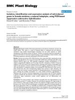
for 15 minutes are plotted in Figure 6.5. It can be seen from this figure that P
increases from a very insignificant value gradually to 0.32 GPa as the annealing
temperature increases from 600 to 800°C. There is a sharp increase in the value of
P to 1.52 GPa when the annealing temperature increases to 900°C, followed by a
drastic reduction to 1.04 GPa at an annealing temperature of 1000°C.
It is reasonable to expect the compressive stress to be lower at low
temperature (i.e. 800
°C) in Sample A due to the small size and the sparse
distribution of the nanocrystals, as observed in the TEM micrograph of Figure
4.2. As temperature increases, the Ge diffusivity increases leading to an increase
Chapter 6 Results & Discussions III
- 132 -
in growth rate and size of nanocrystals. As the nanocrystals become larger in size,
it becomes more difficult for the silicon oxide matrix to accommodate them,
leading to an increase in stress until 900°C.
Figure 6.5: Hydrostatic pressure (P) experienced by Ge nanocrystals
embedded in silicon oxide matrix from Sample A and annealed for
15 and 60 minutes in forming gas (10% H
2
, 90% N
2
) as a function
of annealing temperature.
Beyond 900°C, Ge starts to become molten and its diffusivity increases
tremendously. As temperature continues to increase to 1000°C, the rate of
diffusion of Ge away from the matrix starts to dominate over the nucleation rate
of the nanocrystals. At this stage, more and more Ge atoms will be able to escape
into the Si substrate causing a reduction in the density of the Ge nanocrystals as
shown in Figure 4.4. It is also worthwhile to point out that our HRTEM image of
Sample A annealed at 1000°C (see Figure 6.6(a)) shows that the nanocrystals had
Chapter 6 Results & Discussions III
- 133 -
multiple twinning defects. The formation of such defects can help in alleviating
the compressive stress experienced by the nanocrystals. These factors help to
bring about a net decrease in the compressive stress at 1000
°C despite the large
size of the nanocrystals.
On the other hand, the HRTEM image of Sample A annealed at 900°C
(Figure 6.6 (b)) shows nanocrystals of good crystallinity with no defects. This
means that there is one less route for stress relief for Sample A annealed at 900°C
as compared to the same sample annealed at 1000°C. Note that when annealed at
1000°C, the viscosity of the silicon oxide would decrease and this will also assist
in the stress relief for the nanocrystals. This will also contribute to the reduction
in P for the sample annealed at 1000°C.
Figure 6.6: High magnification TEM micrographs of Sample A annealed at (a)
1000°C and (b) 900°C in forming gas (10% H
2
+ 90% N
2
) for
15minutes.
When the annealing time was increased further to 60 minutes, as can be
seen in Figure 6.5 that, the hydrostatic pressure experienced by the nanocrystals
Chapter 6 Results & Discussions III
- 134 -
increased quite significantly at 800°C. It had been pointed out that, the diffusion
length (
∝ t
1/2
) would be doubled when the annealing time was increased from 15
to 60 minutes. As such, the nanocrystals would grow and increase in number as
the annealing duration increases, which causes an increase in P shown in Figure
6.5. Note that, there is no significant increase in P has been noticed for
temperatures below 800°C due to the fact that nucleation is stifled by the limited
diffusivity of Ge at the relatively lower temperatures.
The increase in Ge diffusion towards the Si substrate, when annealing is
performed at elevated temperatures of 800°C, 900°C and 1000°C for longer
annealing duration can result in Si–Ge bonds being formed at the Si surface. This
accounts for the very clear Raman peaks at 410–440 cm
−1
shown in Figure 6.7.
The intensity of these peaks becomes more prominent as the annealing
temperature increases. As mentioned in Section 4.2, these peaks are due to the
localized Si-Si phonon mode (from the substrate) in the near vicinity of Ge atoms
and they occur due to the diffusion of Ge into the Si substrate.
Chapter 6 Results & Discussions III
- 135 -
Figure 6.7: Raman spectrum showing the low frequency Si peak between 300
to 500 cm
-1
for Sample A annealed for 60 minutes in forming gas
(10% H
2
, 90% N
2
)
At 900°C, two competing factors affecting the stress state of the
nanocrystals seem to be in place when the annealing duration is increased. The
first is the increase in size of the nanocrystals as they grow which would lead to
an increase in the stress while the second is the significant diffusion of Ge into the
Si substrate which would reduce the number of nanocrystals and hence the stress.
The slight increase in the value of P means that the growth of the nanocrystals has
dominated slightly over the diffusion of Ge into the substrate. Therefore, with a
longer annealing time of 60 minutes, the change in the hydrostatic pressure is
obtained in Figure 6.5.
Chapter 6 Results & Discussions III
- 136 -
At 1000°C, an increase in annealing duration will result in even more Ge
diffusing away from the silicon oxide matrix and thus further reducing the number
of nanocrystals. This explains the reduction in the P value when one compares the
samples annealed at 1000°C for 15 and 60 minutes.
6.3.2 Effect of Ge concentration on the stress state of the nanocrystals
It could be seen from Figure 4.7 that, the significant blue shift of the
Raman band for sample C annealed at 1000
°C implies that the Ge nanocrystals
were under the most compressive stress at this temperature. This is in contrast to
the case of sample A where the most compressive stress occurred when the
sample was annealed at 900
°C.
The calculated hydrostatic pressure experienced by the nanaocrystals is
shown in Figure 6.8. It can be seen that, unlike sample A, P of sample B
decreases gradually from 0.39 to 0.18GPa as the annealing temperature increases
from 800 to 900
°C before a significant increase to 1.23GPa at 1000°C. In
addition, for the highest Ge concentration sample (i.e. sample C), P starts from
0.33GPa at 800
°C and decrease to 0.19GPa at 900°C and then increase further to
1.4GPa at 1000
°C.
Chapter 6 Results & Discussions III
- 137 -
Figure 6.8: Hydrostatic pressure (P) experienced by Ge nanocrystals
embedded in a silicon oxide matrix as a function of annealing
temperature for Sample A, B and C annealed for 15 minutes. The
inset is the typical Raman spectra of as-grown and free standing
Ge nanocrystals.
As mentioned in Section 4.3, Figures 4.8 and 4.11 show that, for samples
B and C annealed at 800
°C, even though the size of the nanocrystals is small, they
are closely spaced and the density of the nanocrystals is much higher than those
found in sample A annealed at the same temperature (see Figure 4.2). This
accounts for the higher values of P obtained at 800
°C. As the annealing
temperature increased to 900
°C, the diffusivity of Ge will increase. This, coupled
with a higher Ge concentration in the silicon oxide matrix, will lower the barriers
to nucleation and result in a smaller critical nucleus size for samples B and C as
compared to sample A. The nanocrystals in samples B and C are able to overcome
Chapter 6 Results & Discussions III
- 138 -
kinetic limitations and form facets which minimize interfacial energy. In the
process of faceting, it would be energetically favourable for the nanocrystals to
grow along planes that exerted the least pressure on the silicon oxide matrix thus
minimize stress for the nanocrystals. At 1000
°C, the Ge would become molten
and lose their atomic ordering. This resulted in a significant increase in the
diffusivity of the Ge atoms. Again, due to the high concentration of Ge in the
oxide matrix, nanocrystals will form rapidly which gives rise to a large
compressive stress exerted on the nanocrystals. This large compressive stress will
cause the nanocrystals to adopt a spherical shape in order to minimize the surface-
to-volume ratio of the nanocrystals and thus minimize the strain energy of the
nanocrystals.
It should be noted that, with higher Ge concentration, sample C may start
to facet at a lower annealing temperature as compare to sample B. Therefore, the
observed stress is slightly lower for the sample C than sample B at 800
°C anneals.
However, when the annealing temperature increased to as high as 1000
°C, the
higher Ge supersaturation will result in larger and denser Ge nanocrystal (see
Figures 4.10 and 4.13). This may account for the higher stress experienced by the
nanocrystal in sample C at 1000
°C.
6.4 Summary
Synthesis of Ge nanocrystals via annealing co-sputtered SiO
2
+ Ge films
induces large compressive stresses in the as-grown nanocrystals under certain
conditions of annealing time, annealing temperature, Ge concentration and
Chapter 6 Results & Discussions III
- 139 -
annealing ambient. This compressive stress can be determined quantitatively by
evaluating the Raman peak shift with respect to the peak position of free-standing
nanocrystals. The magnitude of the stress depends strongly on the annealing
temperature, annealing time and the Ge concentration in the co-sputtered films. It
was found that the size, shape, density and quality of the nanocrystals are factors
that will determine the amount of the compressive stress exerted on the
nanocrystals.
Chapter 6 Results & Discussions III
- 140 -
References
[1] S. Takeoka, M. Fujii, S Hayashi, and K. Yamamoto, “Size-dependent
near-infrared photoluminescence from Ge nanocrystals embedded in SiO
2
matrices”, Phys. Rev. B, vol. 58, pp. 7921-7925, 1998.
[2] A. Wellner, V. Paillard, C. Bonafos, H. Coffin, A. Claverie, B. Schmidt
and K. H. Heinig, “Stress measurements of germanium nanocrystals
embedded in silicon oxide”, J. Appl. Phys., vol. 94, pp. 5639-5642, 2003.
[3] W. K. Choi, V. Ng, S. P. Ng, H. H. Thio, Z. X. Shen and W. S. Li,
“Raman characterization of germanium nanocrystals in amorphous silicon
oxide films synthesized by rapid thermal annealing,” J. Appl. Phys., vol.
86, pp. 1398-1403, 1999.
[4] J. V. Borany, R. Grotzschel, K. H. Heinig, A. Markwitz, A.; W. Matz, B.
Schmidt and W. Skorupa, “Multimodal impurity redistribution and
nanocluster formation in Ge implanted silicon dioxide films”, Appl. Phys.
Lett., vol. 71, pp. 3215-3217, 1997.
[5] D. O. Yi, I. D. Sharp, Q. Xu, C. Y. Liao, J. W. Ager III, J. W. Beeman, Z.
Liliental-Weber, K. M. Yu, D. Zakharov, E. E. Haller and D. C. Chrzan,
“Modeling the Stress Evolution of Ion Beam Synthesized Nanocrystals”,
Mater. Res. Soc. Symp. Proc., vol. 821, pp. 8.16.1-6, 2004.
[6] I. D. Sharp, D. O. Yi, Q. Xu, C. Y. Liao, J. W. Beeman, Z. Liliental-
Weber, K. M. Yu, D. Zakharov, J. W. Ager III, D. C. Chrzan and E. E.
Haller, “Mechanism of stress relaxation in Ge nanocrystals embedded in
SiO
2
”, Appl. Phys. Lett., vol. 86, pp. 063107-1-3, 2005.
Chapter 6 Results & Discussions III
- 141 -
[7] I. D. Sharp, Q. Xu, C. Y. Liao, J. W. Ager III, J. W. Beeman, K. M. Yu, D.
Zakharov, Z. Liliental-Weber and E. E. Haller, “Liberation of Ion
Implanted Ge Nanocrystals from a Silicon Dioxide Matrix via
Hydrofluoric Acid Vapor Etching”, Mater. Res. Soc. Symp. Proc., vol.
777, pp. T7.6.1-6, 2003.
[8] F. Cerdeira, C.J. Buchenauer, F. H. Pollack and M. Cardona, “Stress-
Induced Shifts of First-Order Raman Frequencies of Diamond- and Zinc-
Blende Type Semiconductors”, Phys. Rev. B, vol. 5, pp. 580-593, 1972.