Running and adult neurogenesis does septohippocampal sonic hedgehog play a role
Bạn đang xem bản rút gọn của tài liệu. Xem và tải ngay bản đầy đủ của tài liệu tại đây (2.8 MB, 212 trang )
i
RUNNING AND ADULT NEUROGENESIS:
DOES SEPTOHIPPOCAMPAL SONIC HEDGEHOG
P L A Y A R O LE ?
HO NEW FEI
(B.Sc. (Hons.), NUS)
A THESIS SUBMITTED
FOR THE DEGREE OF DOCTOR OF PHILOSOPHY
DEPARTMENT OF PHARMACOLOGY
NATIONAL UNIVERSITY OF SINGAPORE
2008
ii
ACKNOWLEDGEMENTS
This work would not have been possible without the guidance and brilliant
insights of my supervisor, Dr Gavin Stewart Dawe. If I have seen further, it is
by standing on shouders of giants like him. I have also received invaluable
assistance and support from my past and present laboratory members over
the course of my graduate studies, especially from Francis, Rajini, Woon Fei
and Alice. Siew Ping even obligingly offered to count cells for me as a blinded
investigator. I appreciate the kindness of the staff from the Animal Holding
Unit and the Confocal Microscopy Unit at the Clinical Research Centre. I am
also indebted to Dr Sashi Kesavapany for his many fine pointers on western
blotting. I also wish to thank Goh Kaijie for his graphical inputs and Floju for
tenaciously sorting through hundreds of confocal images with me. Many a
times I would have been terribly lost, if not for Lawrence and his countless
stimulating discussions and many late nights spent helping me, all these
years. Last but not least, I am thankful to my family, especially my parents, for
their love. This thesis is dedicated to the memory of my late grandmother.
iii
TABLE OF CONTENTS
RUNNING ADULT NEUROGENESIS: DOES SEPTOHIPPOCAMPAL
SONIC HEDGEHOG PLAY A ROLE? i
ACKNOWLEDGEMENTS ii
TABLE OF CONTENTS iii
ABSTR AC T vi
LIST OF TABLE vii
LIST OF FIGURES viii
1. INTRODUCTION 2
1.1. ADULT NEUROGENESIS
1.1.1. Stages of adult neurogenesis 3
1.1.2. Factors regulating adult neurogenesis 10
1.2. RUNNING AND NEUROGENESIS
1.2.1 Running and cellular plasticity 26
1.2.2. Running and structural/synaptic plasticity 27
1.2.3. Running and learning and memory 29
1.2.4. Factors underlying running-mediated neurogenesis 30
1.2.5. Functional implications of running-mediated neurogenesis 32
1.3. THE HIPPOCAMPUS AND THETA
1.3.1. Functions of hippocampus 36
1.3.2. Structure of hippocampus 37
1.3.3. Theta rhythm 39
1.3.4 The septohippocampal system and theta 40
1.3.5 The septohippocampal system and neurogenesis 41
1.4. HYPOTHESIS 43
2. SEPTOHIPOPCAMPAL CHOLINERGIC NEURONES AND
RUNNING-MEDIATED NEUROGENESIS
2.1. INTRODUCTION 47
2.2. MATERIALS AND METHODS
2.2.1. Animal treatments 52
2.2.2. Immunohistochemistry 53
iv
2.2.3. Microscopy 56
2.2.4. Quantitation of labelled cells 57
2.2.5. Statistical analyses 57
2.3. RESULTS
2.3.1. Cholinergic lesions in the MSDB are partial but selective. 59
2.3.2. Partial cholinergic lesions do not affect baseline progenitor
proliferation but potentiate the running-induced increase 62
2.3.3. Partial cholinergic lesions do not affect progenitor cell survival in
non-runners but reduce cell survival in runners 65
2.3.4. Partial cholinergic lesions do not affect neurogenesis 68
2.4. DISCUSSION 71
3. SHH EXPRESSION IN THE SEPTOHIPPOCAMPAL SYSTEM
3.1. INTRODUCTION 79
3.1.1. Say that again…Sonic hedgehog? 79
3.1.2. Functions of Shh 80
3.1.3. Shh signalling 82
3.2. M AT E R I ALS AND METHODS
3.2.1. Animals 91
3.2.2. RNA extraction and RT-PCR 91
3.2.3. Western blotting 93
3.2.4. Immunoprecipitation 94
3.2.5. Immunofluorescence 95
3.2.6. Colchicine treatment 96
3.2.7. Microscopy 97
3.3. RESULTS
3.3.1. Shh is expressed in the MSDB and hippocampus 98
3.3.2. Shh-N is expressed in neuroneal cell bodies in the MSDB and
has a punctate profile in the DG 100
3.3.3.Shh-N is associated with stem cell markers in the DG neurogenic
niche 101
3.4.DISCUSSION 106
4. ANTEROGRADE TRANSPORT OF SHH IN THE
SEPTOHIPPOCAMPAL SYSTEM
4.1. INTRODUCTION 110
4.2. METHODS
4.2.1. Colchicine treatment and immunohistochemistry 113
v
4.2.2. Retrograde tracing 113
4.2.3. Immunohistochemistry 115
4.2.4. Microscopy and cell counting 115
4.3. RESULTS
4.3.1. Disrupting axonal transport results in Shh-N accumulation in cell
bodies in MSDB and abolishes Shh fibre staining in the DG 117
4.3.2. Shh may be transported from the MSDB to the DG 118
4.3.3. A subpopulation of Shh-immunoreactive cells in the MSDB is
neither cholinergic nor GABAergic. 124
4.4. DISCUSSION 129
5. RUNNING AND SHH SIGNALLING IN THE SEPTOHIPPOCAMPAL
P ATHW AY
5.1. INTRODUCTION 134
5.2. METHODS
5.2.1. Running and cyclopamine injections 137
5.2.2. BrdU labelling 137
5.2.3. Real-time quantitative PCR 138
5.2.4. Western blotting 140
5.2.5. Statistical analyses 140
5.3. RESULTS
5.3.1. Shh signalling is invovled in running-mediated adult hippocampal
progenitor proliferation 141
5.3.2. Running upregulates Shh transcription in the MSDB in spite of
signalling inhibition 145
5.3.3. Running activates transcriptional responses of the Shh-Gli
signalling pathway in the hippocampus 149
5.3.4. Running increases Shh-mediated Gli1 protein expression. 152
5.4.DISCUSSION 154
6. CONCLUSION 159
7. LIST OF PUBLICATIONS .164
8. BIBLIOGRAPHY 165
vi
ABSTR AC T
This study aims to elucidate the molecular underpinnings of running-mediated
neurogenesis. Running has long been associated with hippocampal theta
oscillations critically dependent on medial septum and diagonal band of Broca
(MSDB) afferents.
Specific lesions showed that septohippocampal cholinergic cells were not
responsible for running-mediated neurogenesis (assessed with
bromodeoxyuridine). mRNA and protein expression of a putative candidate
sonic hedgehog (Shh) and its key downstream effectors were observed in the
MSDB and hippocampus. Shh-immunopositive neuronal bodies in the MSDB,
and its presumptive varicosities were present in the hippocampal neurogenic
niche, in close association with stem cell markers. Disruption of axonal
transport enhanced Shh-immunoreactivity in the MSDB, with a concomitant
attenuation in the hippocampus. Retrograde tracing demonstrated that Shh
was expressed mainly in septohippocampal GABAergic projection neurones.
Pharmacological antagonism of Shh signalling, which did not impair baseline
progenitor proliferation, abrogated the running-induced increase. Real-time
PCR and immunoblotting determined that running activates the transcriptional
response downstream of Shh signalling in the hippocampus.
A model is proposed whereby running evokes theta, and the subsequent
release of Shh via septohippocampal GABAergic projections, giving rise to the
increase in hippocampal neurogenesis.
vii
LIST OF TABLES
T ABLE 1 -1. Characteristics of adult born neurones in the SGZ at
different time-points 9
T ABLE 1 -2. Factors regulating Adult Neurogenesis 22
T ABLE 2 -1. Proliferation, survival and phenotypes of BrdU-positive
cells 70
T ABLE 4 -1. Stereotaxic Coordinates of FG injection sites 114
viii
LIST OF FIGURES
1-1. Neurogenesis in the adult rodent brain 3
1-2. Stages of neurogenesis in the SGZ 8
1-3. Major pathways of the hippocampus 38
2-1. Effects of mu p75-S AP on cholinergic neurones 60
2-2. Effects of running on survival of progenitor cells 63
2-3. Effects of running on progenitor proliferation of cholinergic
lesioned animals 66
2-4. Effects of running on neurogenesis 69
3-1. A schematic diagram on the synthesis, modulation and
transduction of Shh activities 88
3-2. Expression of Shh and components of its signal transduction
pathway in the MSDB and hippocampus 99
3-3. Localization of Shh-N in the MSDB and DG 102
3-4. Expression of Shh and its receptor in the DG neurogenic niche.105
4-1. Effects of colchicine treatment in the MSDB and hippocampus.117
4-2. Retrograde labelling of septohippocampal pathway and co-
labelling with Shh in MSDB 120
4-3. Immunohistochemistry of VGLUT1 and VGLUT2 in
septohippocampal pathway 127
5-1 Effects of Shh inhibition on running-mediated progenitor
proliferation…………………………………………………………….143
5-2 Effects of running on Shh synthesis in MSDB 147
5-3 Effects of running on Shh-Gli transcriptional response 150
5-4 Effects of running on protein expression levels of Shh signalling
cascade 153
CONCLUSION 153
1
"…once the development was ended, the founts of growth and
regeneration of the axons and dendrites dried up irrevocably. In adult
centres the nerve paths are something fixed, ended, immutable.
Everything may die, nothing may be generated. It is for science of the
future to change, if possible, this harsh decree.”
Santiago Ramόn y Cajal (1913,
1914/1991) Cajal’s Degeneration and
Regeneration of the Nervous
System, J.DeFilpe and E.G.Jones,
eds. Translated by R.M.May. New
York: Oxford University Press
2
1. INTRODUCTION
1.1 ADULT NEUROGENESIS
For nearly a century neuroscientists embraced the prevailing tenet that unlike
the skin, heart, liver, lungs, blood and other organs, the brain is a closed
system with no regenerative capabilities. A decade ago, however, a
groundbreaking paper established that the adult human brain does indeed
possess the capacity to give rise to new neurones (Eriksson et al., 1998).
This firmly dispels the original dogma and captures the imagination of both
scientists and the public with the possibility that the central nervous system
(CNS) can remodel its circuitry. That certain regions of the CNS can generate
new newborn cells was in fact pointed out decades ago, without much fanfare,
in autoradiographic [
3
H]thymidine studies of rats, cats and song birds (Altman,
1962; Altman and Das, 1965; Kaplan and Hinds, 1977; Paton and Nottebohm,
1984).
The self-renewing cells are not found throughout the brain, but are restricted
to two main germinal areas - the lateral ventricles, which contain
cerebrospinal fluid (Lois and Alvarez-Buylla, 1993), and the hippocampus
(Eriksson et al., 1998; Gould and Cameron, 1996; Gould et al., 1999b) , a
region important for learning and memory (Squire et al., 2004). Animal models
show that newly generated precursors have the ability to migrate: after a spell
of proliferation the progenitors of the subventricular zone (SVZ) travel rostrally
to the olfactory bulb to complete formation into interneurones, and those
3
found in the subgranular zone (SGZ) of the dentate gyrus (DG) will move
radially into the granule cell layer to continue their differentiation into dentate
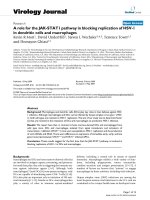
granule cells (Alvarez-Buylla et al., 2002; Gage, 2002) (FIGURE 1-1). This
thesis will centre on adult neurogenesis in the DG of the hippocampus per se.
FIGURE 1-1 Neurogenesis in the adult rodent brain (adapted fromGage,
2002). Arrows point to the two neurogenic regions: the subgranular zone
(SGZ) and subventricular zone (SVZ).
1.1.1 Stages of adult neurogenesis
Neurogenesis is a multi-step process, orchestrated at every phase by an
intricate interplay of environmental cues (such as interacting cells, growth
factors, axon guidance molecules, etc.) present in the microenvironment
where the neural precursors reside. The specific pockets of cellular
rejuvenation are termed as neurogenic niches.
Precursor cells along each stage of neurogenesis can be divided into various
cell types, largely identified by their antigenic characteristics. Recent
Olfactory
bulb
Lateral
Ventricle
s
Hippocampus
Rostral
Migratory
Stream
SGZ
SVZ
4
advances in techniques like retroviral labelling with green fluorescence protein
(GFP) also allow tracking of the maturation progress of cells over time.
The birth of new neurones does not occur in batches like a factory assembly
line. The creation, maturation and eventual survival of an individual neurone in
the SGZ are unique events at any one point of time. To sketch an outline of
the developmental process, multipotent neural stem cells first go through
intermittent cycles of division, giving rise to rapidly dividing precursor cells of
limited renewal potential, which then go on to differentiate into various
lineages. Half of the immature neurones perish before successfully migrating
and evolving into fully functional neurones (FIGURE 1-2). The sustained
production and elimination of cells in the DG are a testament of the brain’s
dynamic ability to remodel discrete networks throughout the entire lifespan.
The defining characteristics of the cells at differential time-points are charted
in Table 1-1.
1.1.1.1 Type I cells
Type I cells are the prototype neural stem cells: they are multipotent (having
the potential to differentiate into various lineages e.g. neurones, astrocytes or
oligodendrocytes) and self-renewing (possessing the ability to produce
identical daughter cells) (Seri et al., 2001). These radial glia-like cells share
morphological similarities with astrocytes. They have triangular somas in the
SGZ with long apical processes across the granule cell layer (Filippov et al.,
2003), and are immunopositive for an intermediate filament marker, glial
fibrillary acidic protein (GFAP), which has long been used to identify
5
astrocytes. They also possess electrophysiological characteristics similar to
astrocytes with delayed rectifying currents and low input resistance (Filippov
et al., 2003; Fukuda et al., 2003). However, they do not express the calcium
binding protein S100β, another marker for astrocytes (Steiner et al., 2004).
Type I cells receive no synaptic input despite expressing GABA
A
and
glutamate receptors (Wang et al., 2005).
1.1.1.2 Type II cells
The most proliferative among all cell types, Type II cells serve as the
transition phase between multipotency and lineage specialization (Steiner et
al., 2006a). The cell bodies of type II cells are also in the SGZ, with their short
plump processes oriented tangentially (Filippov et al., 2003; Kronenberg et al.,
2003; Suh et al., 2007). Type II cells have higher input resistance than Type I
cells (Fukuda, 2003). The progressive development of these progenitors can
be subdivided into 2 phases: Type IIa and Type IIb, based on their
immunoreactivity to specific cell markers. It is believed that Type IIb cells are
lineage committed (Steiner et al., 2006a). The initial inputs to Type II cells are
excitatory GABAergic synapses (Tozuka et al., 2005; Wang et al., 2005).
1.1.1.3 Type III cells
The expansion of the pool of these neuroblasts is not as prolific as the Type II
cells. Type III cells display antigenic characteristics typical of a neurone, and
do not express any glial cell markers. Radial migration into the granule cell
area commences in this phase in which the cells proceed to their postmitotic
development into neurones (Brandt et al., 2003).
6
1.1.1.4 Immature neurones
No longer in the neurogenic milieu of the SGZ, the new immigrant cells in the
granule cell layer now face a harsh selection process in an unfamiliar
environment. Cell death occurs at a constant and relatively high rate, and
about 50% of the 1- to 4- week old newborn neurones perish (Biebl et al.,
2000; Dayer et al., 2003). Programmed cell death plays a regulatory
mechanism here, by eliminating excess new neurones to ensure a prescribed
granule cell layer size and to determine that the eventual selected population
will form proper neuroneal circuits (Kuhn et al., 2005). This apoptotic process
does not affect preneuroneal progenitor cells (Kuhn et al., 2005).
The young granule cells possess different membrane properties from mature
granule cells such as very high input resistance and greater paired-pulse
facilitation, which is indicative of an increased probability of vesicle release
(Schmidt-Hieber et al., 2004). These membrane properties make the young
neurones more excitable than their neighbouring mature cells. The newly
minted dendrites of the new neurones project out into the molecular layer
(Wang et al., 2000) guided by scaffolds formed by radial processes of glia
(Shapiro et al., 2007). They receive synaptic inputs through axosomatic,
axodendritic, and axospinous synapses (Toni et al., 2007; van Praag et al.,
2002). GABAergic inputs are now inhibitory, and the first glutamatergic inputs
appear around this period (Ge et al., 2006). The changing synaptic
connections further mature the neurone functionally and are crucial for the
integration of young cells into the existing network (Ge et al., 2006).
7
1.1.1.5 Fully functional neurones
Having survived the period of high susceptibility to apoptosis, cell death
appears to halt for the approximately 1-month old postmitotic neurones (Dayer
et al., 2003). These fully mature cells are now part of the principal cells of the
DG and are physiologically indistinguishable from their neighbours 7 weeks
after cell division (van Praag et al., 2002). It was found from comparative
electrophysiological recordings that similar to granule cells of the embryonic
brain, adult born neurones have excitatory glutamatergic and inhibitory
GABAergic inputs, and can fire action potentials in response to excitation
(Laplagne et al., 2006).
These new neurones preferentially contact pre-existing boutons involved in
synapses with other neurones but form synapses with boutons devoid of other
synaptic partners as they mature over the next few weeks. The connectivity
continues to change until at least 2 months indicating that full maturation of
the connectivity of the adult-born neurone is reached between 60-180 days
after cell division (Toni et al., 2007). Axonal outgrowth occurs later than the
dendritic projections into the cellular layer (Shapiro et al., 2007) and projects
into the hippocampal CA3 regions (Hastings and Gould, 1999; Markakis and
Gage, 1999).
8
FIGURE 1-2 Stages of neurogenesis in the SGZ (adapted from Duan et al.,
2008). The newborn cell residing in the subgranular zone (SGZ) will migrate
across the granule cell layer (GCL), and extend its newly formed dendrites out
into the molecular layer (ML).
SGZ
Hilus
GCL
ML
Type III
Type II
Type I
Immature
Neurone
Mature
Neurone
9
Cell type
Type I
Type IIa
Type IIb
Type III
Immature
neurone
Mature neurone
Cell age
1-3 days
2-3 weeks
>4 weeks
Stage of cell
cycle
Quiescent
Mitotic
Postmitotic
Defining
characteristic
Multipotent radial
glia -like stem cell;
rare and slowly
proliferating;
Present in SGZ
Highest proliferative
rate among all cell
types but limited self-
renewal
Highly proliferative
but limited self-
renewal.
Differentiation into
various lineages
Migrates to
granule cell layer
50% die by
apoptosis
Forms functional
synapse with
other neurones
Markers
GFAP+
DCX-
DCX-
DCX+
DCX+
DCX+
Nestin+
Nestin+
Nestin+
Nestin-
NeuN+
NeuN+
PSA-NCAM-
PSA-NCAM+
PSA-NCAM+
Prox1+
Prox1+
Prox1+
Sox2+
Sox2+
Sox2+
Sox2-
Calretinin+
Calbindin+
BLBP+
BLBP+
BLBP+
BLBP-
TuJ1+
TuJ1+
NeuroD1-
NeuroD1+
NeuroD1+
Map2ab+
Synaptic
inputs
-
Excitatory GABAergic
Initially inhibitory
GABAergic, then
glutamatergic
all
Input
resistance
<100 MΩ
~50 –8500 MΩ
~5000–10000 MΩ
>1500 MΩ
~300 MΩ
Voltage-gated
currrents
A type K+
K+, small Na+
K+, small Na+, T-
type Ca2+
K+, Na+, T-type
Ca2+
K+, Na+
TABLE 1-1 Characteristics of adult-born neurones in the SGZ at different time-points
Abbreviations and key references: β-tubulin (TuJ1) (Parent et al., 1997); Brain lipid binding factor (BLBP) (Steiner et al., 2006a); Calbindin
(Sloviter et al., 1989); Calretinin (Brandt et al., 2003); Doublecortin (DCX) (Filippov et al., 2003; Plumpe et al., 2006); Glial fibrilliary acidic
protein (GFAP) (Filippov et al., 2003); Microtubule associated protein 2ab (Map2a) (Brazel et al., 2005; Steiner et al., 2006a); Nestin (Filippov
et al., 2003; Lendahl et al., 1990; Mignone et al., 2004); Neurogenic differentiation 1(NeuroD1) (Steiner et al., 2006b); Polysialic acid neural cell
adhesion molecule (PSA-NCAM) (Seki, 2002a, b; Seki and Arai, 1993); Prospero-related homeobox1 (Prox1) (Brandt et al., 2003); SRY(sex-
determining region Y)-box 2 (SOX2) (Brazel et al., 2005; Steiner et al., 2006a; Suh et al., 2007)
10
1.1.2 Factors regulating neurogenesis
For a seemingly restricted region, the permissive SGZ niche is susceptible to a
host of regulatory agents that affects neurogenesis at every stage. Being
vestiges of the embryonic brain, it is fairly straightforward to imagine niches as
microenvironments where developmental neurogenic qualities are retained, and
where original neuromodulators are still at work.
Most studies investigating the mechanisms behind neurogenesis have been
accomplished utilizing the thymidine analog bromodeoxyuridine (BrdU) as an in
vivo marker of proliferating cells. BrdU can be visualized using
immunohistochemical techniques and quantitatively assessed (Gould and Gross,
2002). The colocalization of BrdU-labelled cells with cell type-specific markers
can be verified by orthogonal reconstruction of different planes captured by
confocal microscopy (Gould and Gross, 2002).
In the context of this discourse, modulators of neurogenesis are broadly
subdivided into (i) cellular and molecular factors, and (ii) physiological and
behavioural factors. A list of these factors is given in TABLE 1-2.
1.1.2.1 Cellular and molecular factors
1.1.2.1.1 Glial cells
There is increasing documentation to suggest that glial cells, originally regarded
as supporting cells, are instrumental in regulating neurogenesis (Ma et al., 2005).
11
Astrocytes are the most abundant of all glia. When extracted from the
hippocampus and cultured, astrocytes were shown to spur the growth of
progenitors and subsequently commit these progenitors to a neuroneal lineage
(Song et al., 2002). Hippocampal astrocytes were also able to promote synapse
formation of neurones derived from adult neural stem cells (Song et al., 2002).
This is because astrocytes provide a lattice for the growth of axons and dendrites
from newly generated neurones, as revealed through structural studies (Horner
and Palmer, 2003).
Microglia, another non-neuroneal cell normally activated during CNS
inflammation, is proposed to regulate the pro- and anti-neurogenic effects of
immune cytokines in the DG niche (Battista et al., 2006). Microglia activation
correlates with the presence of an anti-inflammatory cytokine transforming
growth factor-β (TGFβ) and an increase in progenitor proliferation (Battista et al.,
2006). Exposure of microglia to other cytokines such as interleukins also induces
neurogenesis (Butovsky et al., 2007).
1.1.2.1.2 Growth factors
A growing body of evidence suggests a primary role for peptide growth factors
such as basic fibroblast growth factor (FGF2), insulin-like growth factor-I (IGF1),
granulocyte-colony stimulating factors (G-CSF), vascular endothelial growth
factor (VEGF), erythropoietin, epidermal growth factor (EGF) and TGFβ in
influencing neurogenesis. These ligands are detected in early stages of
12
development, and their expression persists postnatally into adulthood in the
hippocampal DG (Bondy and Lee, 1993; Ozawa et al., 1996).
Specifically, FGF2 has been widely used to expand cultured neural progenitor
cells from fetal and adult brains. In primary cultures of DG granule cells from
neonatal rats, addition of FGF2 enhanced neuroneal survival and differentiation
(Lowenstein and Arsenault, 1996b). FGF2 also increased axon number and
length, and boosted migration (Lowenstein and Arsenault, 1996a). Infusions of
FGF2 into the ventricles of middle aged rats increased neurogenesis and
augmented dendritic growth (Rai et al., 2007). Some reports indicate that FGF2
inhibits neuroneal lineage determination and hence maintains the progenitor pool
in a proliferative state (Chen et al., 2007). Another growth factor IGF1 has been
shown to generate new neurones from adult hippocampal progenitors in vitro
(Aberg et al., 2000; Anderson et al., 2002). The angiogenic factor VEGF can
stimulate cell genesis in cortical cultures, and increase the overall production of
neurones (Jin et al., 2002). In vivo experiments also show that
intracerebroventricular injections of VEGF into the adult rat brain increased SGZ
progenitor proliferation (Jin et al., 2002).
The source of these growth factors may or may not be intrinsic to the neurogenic
niche. Underlying the region is a rich network of blood vasculature, where tight
clusters of proliferating precursors, committed progenitors, neurones and glial
cells are grouped (Palmer et al., 2000). The growth factors may derive from the
13
circulatory system. In vitro, soluble factors secreted by the vascular endothelial
cells, components of blood vessels, promote self-renewal and neurogenesis in
fetal neural stem cells (Shen et al., 2004).
The survival of newly generated neurones may also involve neurotrophins such
as brain-derived neurotrophic factor (BNDF), nerve growth factor (NGF) and
neurotrophin 3 (NT3). In vitro, NT3 but not BDNF significantly increases the
number of newborn neurones (Babu et al., 2007). ICV infusions of NGF increase
the proportion of both BrdU-positive and DCX-positive cells two weeks later
(Frielingsdorf et al., 2007). In BDNF heterozygous mice (BDNF
+/-
) and trkB
(receptor of BDNF) dominant null mice, the number of new neurones born is
considerably less (Sairanen et al., 2005).
1.1.2.1.3 Neurotransmitters
Afferents from other parts of the brain extend to postsynaptic neurones in the
DG, releasing chemical messengers that facilitate neurognenesis. A couple of
amino acid neurotransmitters, namely γ-aminobutyric acid (GABA) and
glutamate, are the major forces behind excitatory-neurogenesis coupling.
In the embryonic brain, GABA initially acts as an excitatory molecule. GABA
binds mostly to GABA
A
receptors present in the precursor cell, which has an
elevated intracellular chloride (Cl
-
) concentration, and hence a lower resting
membrane potential. This leads to an efflux of Cl- ions and depolarization, and
14
subsequent activation of voltage-dependent calcium channels (Ben-Ari, 2002).
Later, due to a drop in intracellular Cl
-
concentration in the more mature cell,
GABA switches from being excitatory to inhibitory (LoTurco et al., 1995).
Drawing parallels from the embryonic brain, Type II progenitors in the SGZ
similarly receive GABAergic inputs (Tozuka et al., 2005). By triggering
spontaneous GABAergic synaptic events, Type II cells are depolarized, causing
increased intracellular calcium concentration and induction of NeuroD expression
(Tozuka et al., 2005). NeuroD is a transcription factor that drives neuroneal
differentiation (Liu et al., 2000). Addition of GABA
A
receptor antagonists elevates
progenitor proliferation, while GABA
A
receptor agonists elicit the opposite effect,
increasing differentiation of newly born neurones, further cementing the evidence
that GABAergic inputs promote activity-dependent neuroneal differentiation
(Tozuka et al., 2005). Other reports show that injection of GABA
A
receptor
agonist into the rodent brains do not affect the survival of newborn cells (Karten
et al., 2006), but rather increase dendritic length and complexity (Ge et al., 2006).
Initial GABA-induced depolarization is crucial for ensuing inhibitory GABAergic
and excitatory glutamatergic synaptic inputs in newly generated neurones (Ge et
al., 2006).
Glutamatergic synapses are formed after GABAergic synapses in the embryonic
brain (Ben-Ari et al., 2007). The dentate granule cells receive most of the
excitatory glutamatergic inputs from the entorhinal cortex. In adult rodents, the
15
activation of N-methyl-d-aspartate receptors (NMDAR) by the agonist NMDA
resulted in a drop in cell division in the SGZ. In contrast, intraperitoneal injections
of NMDA receptor antagonist led to an increase in cell birth in both young adult
(Cameron et al., 1995) and middle-aged rats (Nacher et al., 2003). NMDA
receptor subunits NR1 and NR2B are expressed in Type I precursor cells and
immature neurones in the DG (Nacher et al., 2007). An elegant experiment in
which retrovirus-mediated gene knockout of NMDAR in a single-cell reduces
neuroneal survival, only to be rescued by NMDAR antagonist application that
blocks receptors of surrounding functional neurones demonstrates that
glutamatergic inputs may be important for extending the lifespan of newly
generated neurones (Tashiro et al., 2006).
The regulatory effects of acetylcholine amino acid from cholinergic inputs to the
DG will be elaborated more in Chapter 2 of this dissertation.
1.1.2.1.4 Steroid hormones
Many studies have revealed that glucocorticoid stress hormones are major
dampeners of progenitor proliferation (Cameron and Gould, 1994). Removal of
circulating adrenal steroids by adrenalectomy reverses the stress-induced
decline in neurogensis in DG (Cameron and McKay, 1999; Tanapat et al., 2001).
Sex hormones, another class of steroids, generate and sustain new cells
differentially in adult female and male rodents. One of the earlier observations
16
comes from female rats, which possess a greater number of newborn cells in the
DG compared to male rats, and which the cell count fluctuates at different
periods of the oestrus cycle (Tanapat et al., 1999). Cell proliferation is decreased
by ovariectomy but can be reversed by progesterone (Tanapat et al., 2005).
Acute estrogen treatment likewise induces cellular proliferation (Tanapat et al.,
2005). Estrogen receptor agonists also enhances cell genesis (Mazzucco et al.,
2006). Interestingly, estradiol too stimulates progenitor proliferation in middle
aged male mice (Saravia et al., 2007). Unlike estrogen, androgens targets
neurogenesis at a later time point. Cell survival was decreased for castrated rats,
but prolonged in male rats injected with testosterone and one of its derivatives,
dihydrotestosterone (Spritzer and Galea, 2007).
1.1.2.1.5 Morphogens
Properties of the embryonic brain are conserved in specialized niches. As such,
developmental morphogens such as Notch, bone morphogenetic proteins
(BMPs), Noggin, Sonic hedgehog (Shh), Wingless-type MMTV integration (Wnt)
have all been implicated in the regulation of neurogenesis (Babu et al., 2007;
Breunig et al., 2007; Fan et al., 2004; Lai et al., 2003).
For instance, Notch1 signalling acts like a switch between Type I, Type IIa and
Type IIb cells (Breunig et al., 2007) in postnatal mice. Another developmental
protein, BMP4 and its signalling antagonist Noggin are expressed in the SGZ of
adult DG (Fan et al., 2004). Antisense Noggin infusion into the ventricles reduced
17
DG progenitor proliferation (Fan et al., 2004). Another member of the BMP
family, BMP2 inhibited neurogenesis in monolayer precursor cell culture from
adult mouse DG (Babu et al., 2007).
Shh is a potent mitogen of multipotent adult hippocampal progenitor cells in vitro
(Babu et al., 2007; Lai et al., 2003). In vivo, viral delivery of Shh in the
hippocampus increases progenitor division and subsequently the number of
newborn neurones in the granule cell layer (Lai et al., 2003) whereas
pharmacological blockade of Shh signalling reduces proliferation (Banerjee et al.,
2005). Wnt signalling affects neuroneally restricted Type IIb precursors (Pozniak
and Pleasure, 2006). Wnt3 proteins are secreted by astrocytes in the DG hilus
and cause increases in the total number of immature neurones (Lie et al., 2005).
1.1.2.2 Behavioural and physiological factors
1.1.2.2.1 Aging
Our brains, like other parts of the body, deteriorate over time. Not surprisingly,
neurogenesis decreases with increasing age, attributed by different groups either
to slower precursor proliferation, migration or differentiation (Hattiangady and
Shetty, 2008; Kempermann et al., 1998b; Kuhn et al., 1996);. The turnover of the
newly generated cells, characterized by rates of apoptosis, also slows down with
increasing age (Heine et al., 2004). The observations of age-related decline in
neurogenesis were replicated outside laboratory settings, in different species of