Structural and functional genomics study of singapore grouper iridovirus 1
Bạn đang xem bản rút gọn của tài liệu. Xem và tải ngay bản đầy đủ của tài liệu tại đây (880.26 KB, 100 trang )
1
Chapter 1
Literature Review
2
1.1 Introduction to virus
In 1898, Friedrich Loeffler and Paul Frosch found evidence that the cause of foot-and-mouth
disease in livestock was an infectious particle smaller than any bacteria. This was the first clue to
the nature of viruses, genetic entities that lie somewhere in the grey area between living and non-
living organisms.
A virus (from the Latin virus meaning toxin or poison) is a sub-microscopic infectious agent that
is unable to grow or reproduce outside a host cell. Each viral particle, or virion, consists of
genetic material, DNA or RNA, within a protective protein coat called a capsid. Some viruses
have more complex structures with tail or envelop (Emiliani, 1993).
Viruses depend on the host cells that they infect to reproduce. A virus can insert its genetic
material into its host, literally taking over the host’s DNA replication and protein expression
machinery. Some viruses may remain dormant inside host cells for a long period of time, causing
no obvious change in their host cells (lysogenic phase). But when a dormant virus is stimulated,
it enters the lytic phase: new viruses are formed, self-assemble, eventually rupturing and killing
the host cell before infecting other cells (Emiliani, 1993).
Viruses can infect all organisms from bacteria to plants and animals and cause a number of
severe diseases in eukaryotes. Antibiotics have no effect on viruses, but antiviral drugs have been
developed to treat life-threatening infections.
3
1.2 Overview of the Iridoviridae family
1.2.1 Characteristics of the Iridoviridae family
Iridoviruses have been found to infect invertebrates (insects) and poikilothermic vertebrates,
including amphibians, reptiles and fishes. This virus family has three distinct features including
the virus morphology, the cytoplasmic location of virion particles and the genomic organization.
Iridoviruses are a family of large viruses (120- 300 nanometers in size) that contain linear,
double-stranded DNA as their genetic material and have an icosahedral (20-sided) capsid (Figure
1). An iridovirus virion is composed of three concentric domains; an outer proteinaceous capsid,
an intermediate lipid membrane with associated polypeptides, and a central core containing
DNA-protein complexes. Some, but not all, viruses possess an outer envelop acquired by
budding through the host membrane. Fibrillar structures have also been observed protruding
from capsid subunits of Lymphocystis disease virus 1, Megalosystisivirus and Chloriridovirus
but not from Frog virus 3. A common feature of all iridoviruses is the presence of a major capsid
protein of around 50 kDa that accounts for up to 45% of total virion protein (Williams et al.,
2006).
4
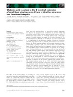
Figure 1: Diagram of icosahedral capsid of Sericesthis Iridescent Iridovirus.
Trisymmetron are shown in white subunits, disymmetrons in black and pentasymmetrons in
grey. The geometrical edges of the icosahedral are picked out in broken lines (Wrigley, 1969).
5
Iridovirus infections result in the appearance of large, morphologically distinct viral assembly
sites within the cytoplasm. These sites serve as a concentration point for viral proteins and DNA
and are the site of virion assembly (Williams et al., 2006). The viral particles accumulate within
the cytoplasm in large crystalline arrays. Light reflected from the surface of this special
arrangement interferes with newly arriving light, causing Bragg reflection (Klug et al., 1959)
resulting in “rainbow-like” iridescence. The name Iridoviridae was originally derived from Iris,
who was the Greek goddess of the rainbow. However, the iridescent phenomenon takes place
only in invertebrate iridoviruses, not in vertebrate iridoviruses.
In addition to their distinctive size and cytoplasmic location, iridoviruses are distinguished from
other virus families by their genomic organization. The iridovirus genome is circularly
permutated and terminally redundant. This structure is a result of the resolution of genome
concatamers during DNA replication (Williams et al., 1996).The large concatameric DNA is
moved to the assembly site and packaged into the viral capsid through a “headful” mechanism
until the head of the virus is full (Goorha and Murti, 1982).
During replication, multiple copies of a hypothetical viral genome form a long concatamer. The
resolution of this concatamer results in packages of DNA that contain a complete genome and
duplicated copies of some genes as well (terminal redundancy). The ends of each of these
packaged DNA fragments differ from one virus particle to the next (cyclic permutation). This
genomic structure has been found in all iridoviruses so far studied (Williams et al., 2006).
6
1.2.2 Classification of the Family Iridoviridae
To date, more than 100 species of iridoviruses have been discovered in a wide variety of
invertebrates and vertebrates. It is necessary to classify them into different genera based on their
common characteristics including the sources of host organisms, genetic properties, and
morphological evidences (Table 1).
The family Iridoviridae is currently subdivided into five genera: Iridovirus, Chloriridovirus,
Lymphocystivirus, Megalocytivirus and Ranavirus (Williams 2006). The first two genera can
infect a large range of insects such as flies, silkworms (for Iridovirus) and mosquitoes (for
Chloriridovirus). The last 3 genera contain veterbrate viruses that infect poikilothermic
vertebrates including fishes, amphibians and reptiles. Ranavirus is a large genus, in which frog
virus 3 contains at least 15 isolates including Box turtle virus 3, Bufo bufo United Kingdom
virus, Bufo marinus Venezuelan iridovirus 1, Lucke triturus virus 1, Rana temporaria United
Kingdom virus, Redwook Park virus, Stickleback virus, Tadpole virus 2, Tiger frog virus,
Tortoise virus 5, Largmouth bass virus, Doctor fish virus and Guppy virus 6 (Williams et al.,
2006).
7
Table 1: Current classification of the Iridoviridae family (Williams et al., 2006)
Genus Distiguishing features Host species Members of genus
Iridovirus DNA is not methylated Insects Invertebrate iridescent virus 1
~ 212 kbp Crustaceans Invertebrate iridescent virus 6
Virion diameter possibly mollusks
~ 120-130 nm
Chloriridovirus
DNA is not methylated Mosquitoes Invertebrate iridescent virus 3
~ 135 kbp Diptera
Virion diameter
~ 180 nm
Ranavirus
DNA is methylated* bony fish, Frog virus 3
~ 105 kbp reptiles, Frog virus 1, 2, 5-24
Virion diameter amphibians Frog virus L2, L4, L5
~ 150 nm Tadpole edema virus
Lucke triturus virus LT1-LT4
Newt virus T6-T20
Xenopus virus T21
Ambystoma tigrinum
Tiger frog virus
Grouper iridovirus
Singapore grouper iridovirus
Lymphocystivirus
DNA is methylated Marine and fresh Lymphocystis disease virus 1
~ 103-186 kbp water fishes Lymphocystis disease virus 2
Virion diameter world wide Lymphocystis disease virus
~ 200- 300 nm China
Megalocystivirus
DNA is methylated Marine fishes Infectious spleen and kidney
~ 105- 118 kbp in SE Asia necrosis virus
Virion diameter
~ 150 nm Rock bream iridovirus
Orange spotted grouper
Iridovirus
Sea bass iridorivirus
Red sea bream iridovirus
* Singapore grouper iridovirus and Grouper iridovirus appears to lack a DNA methyltransferase
8
1.3 Singapore Grouper Iridovirus: significance and research progress
1.3.1 Significance of SGIV
Singapore Grouper Iridovirus (SGIV), a member of Ranavirus genus, is an important pathogen
which causes “Sleepy Grouper Disease” (SGD) in grouper fish (Chua et al., 1994, Qin et al.,
2001, Song et al., 2004). The severe disease, with symptoms of enlargement of cells, necrosis of
the renal and splenic hematopoietic tissues, could lead to 30% to 100% mortality (Qin et al.,
2001). The SGD outbreaks in 1992 resulted in losses of 50% of Singapore brown- spotted
grouper stock ( Chua et al., 1994). This virus threatens the aquaculture economic in Singapore
and South East Asia as well.
SGIV genome was fully sequenced with many of the open reading frames (ORFs) are novel
with unknown functions (Song et al., 2004). However, with the availablity of a grouper cell line
(Chew-Lim et al., 1994), the functional and structural genomics studies could provide a new
insight into molecular biology of the virus and be meaningful for drug design.
1.3.2 Reseach progress on SGIV
1.3.2.1 Isolation and propagation of SGIV
Study of grouper diseases can be traced back to an investigation on a mass mortality in marine
cage-cultured sea perch, Lates calcarifer, and grouper, E. tauvina in the Johore Straits about
9
twenty year ago (Nash et al., 1987) and in Singapore in 1992(Chua et al., 1994). A large number
of infected fish suffered from severe hemorrhagic ulcerative dermatitis. The spleens of the
infected fish were two to three times larger than those of the normal ones due to the intrusion of
viruses (Chua et al., 1994). The supernatants of infected tissue homogenates were then
inoculated onto confluent monolayers of grouper cell line, with good resultant titers. This novel
iridovirus has been successfully isolated from infected grouper- Epinephelus tauvina and
designated as Singapore grouper iridovirus (SGIV) (Qin et al., 2003). The grouper embryonic
egg (Epinephelus tauvina) cell line, developed by the Agri-Food and Veterinary Authority of
Singapore (Chew-Lim et al., 1994), was used as a souce to propagate SGIV.
1.3.2.2 Structure of SGIV
Sucrose gradient ultracentrifugation has been developed for the purification of SGIV from
infected grouper cell line (Qin et al., 2003). Using this approach, most of the virus was
suspended at the boundary layer between 40% and 50% sucrose (an equilibrium density
banding). The virus was aspirated and examined under electron microscopy after negative
staining. The viral particle revealed a three-layer membrane structure with an inner electron-
dense core. The outline of the SGIV was also determined by negative staining and observed by
electron microscopy under which the average size was estimated as 200±13nm. The SGIV
formed a well-defined hexagonal contour, suggesting that the three-dimensional structure of the
SGIV is an icosahedral particle (Qin et al., 2001).
10
1.3.2.3 Classification of SGIV
In Iridoviridae family, major capsid protein (MCP) is one of the highly conserved genes but
sufficiently diversed to distinguish closely related iridorivus isolate (Tidona et al., 1998). Owing
to the special characteristic, a partial DNA sequence of the SGIV MCP has been successfully
amplified by a PCR technology (Qin et al., 2001). Compared with other MCP sequences, SGIV
was easily classified into the genus Ranavirus, under the family Iridoviridae.
1.3.2.4 Physical properties of SGIV
One important aspect of the SGIV is its physicochemical properties which has been fairly well
established (Qin et al., 2001). The SGIV isolate, whose infectivity maintained at a high titer of
10
6.0
TCID
50
mL
-1
, propagated continuously in a grouper embryonic cell line. Nevertheless, the
infectivity dropped dramatically when treated by high temperature at 56 ºC for 30 min. Under an
acidic environment with 0.1 M citrate buffer (pH 3.0), the SGIV almost lost all its infectivity in
culture media. The titer was also reduced dramatically from 10
7.0
to 10
3.0
TCID
50
mL
-1
with
ether. The SGIV was affected with treatment of low concentration of 5-iodo-2-deoxyuridine
(IUdR, 10 µM), suggesting that the virus possessed a DNA genome. Elucidation of
physicochemical properties of the SGIV has facilitated us to monitor the fish disease. Besides, all
the above characteristics provide the evidence for the classification of SGIV within the virus
kingdom. However, the conclusive evidence for classifying it as a member of the family
Iridoviridae is the genetic structure of the virus.
11
1.3.2.5 Genome sequence and proteomics analysis of SGIV
The complete genome of SGIV was sequenced using random shotgun and restriction
endonuclease genomic approaches. The genome sequence was deposited at NCBI data base, and
the accession number is AY521625. The entire SGIV genome consists of 140,131 nucleotide
base pairs with 162 ORFs (Song et al., 2004). Using peptide mass finger prints generated from
MALDI-TOF MS, 77 of the ORFs exhibited homologies to known viruses, 23 of which matched
functional iridovirus proteins. In addition, 26 proteins of this virus were identified for the first
time , twenty of these represented novel or previously unidentified genes, which were further
confirmed by reverse transcription-PCR, followed by DNA sequencing of the respective RT-
PCR products (Song et al., 2006).
Another proteomics investigation using 1-DE-MALDI and LC-MALDI workflows resulted in a
more comprehensive identification of the SGIV proteome with another newly 25 SGIV proteins
identified (Song et al., 2006). Although a total of 51 SGIV proteins have been identified, the
translational products of the remaining 111 ORFs are unknown (Song et al., 2006).
1.3.2.6 Temporal and differential stage gene expression of SGIV
A DNA microarray was generated for the SGIV genome to analyze the expression of its
predicted ORFs. The noninfected and infected cells at different time course of SGIV infection
were collected and treated with cycloheximide and aphidicoline to study the temporal gene
expression and to classify them into different-stage viral genes such as Immediate Early, Early
12
and Late genes. The DNA microarray data was verified and consistent with real-time RT-PCR
studies (Chen et al., 2006). These results should provide important insights into the replication
and pathogenesis of iridoriviruses.
1.4 Introduction to Ubiquitin and Ubiquitin-like protein
1.4.1. Ubiquitin
Ubiquitin (Ub) is a small and highly conserved protein with 76 amino acids (Schlesinger et al.,
1975). Its main role is to label proteins, including misfolded, damaged or malfunctioned proteins
that are tageted for proteolytic degradation. However, ubiquitin also has nonproteolytic function
by reacting with other proteins to modify the protein structures. With or without protein
degradation, the ubiquitin system is involved in the regulation of a number of cell signaling
pathway (Herrmann et al., 2007).
Ub is known to function in Ubiquitin proteosome sytem (UPS) (Figure 2), which plays a key role
in protein degradation of a variety of basic cellular processes such as cell cycle, cell division,
transcription regulation (Schwartz 1999) and apoptosis (Jentsch and Pyrowolakis, 2000). In this
pathway, Ub is activated by activating enzyme E1, transferred to conjugating enzyme E2,
followed by ligase enzyme E3. Ub is then conjugated to specific substrate or next ubiqiuitin
moiety to generate the polyubiquitin chain. This polyubiquitin chain serves as a signal for protein
degradation by 26S proteasome (Ciechanover, 1998).
In addition, Ub is able to modify proteins by monoubiquitination independent of proteolysis.
Monoubiquitination modifies histone proteins to control gene expression (Robzyk and Osley,
2000; Pham and Sauer, 2000), regulates the membrane transport endocystosis (Nakatsu et al,
13
2000; Shih et al, 2000) and is involved in the budding of retrovirus from the plasma membrane
(Hicke, 2001).
Figure 2: Schematic representation of the UPS pathway (Belz et al., 2002)
14
1.4.2. Ubiquitin-like proteins
In the past few years, a suprising number of ubiquitin-like proteins (UBL) or molecules have
been identified, which can be divided into two separate classes: ubiquitin-like modifiers (ULM)
and ubiquitin-domain proteins (UDP).
ULMs have very little homologous sequences but surprisingly a common 3D structure, the
ubiquitin fold and C-terminal di-glycine residues. They conjugate to proteins and function in a
‘ubiquitin-like’ manner (Kerscher et al., 2006). At least 10 different ULMs exits in mammals. Of
these, SUMO (small-ubiquitin-related modifier) and RUB1 (related-to-ubiquitin 1) pathways
have received the most intense scrutiny. On the other hands, UDPs bear a sequence domain that
is similar to ubiquitin, but are not conjugated to proteins. Instead, they serve as adaptor function,
binding noncovalently to ubiquitin or ULMs via an “ubiquitin-interaction motif” or ubiquitin-
associated (UBA) domain. The first UDP identified was the Rpn10 subunit of the 19S
proteasome, which allows the direct recognition of polyubiquitinated proteins by the 26S
proteasome. Other UDPs function as cofactors or adaptors involved in escorting a subset of
polyubiquitinated proteins to the 26S proteasome (Herrmann et al., 2007).
1.5 Introduction to NMR spectroscopy
In 1946, two research groups, Purcell (Massachusetts Institute of Technology) and Bloch
(Stanford University) reported for the first time the nuclear magnetic resonance (NMR)
phenomenon. In 1953, Overhauser defined the concept of nuclear overhauser effect (NOEs),
which formed the basis for the structural determination by NMR. After three decades, the first
protein structure was solved using NMR spectroscopy by Ernst and Wuthrich. Since then, NMR
15
spectroscopy has become an alternative method to X-ray crystallography for the structural
determination of small to medium sized proteins (less than 25 kDa) in aqueous or micellar
solutions. Notably, in 2006, Yang and his group ( National University of Singapore) has
developed a new strategy for structure determination of large proteins up to 60 kDa (Xu et al.,
2006). Recent progress in computational and experimental NMR techniques has improved the
efficiency of biological research (Bax, 2003).
A simple one-dimensional (1D) proton experiment is the most basic spectrum in NMR
spectroscopy that contains a vast amount of information. It is able to show the folding status of
the proteins, whether a protein is folded or unfolded. This is very important for any further
functional or structural studies on the protein because only folded proteins retain their functional
activities and the three dimensional structures (Rehm et al., 2002). Unfortunately, 1D spectra of
protein molecules that contain overlapping signals from many hydrogen atoms due to the
differences in chemical shifts are often smaller than the resolving power of the experiments
(Freeman and Anderson, 1962).
Two-dimensional (2D) experiments has been greatly improved in resolution. The simplest and
most powerful 2D experiment is the heteronuclear single-quantum coherence (HSQC), in which
15
N-labeled protein samples are used. The HSQC shows one peak for every proton bound
directly to a nitrogen atom and thus exactly one signal per residue in the protein. However, this
is not correct for Proline, Asparagine and Glutamine. HSQC is devoid of Proline backbone
amide but displays additional peak for side chain signals of Asparagine and Glutamine. In
16
addtion, 2D NMR data is not sufficient to determine structure of proteins with large M.W due to
signal overlapping and faster signal relaxation (Kalic et al., 2000).
To solve the complex problems of 2D NMR, three-dimensional (3D) NMR spectroscopy is a
logical approach to tremendously increase the effective resolution (Fesik and Zuiderweg, 1990).
The heteronuclear 3D experiments involve in at least two types of nuclei. The experiment can
correlate various nuclei either through scalar coupling (COSY, TOCSY, HMQC and HSQC) or
through space (NOESY). The 3D NMR experiments consist of two 2D experiments after another
such as NOESY-HSQC, TOCSY-HSQC (Clore and Gronenborn, 1991).
The final result of the sequence-specific assignment of NMR signals is a list of distance
constraints from a specific hydrogen atom in one residue to hydrogen atoms in the same or
different residue. This list immediately identifies the secondary structure elements of the protein
molecule because both α helices and β sheets are very distinct sets of interactions of less than 5
A
o
between hydrogen atoms in their amino acid residues. It is therefore possible to calculate
models of three dimensional structure of protein. Eventually, a set of possible structures (usually
more than 10) rather then a unique structure will be determined (Branden and Tooze, 1999).
1.6 Introduction to Isobaric Tags for Relative and Absolute Quantification
1.6.1 Proteomics and Mass spectrometry
Proteome of an organism is the set of proteins produced during its life. Proteomics is the large
scale study of proteins. The goal of proteomics is a comprehensive, quantitative description of
17
protein expression and its changes under the influence of biological perturbations such as disease
or drug treatment (Anderson & Anderson, 1998). Proteomics can be seen as a mass-screening
approach to molecular biology, which aims to document the overall distribution of proteins in
cells, identify and characterize individual proteins changes, and ultimately elucidate the
functional relationships (Twyman, 2004). There are many different proteomics branches, for
example protein separation ( 1D gel, 2D gel, liquid chromatography-LC), protein modification,
protein quantification ( ICATs, iTRAQ), and protein identification (mass spectrometry), etc.
Mass spectrometry (MS) is an analytical tool used for measuring the molecular mass of a sample.
A mass spectrometer consists of three fundamental parts: the ionisation source, the analyzer and
the detector. The sample is introduced into the ionization source to become ionized that is easier
to be manipulated than neutral molecules. These ions are extracted into the analyzer, separated
according to their mass m-to-charge ratios and detected by the detector. The signal is sent to a
data system and presented in the format of a spectrum. There are several methods of ionization,
the two most common methods are Electrospray Ionization (ESI) and Matrix Assisted Laser
Desorption Ionization (MALDI) ( />The MS applications are diverse in both routine work and research. In the proteomics field, MS
is used to accurately measure the molecular mass of proteins and oligonucleotides to determine
the sample’s purity, identify amino acid sequence, characterize oligonucleotides, detect post-
translational modification; to monitor reactions of enzymes, chemical modifications, protein
digestion; and to study protein folding, protein-ligand complex formation and macromolecular
structure determination ( />18
Tandem mass spectrometry or MS/MS is used to study the structural and sequence information
from MS. MS/MS also enables specific compounds to be detected in complex mixtures. A
tamdem mass spectrometer is a mass spectrometer that has more than one analyser, usually two.
( />1.6.2 Isobaric Tags for Relative and Absolute Quantification (iTRAQ)
iTRAQ is a stable isotope method for relative and absolute protein quantitation using mass
spectrometry. The core of this methodology is a multiplex set of isobaric reagents which are
amine-specific and allow for the identification and quantitification of up to four different
samples simultaneously (Ross et al., 2004). In the 4-plex iTRAQ, the reagents designed as
isobaric tags consist of a charged reporter, a peptide reactive group and a neutral balance portion
to maintain an overall mass of 145 Da (Figure 3). The charge reporters, from 114 to 117 Da, are
unique to each of the four reagents.
These unique reagents, upon MS/MS fragmentation give rise to four unique reporter ions (m/z=
114-117) that are used to quantify their respective samples.The peptide reactive group was
designed to react with all primary amines to label all peptides of different samples thus
enhancing peptide coverage for any given protein.
Each individual sample is reduced, alkylated and digested with trypsin. The resulting peptide
pools are respectively labeled with one member of multiplex set, then combined and
subsequently analysed by LC-MS/MS (Liquid Chromatography/ Mass Spectrometry/ Mass
Spectrometry) (Figure 4). Quantitation is achieved by comparison of the peak areas and the
19
resultant peak ratios for the four MS/MS reporter ions, which range from 114 to 117 Da (Zieske,
2006).
The advantages of this method are the increased confidence and higher quality data because all
trypic peptides are labeled, there is no loss of information from samples involving post-
translational modifications. The new class of isobaric reagents enhance MS/MS fragmentation
thus giving more confident identification than previously encounted (Zieske, 2006). Finally, the
multiplex capacity of these reagents allows information for replication within certain LC-MS/MS
experimental regimes, providing additional statistical validation within any given experiment.
Compare to other methods such as ICAT (Isotope Coded Affinity Tags) and DIGE (Different
Gel Electrophoresis), iTRAQ is more sensitive for quantitation but more susceptible to errors in
precursor ion isolation (Gan et al., 2006). Furthermore, this multiplex protein quantitation
requires more mass spect time because of the increased number of peptides (Pierce et al., 2007).
Recently, the novel 8 channel iTRAQ are available with eight specific reagents (Figure 5)
(Pierce et al., 2007). This new generation of iTRAQ reagents greatly enhances the reproducible
information, thus higher confidence identification data.
20
Figure 3 Diagram of the iTRAQ reagent for 4-plex iTRAQ.
Each reagent consists of a charged reporter, a peptide reactive group and a neutral balance
portion to maintain an overall mass of 145 Da. The charge reporters, from 114 to 117 Da, are
unique to each of the four reagents (Zieske, 2006).
21
Figure 4: The general iTRAQ workflow for four different samples.
Each sample is reduced, alkylated, digested with trypsin, then combined and subsequently
analysed by LC-MS/MS (Zieske, 2006).
22
Figure 5 : Diagram of iTRAQ reagents for 8-plex iTRAQ system
Each reagent consists of a charged reporter, a peptide reactive group and a neutral balance
portion to maintain an overall mass of 305 Da. The charge reporters, from 113 to 121 Da, are
unique to each of the eight reagents (Pierce et al., 2007).
23
1.7 Introduction to Morpholino oligonucleotides technology
1.7.1 Gene knock-down
Gene knock-down refers to a technique in which an organism is genetically modified to have
reduced expression of one or more genes through the insertion of an agent such as a short DNA
or RNA olionucleotide with a sequence complementary to an active gene or its mRNA
transcripts.
There are three major gene knock-down types: 1) phosphorothiotate-linked DNA (S-DNA); 2)
short interfering RNA (siRNA); and, 3) Morpholino. The structures of these 3 types of gene
knock-down are illustrated in Table 2 (Summerton, 2007).
1.7.2 Gene knock-down by Morpholino
1.7.2.1 What is Morpholino?
The word "morpholino" can occur in other chemical names, referring to chemicals containing a
six-member morpholine ring. This work discusses only the Morpholino antisense
oligonucleotides.
Morpholinos or morpholino antisense oligonucleotides or oligos are called MO in short. MO is a
gene knock-down agent which consists of short chains of about 25 morpholino subunits. Each
morpholino subunit contain a nucleotide base, a morpholine ring and a non-ionic
phosphorodiamidate inter-subunit linkage (Table 2) (Summerton, 2007).
24
1.7.2.2 Mechanism of MO gene knock-down
Morpholinos act via a steric blockage mechanism (RNAse H-indepedent) and with their high
mRNA binding affinity and exquisite specificity they yield reliable and predictable results. They
either can block translation initiation in the cytosol (by targeting the 5' UTR through the first 25
bases of coding sequence), modify pre-mRNA splicing in the nucleus (by targeting splice
junctions) or block miRNA activity (Summerton, 2007).
1.7.2.3 Multiple Advantages of MO
Morpholinos appear to be completely stable in biological systems. Oligomers, possessing the
Morpholino phosphorodiamidate backbone, were evaluated for resistance to a variety of enzymes
and biological fluids. A 25-mer was incubated with nucleases, proteases, esterases, and serum,
and the reaction mixtures were directly analyzed by MALDI-TOF mass spectrometry. The 25-
mer was completely resistant to 13 different hydrolases serum and plasma. The excellent
resistance of Morpholino phosphorodiamidates to enzymatic attack indicates their suitability for
in vivo use (Hudziak et al., 1996).
Relative to S-DNAs, Morpholinos have a much higher affinity for their complementary RNA
sequences, and in fact Morpholinos bind RNA with a higher affinity than DNA binds to RNA
and much higher affinity than S-DNA for RNA (Summerton et al., 1999). Morpholinos have a
minimum inhibitory length (MIL) of about 14 to 15 bases. This means that a Morpholino of this
length, or a longer Morpholino having at least a 14 to 15 contiguous base match to a
25
complementary RNA sequence, is effective to inhibit the expression of its targeted RNA, either
via blockage of splicing of the initial RNA transcript in the nucleus or via blockage of translation
of the mature mRNA in the cytosol (Summerton, 2004).
Morpholinos show excellent solubility in aqueous solution (typically in excess of 100 mg/ml)
due to their exceptional base stacking properties, in sharp contrast to other non-ionic structural
types which are generally plagued by poor aqueous solubility (typically several hundred fold
lower than for Morpholinos) (Summerton and Weller, 1997).
Morpholinos are free of the widespread off-target effect (non-antisense effects) and do not
induce innate immune responses. Probably because of their highly unnatural backbone structure
and the lack of charge on the backbone, Morpholinos appear not to interact to any significant
extent with proteins. In addition, MOs exhibit no significant binding to macromolecular
components of blood and serum. The fact that Morpholinos are not degraded in biological
systems may also contribute to their lack of off-target effects. This is because they have no
opportunity to generate degradation products which might be toxic to cells (Summerton, 2007).
It can be said that, because of their freedom from off-target effects, exquisite sequence
specificity, complete stability in biological system and highly predictable targeting, MO is an
excellent approach for gene knock-down studies (Summerton, 2007).