Anti inflammatory effects of inhibitors of the tyrosine kinase signaling cascade in animal models of asthma
Bạn đang xem bản rút gọn của tài liệu. Xem và tải ngay bản đầy đủ của tài liệu tại đây (2.98 MB, 172 trang )
1. INTRODUCTION
1
1.1 Asthma
1.1.1. Pathophysiology of asthma
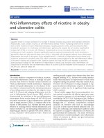
Allergic asthma is a chronic airway disorder characterized by airway
inflammation, mucus hypersecretion and airway hyperresponsiveness (Busse
and Lemanske, 2001) (Figure 1.1). It is attributable to the coordinated and
sustained activation of inflammatory cells including mast cells, T-helper 2 cells, B
cells, macrophages and eosinophils, and synthesis of a variety of pro-
inflammatory mediators (Maddox and Schwartz, 2002; Hamid et al., 2003). Acute
bronchoconstriction is triggered by the release of bronchoconstrictors including
histamine, cysteinyl-leukotrienes (CysLTs) and platelet-activating factor (PAF)
from mast cells upon allergen-induced cross-linking of IgE-bound high-affinity Fc
receptors (FcεRI) (Busse and Lemanske, 2001). Airway inflammatory responses
are contributed by T-helper type 2 cells (Th2 cells), together with other
inflammatory cells such as mast cells, B cells and eosinophils, and inflammatory
cytokines and chemokines (Busse and Lemanske, 2001; Herrick and Bottomly,
2003). Upon activation, Th2 cells produce cytokines such as IL-4, IL-5 and IL-13.
IL-4 is essential for B cell maturation and IgE synthesis, and plays an important
role in the initiation of Th2 inflammatory responses (Li-Weber and Krammer,
2003). IL-5 is pivotal for the growth, differentiation, recruitment and survival of
eosinophils (Greenfeder et al., 2001). IL-13 plays a prominent role in the effector
phase of Th2 responses, such as eosinophilic inflammation, mucus secretion,
and AHR (Wynn, 2003; Taube et al., 2002; Hershey 2003). On the other hand,
2
Leukotriene C
4
,
PAF
Histamine
Tryptase
IL-4, IL-5, etc
Late Responses
Airway inflammation
AHR
Mucus Hypersecretion
Edema
IL-3,IL-5,
GM-CSF,
RANTES
MBP, ECP
Leukotrienes, PAF
IL-4, IL-5, GM-CSF
MIP-1
α
, RANTES
TGF
α
,
PDGF
Eosinophils
APC
Antigen
IgE
Y
IL-4
IL-13
Th2
B
IL-5
Smooth Muscle Cells
Mucus
Epithelium
Early Responses
Acute bronchoconstriction
Edema
Y
Y
Mast Cell
Fc
ε
RI
Figure 1.1
Pathogenesis of asthma.
Endothelium
VLA-4
VCAM
eotaxin
3
chemokines such as RANTES and eotaxin are central to the delivery of
eosinophils to the airways. The specific transendothelial migration of eosinophils
is regulated by the interaction of adhesion molecules such as VLA-4 and its
ligand VCAM-1 (Lukacs, 2001). Airway eosinophilia together with effector
cytokines such as IL-13 may ultimately contribute to AHR in asthma (Wills-Karp,
1999).
1.1.1.1. Mast cells
Mast cells are derived from bone marrow and enter the circulation as CD34
+
mononuclear cells. They then migrate to mucosal and submucosal sites in the
airway, and undergo tissue-specific maturation which depends on the T cell-
derived IL-4 (Busse and Lemanske, 2001).
Inhaled allergen enters the body via airway mucosal surfaces, and is taken
up by antigen-presenting cells (APCs). These APCs then migrate to draining
lymph nodes, where they present the processed antigen to T and B cells.
Interactions among these cells elicit responses that are influenced by cytokines
and the presence or absence of costimulatory molecules. IL-4 and IL-13 provide
the first signal to B cells to switch to the production of the IgE isotype. The
second signal is delivered when CD40 on B cells binds to its ligand on T cells.
Once formed, IgE antibody circulates in the blood and eventually binds to high-
affinity IgE receptors (FcεRI) on mast cells thus sensitizing them (Busse and
Lemanske, 2001).
Crosslinking of FcεRI with IgE and antigen is the triggering event of the
activation of protein-tyrosine kinases (PTK), including those of the Src, Syk and
4
Tec families (Scharenberg and Kinet, 1994; Vangelista, 2003). Current
understanding of the activation sequence is that Lyn-a Src-family PTK that is
expressed predominantly in mast cells and is associated constitutively with the β-
subunit of FcεRI is activated by FcεRI aggregation and then phosphorylates
tyrosine residues in the immunoreceptor tyrosine – based activation motifs
(ITAMS) of the β- and γ-subunits of the receptor. Phosphorylated ITAMs of the β-
and γ-subunits recruit additional Lyn and Syk, respectively, through interactions
with the Src-homology 2 (SH2) domains encoded in the PTKs. Syk is then
activated through conformational change and tyrosine phosphorylation by Lyn.
Active Syk then phosphorylates many substrates downstream, including LAT
(linker for activation of T cells), SLP76 (SH2-domain-containing leukocyte protein
of 76 kDa) and VAV, which leads to the activation of several signalling pathways,
such as those through PI3K, phospholipase Cγ (PLCγ), and MAPK (Sanchez-
Mejorada and Rosales, 1998; Kinet, 1999). The activation of these pathways
leads eventually to mast cell degranulation, synthesis and release of lipid
mediators (e.g. CysLTs and PAF), and the production and secretion of cytokines,
chemokines and growth factors, which cause immediate bronchoconstriction,
mucosal edema and hypersecretion (Busse and Lemanske, 2001).
1.1.1.2. Eosinophilia
The eosinophil is the principal effector cell for the pathogenesis of allergic airway
inflammation via the secretion of inflammatory mediators such as leukotrienes
and granule products including reactive oxygen species and cytotoxic granule
and vesicular proteins: major basic protein (MBP), eosinophil cationic protein
5
(ECP), eosinophil peroxidase, and eosinophil-derived neurotoxin, as well as
cytokines and chemokines (Giembycz and Lindsay, 1999).
Eosinopoiesis begins in the bone marrow and is regulated by IL-3, IL-5 and
granulocyte-macrophage colony-stimulating factor (GM-CSF) (Giembycz and
Lindsay, 1999). IL-5 is critical for regulating the growth, activation, and survival of
eosinophils and cooperates with eotaxin to selectively regulate tissue
eosinophilia (Adachi and Alam, 1998; Choi et al., 2003). IL-5 not only induces
terminal differentiation of immature eosinophils (Yamaguchi et al., 1988a) but
also stimulates the release of eosinophils into the circulation and prolongs their
survival (Yamaguchi et al., 1988b; Palframan et al., 1998). Moreover, IL-5 has
been shown to play an important role in mediating eosinophil adhesion
(Sanmugalingham et al., 2000) and migration (Schweizer et al., 1996). IL-5
exerts its actions by binding to IL-5 receptor on the cell surface. The receptor for
IL-5 belongs to the hematopoietin receptor superfamily and is comprised of an IL-
5-specific α chain and the common β chain that is shared with IL-5, IL-3 and GM-
CSF for signal transduction (Adachi and Alam, 1998). To date, there are at least
3 principal signaling pathways that have been described upon IL-5 receptor
activation on eosinophils: the Janus kinase (JAK)/signal transducer and
activation of transcription (STAT) pathway, the MAPK pathways, and PI3K
pathway (Martinez-Moczygemba and Huston, 2003). IL-5 receptor binding leads
to activation of the receptor-associated JAK2 kinase (Quelle et al., 1994), STAT1
and STAT5 (Adachi and Alam, 1998) and Src family of kinases, such as Lyn
(Pazdrak et al., 1995; Yousefi et al., 1996), Syk (Yousefi et al., 1996), and Btk
6
(Sato et al., 1994). It has been demonstrated that Lyn, Syk and JAK2 are
important for eosinophil survival (Yousefi et al., 1996; Ishihara et al., 2001).
However, Lyn and JAK2 appear to have no role in eosinophil degranulation or
expression of surface adhesion molecules whereas Raf-1 kinase has been
shown to be critical for eosinophil degranulation and adhesion molecule
expression (Pazdrak et al., 1998). This is consistent with the studies showing the
involvement of Ras-Raf-1-MEK-MAP kinase pathway in the IL-5 induced
intracellular signal transduction (Pazdrak et al., 1995; Coffer et al., 1998) and
survival (Hall et al., 2001) in eosinophils. PI3K has been shown to be involved in
IL-5 stimulated eosinophil mobilization for the bone marrow (Palframan et al.,
1998)
Eosinophil transmigration into the airways is a multistep process that is
orchestrated by Th2 cytokines such as IL-4, IL-5 and IL-13, and coordinated by
specific chemokines such as eotaxin in combination with adhesion molecules
such as VCAM-1 and VLA-4 (Busse and Lemanske, 2001; Lukacs, 2001; Jia et
al., 1999). Cell rolling, which is mediated by P-selectin on the surface of
eosinophils is the first step in this process. Cell rolling activates eosinophils and
requires the participation of the β
1
and β
2
classes of integrins on the eosinophil
surface (Busse and Lemanske, 2001). Eosinophils express the α
4
β
1
integrin (also
known as very late antigen-4, VLA-4), which binds to its ligand, VCAM-1 on the
endothelium (Nagata et al., 1995; Matsumoto et al., 1997; Yamamoto et al.,
1998). Interactions between the β
2
integrins on eosinophils and intracellular
adhesion molecule 1 (ICAM-1) on vascular endothelium also appear to be
7
important for the transendothelial migration of eosinophils (Yamamoto et al.,
1998; Jia et al., 1999).
The chemokines such as RANTES, macrophage inflammatory protein 1α
(MIP-1α), and the eotaxins are central to the delivery of eosinophils to the airway
(Lukacs, 2001). Eotaxin was distinguished from all other chemokines because it
was found to be a potent eosinophil-selective chemoattractant and activator
(Elsner et al., 1996; Palframan et al., 1998; Rothenberg, 1999; Conroy and
Williams, 2001; Pease and Williams, 2001). Eotaxin was initially discovered to be
potent in stimulating eosinophils in vitro and in vivo in guinea pigs (Griffith-
Johnson et al., 1993; Jose et al., 1994). Subsequently, it has been cloned in
other species such as mice (Gonzalo et al., 1996), rats (Williams et al., 1998; Ishi
et al., 1998) and human beings (Ponath et al., 1996; Garcia-Zepeda et al., 1996;
Kitaura et al., 1996) and, meanwhile, two more functional homologues of eotaxin
have been termed eotaxin-2 (Forssmann et al., 1997; White et al., 1997) and
eotaxin-3 (Shinkai et al., 1999; Kitaura et al., 1999), although they lack sequence
similarity to eotaxin. Eotaxin has been shown to be synthesized by many cell
types in the lung, including airway epithelial cells, airway smooth muscle cells,
vascular endothelial cells and macrophages, as well as eosinophils themselves
(Humbles et al., 1997; Ying et al., 1997; Lamkhioued et al., 1997). In line with the
study which shows that eotaxin production is T-cell-dependent in a mouse
asthma model (Maclean et al., 1996), Th2 cytokines such as IL-4 and IL-13 have
been shown to induce eotaxin production. Sanz and co-workers showed that
intradermal IL-4 induced eosinophil accumulation in the rat was mediated partly
8
by endogenously generated eotaxin (Sanz et al., 1998). Similarly, eotaxin mRNA
expression in a lung granulomas model was inhibited by an anti-IL-4 antibody
(Ruth et al., 1998). On the other hand, IL-13 has been shown to be more potent
than IL-4 in inducing eotaxin expression by lung epithelial cells and promoting
lung eosinophilia in vivo (Li et al., 1999) as well as induces mucus
hypersecretion, subepithelial fibrosis and bronchial hyperreactivity (Zhu et al.,
1999). The eotaxins signal exclusively via a single receptor, CCR3, which
accounts for eotaxin’s cellular selectivity (Kitaura et al., 1996; Ponath et al., 1996;
Daugherty et al., 1996). CCR3 is a seven-transmembrane-spanning G-protein-
linked receptor (Sallusto et al., 2000; Fernandez and Lolis, 2002) primarily
expressed on eosinophils (Ponath et al., 1996), basophils (Uguccioni et al.,
1997), mast cells (Romagnani et al., 1999), and a subpopulation of Th2 cells
(Sallusto et al., 1997). The binding of eotaxin to CCR3 receptor induces a series
of biochemical changes (Mellado et al., 2001), including activation of Gi proteins,
transient calcium mobilization (Ponath et al., 1996; Kitaura et al., 1996;
Daugherty et al., 1996), MAPK activation (Alam et al., 1999; Boehme et al., 1999;
Kampen et al., 2000; Tachimoto et al., 2002), and actin polymerization (Boehme
et al., 1999) that is associated with chemotaxis and granule release.
1.1.1.3. T cells and Th2 cytokines
Cumulative evidence shows that T-helper type 2 cells (Th2 cells) are the main
orchestrators of allergic airway inflammation (Herrick and Bottomly, 2003; Larche
et al., 2003). T cells arise from bone marrow-derived progenitor cells that
undergo maturation in the thymus where they become thymocytes. After
9
presentation of a foreign antigen peptide by activated dendritic cells (DC),
thymocytes start to secrete IL-2, then undergo rapid proliferation and
differentiation. Thymocytes differentiate into phenotypically distinct types of T
cells based on the specificity of the T cell receptor (TCR) for antigen (Werlen et
al., 2003). Thymocytes expressing a TCR specific for major histocompatibility
complex (MHC) class I differentiate into CD8 cytotoxic T cells and thymocytes
expressing a TCR specific for MHC class II differentiate into CD4 helper T cells –
a process known as CD4/CD8 lineage commitment (Sezda et al., 1999; Hedrick,
2002; Kioussis, 2002). The duration of activation of both Ca
2+
/calmodulin-
dependent calcineurin and the ERK pathway appears to be crucial for CD4/CD8
lineage commitment (Adachi and Iwata, 2002). T helper cells (Th cells) further
differentiate into Th1, characterized by the secretion of IL-12 and interferon
(IFN)-γ, and Th2 cells, which were characterized by the secretion of IL-4, IL-5
and IL-13 (Rogge, 2002; Gor et al., 2003). Th cell differentiation can be driven in
vitro by stimulating unpolarized T cells with antigen or other TCR ligands in the
presence of appropriate cytokines (IL-12 for Th1 and IL-4 for Th2), suggesting
that it is the combination of TCR and cytokine stimulation act in synergy to induce
cellular differentiation (Ansel et al., 2003). Transcription factor T-bet and GATA-3
appear to be the key regulators of Th1 and Th2 differentiation, respectively
(Ansel et al., 2003).
T cell development and differentiation share a common requirement for
signals emanating from the TCR (Werlen et al., 2003). TCR complex consists of
ligand-binding αβ chains, signal-transducing CD3 molecule (γε dimer and δε
10
dimer) and ξ chain dimer. TCR activation results in tyrosine phosphorylation of
ITAMs located in the CD3 molecule by Lck and Fyn, two Src family kinases.
Phosphorylated ITAMs then recruit and activate ZAP-70, a member of the Syk
family kinase. Subsequently, these activated tyrosine kinases phosphorylate a
plethora of downstream signaling molecules such as PLCγ1 and adaptor
molecules such as linker for activation of T cells (LAT) and SLP-76, which then
activate downstream signaling pathways such as PI3K and MAPK pathways for
effector responses (Hussain et al., 2002; Nel, 2002; Samelon, 2002; Wong and
Leong, 2003).
Th2 cells mainly contribute to asthma pathophysiology by producing an
array of Th2 cytokines such as IL-4, IL-5 and IL-13 (Romagnani, 2001; Hedrick,
2002).
IL-4 plays an important role in the initiation of Th2 inflammatory responses
(Herrick and Bottomly, 2003). IL-4 induces B cell growth, differentiation and
secretion of immunoglobulin (Ig) E and IgG4 (IgG1 in the mouse). It has been
shown to be the most potent cytokine mediating IgE synthesis (Finkelman et al.,
1988; Pene et al., 1988). In addition, IL-4 has been shown to be able to induce
the rolling on and adhesion to endothelial cells of circulating eosinophils
(Bochner and Schleimer, 1994). IL-4 blocking antibodies inhibit allergen-induced
AHR, goblet cell metaplasia, and pulmonary eosinophilia in a murine model of
asthma (Gavett et al., 1997). Th2 cells are the main sources of IL-4 production,
although various other cells including basophils, mast cells as well as eosinophils
also produce IL-4 (Dubucquoi et al., 1994; Seder, 1994). IL-4 exerts its effect
11
through its IL-4R complex which consists of the IL-4Rα and the common gamma
chain (γc). IL-4Rα binds to IL-4 with high affinity and heterodimerizes with a
second chain (γc or IL-13Rα1) to produce biological effects. γc Chain only
modestly increases the affinity of the IL-4R complex for IL-4, yet it is required for
the activation of IL-4R (Nelms, 1999). IL-4Rα chain also functions as a
component of the IL-13 receptor (IL-13R), which may explain the overlap effects
between these two Th2 cytokines (Obiri et al., 1995; Miloux et al., 1997; Murata
et al., 1998). Neither the IL-4Rα nor the γc chain has endogenous kinase activity;
therefore, like other members of the hematopoietin receptor family, IL-4R
requires receptor-associated kinases for the initiation of signal transduction
(Nelms, 1999). The IL-4Rα chain cytoplasmic region has three functionally
distinct domains: (a) an interaction domain for JAK: IL-4Rα chain is usually
associated with JAK1 while γc chain is associated with JAK3 (Miyazaki et al.,
1994; Russell et al., 1994); (b) a domain containing conserved Tyr residue for
activation of proliferation pathways (Deutsch et al., 1995); and (c) the domain
comprises sequences from C-terminal to residue 557 which is critical for
transducing signals leading to activation of IL-4-induced gene expression (Ryan
et al., 1996). The engagement of IL-4R activates signaling pathways to elicit IL-4-
induced diverse biological effects. PI3K and MAPK pathways have been
observed to be involved in IL-4-induced cellular proliferation (Hershey, 2003).
IRS-1/2 (insulin receptor substrate-1/2) signaling pathway has been observed to
be upstream of these two pathways in IL-4-induced cellular proliferation (Wang et
al., 1993; Sun et al., 1995). Inhibiting PI3K by Wortmannin blocked the ability of
12
IL-4 to prevent apoptosis in haematopoietic cells through the production of
phosphoinositides and the subsequent activation of kinases critical for cell
survival (Zamorano et al., 1996). Nevertheless, MAPK pathway activation by IL-4
may depend on cell type since IL-4 induced MAPK activation has only been
shown in certain cell types (Wery et al., 1996) while not in others (Welham et al.,
1994). STAT-6 is the primary STAT activated in response to IL-4 stimulation and
acts as a direct connection between IL-4 receptor and the transcription apparatus
(Jiang et al., 2000). Upon the IL-4R engagement, JAK1 and JAK3 are activated
and specific tyrosine residues in the receptor cytoplasmic region are
phosphorylated. STAT-6 is then recruited to the phosphorylated receptor through
its SH2 domain, enabling the activated kinases to phosphorylate STAT-6 at a C-
terminal tyrosine residue (Mikita et al., 1996). Once phosphorylated, the STAT-6
disengages from the receptor and forms homodimers. The dimerized STAT-6
complex is translocated to the nucleus where they bind to specific DNA motifs in
the promoter of responsive genes (Ihle, 1996; Nelms, 1999).
IL-5 plays an essential role in eosinophil growth, differentiation, activation
and survival. The functional role of IL-5 in allergic asthma has been described in
details in section 1.1.1.2. Please refer to that section for IL-5 functions.
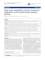
IL-13 plays a prominent role in the effector phase of Th2 responses, such as
eosinophilic inflammation, mucus secretion, and AHR by activating a wide variety
of cell types that are relevant to the pathogenesis of asthma (Grunig et al., 1998;
Taube et al., 2002; Wynn, 2003), as shown in Table 1. These alterations are the
result of IL-13 binding to the multimeric IL-13 receptor (which is made up of IL-
13
Table 1. Actions of IL-13 on haematopoietic and nonhematopoietic cells
Cells
Function Reference(s)
Human B cells promoting B-cell proliferation
inducing class switching to IgG4
and IgE
inducing expression of the FcεRII
and MHC class II
Oettgen et al., 2001
Chomarat and
Banchereau, 1998
Monocytes
and
macrophages
enhancing the expression of
integrins, including CD11b,
CD11c, CD18, and CD29
inducing MHC class II and
CD23 expression
Inhibiting the production of
prostaglandins,
reactive oxygen,
nitrogen intermediates, and
IL-1, IL-6, IL-8, TNF-α, and IL-
12
Zurawski and de
Vries, 1994
De Vries, 1998
Endo et al., 1996
Doherty et al., 1993
Sozzani et al., 1995
De Vries, 1998
Eosinophils promoting eosinophil survival,
activation, and
recruitment
Horie et al., 1997
Luttmann et al., 1996
Pope et al., 2001
Mast cells activating mast cells
promoting IgE synthesis
De Vries, 1998
Endothelial
cells
inducing VCAM-1 expression Bochner et al., 1995
Airway smooth
muscle cells
enhancing proliferation
increasing cholinergic-induced
contraction
Wills-Karp, 2001
Fibroblasts inducing type I collagen
synthesis
Roux et al., 1994
Epithelial cells potently inducing eotaxin
expression
altering mucocilliary
differentiation
resulting in goblet cell
metaplasia
Li et al., 1999
Laoukili et al., 2001
Zhu et al., 1999
14
4Rα, IL-13Rα
1
and IL-13Rα
2
). IL-13 has two cognate receptors, IL-13Rα1
and IL-13Rα2 (Hershey, 2003). IL-13Rα1 binds to IL-13 with low affinity by itself,
while binds to IL-13 with high affinity when paired with IL-4Rα and forms a
functional IL-13 receptor (Miloux et al., 1997; Wynn, 2003). This receptor
complex is also utilized by IL-4, serving as an alternative receptor to IL-4
(Hershey, 2003). In vitro studies show that IL-13Rα
2
might be a decoy receptor,
which downregulates IL-13 signaling (Donaldson et al., 1998; Kawakami et al.,
2001; Daines et al., 2003). Since IL-4 and IL-13 share common subunits of
receptors, they share signaling pathways accordingly (Welham et al., 1995). The
binding of IL-13 to IL-13R complex induces the activation of JAK1 and Tyk2.
Activated JAKs phosphorylate the cytoplasmic tyrosines in IL-4Rα, which then
recruit STAT6 to the receptor, followed by STAT6 phosphorylation and activation
(for details of STAT6 activation please refer to IL-4 signaling pathways).
1.1.1.4. B cells and immunoglobulins
B cells mainly contribute to pathogenesis of asthma by producing
immunoglobulins, and IgE has been associated with mast cell activation and
AHR in humans (Kalesnikoff et al., 2001; Oettgen and Geha, 2001).
B cell development occurs through several discrete stages and at many
anatomical locations including bone marrow, fetal liver, peritoneum, and spleen
(Hardy and Hayakawa, 2001). B-cell receptor (BCR) instructs B cells
development and mediates the response to the antigen (Niiro and Clark, 2002).
The BCR complex is made up of antigen-binding component membrane
immunologlobulin (mIg), associated with two signaling transduction components,
15
Igα (CD79a) and Igβ(CD79b) (Gauld et al., 2002). After BCR ligation by antigen,
Src-family kinase Lyn is activated. Lyn then phosphorylates ITAMs in the
cytoplasmic tails of Igα and Igβ, which recruit and activate of Syk and the Tec-
family kinase Btk (Niiro and Clark, 2002). Some non-enzymatic adaptors, such as
B-cell linker (BLNK) (Fu et al., 1998), B-cell adaptor for PI3K (BCAP) (Okada et
al., 2000), and B-lymphocyte adaptor molecule of 32 kDa (BAM32) (Niiro et al.,
2002), fine-tune BCR signals by efficiently connecting the kinases with the
effectors. Phospholipase Cγ2 (PLCγ2) and PI3K are two important downstream
effectors of BCR signaling (Marshall et al., 2000; Niiro and Clark, 2002). Btk,
together with Syk, phosphorylates and activates PLCγ2. Activation of PLCγ2
leads to the release of intracellular Ca
2+
and activation of PKC, which
subsequently induce the activation of MAPKs, and transcription factors, including
NFκB and nuclear factor of activated T cells (NFAT) (Niiro and Clark, 2002). PI3K
phosphorylates phosphatidylinositol-4,5-bisphosphate (PtdInsP
2
) to produce
phosphatidylinositol-3,4,5-triphosphate (PtdIsnP
3
), which recruits some BCR
signalling molecules to the membrane through pleckstrin homology (PH) domains
and activates downstream kinases such as Akt (Okkenhaug and
Vanhaesebroeck, 2003). The Vav family of Rho-family GTPase, is also critical for
BCR signalling. Vav activates RAC1 and regulates cytoskeletal structures and
BCR-induced proliferation. Vav might function both upstream and downstream of
PI3K in B cells (Gauld et al., 2002; Niiro and Clark, 2002).
16
1.1.1.5. Airway mucus hypersecretion and goblet cell hyperplasia
Airway mucus hypersecretion is a prominent feature of asthma. Excessive
production of mucus causes plugging in the airways that might lead to airway
obstruction (Lundgren and Shelhamer, 1990). Mucus hypersecretion is a
complex pathologic process that involves goblet cell hyperplasia and
degranulation, microvascular remodeling and leakage, and chemoattraction of
inflammatory cells (Fahy, 2002). The major constituents of airway mucus are
termed as mucins. Mucins are large complex molecules consisting of a peptide
backbone and numerous oligosaccharide side chains which represent the
products of mucin genes (MUC genes) and glycosyltransferase genes,
respectively (Fahy, 2002). At least 12 human MUC have been identified, 7 of
which are expressed in human airways (Fahy, 2002). Nevertheless, only
MUC5AC and MUC5B have been convincingly shown to be the major mucins
secreted in the airway (Chen et al., 2001; Fahy, 2002).
A variety of inflammatory mediators, including histamine, leukotrienes, and
PAF have been shown to stimulate mucus secretion (Lundgren and Shelhamer,
1990; Nadel, 1991; Cohn et al., 1999). CysLTs has been shown to be important
for both early-phase (1 hour after the allergen challenge) and late-phase (6 hour
after allergen challenge) in a rat asthma model (Shimizu et al., 2003). Histamine
is mainly involved in the early-phase mucus secretion through the H
1
-receptor of
cholinergic nerve terminals, whereas infiltrating cells (eosinophils and
neutrophils) play a more critical role in late-phase mucus secretion (Shimizu et
al., 2003). On the other hand, Th2 cytokines, such as IL-4 (Temann et al., 1997),
17
IL-9 (Temann et al., 1998) and IL-13 (Zhu et al., 1999), have been shown to
influence mucus secretion. In a mouse asthma model, it has been shown that IL-
4 Rα is essential for Th2-induced airway mucus production while IL-5,
eosinophils, and mast cells are not critical for the mucus production (Cohn et al.,
1999). Epidermal growth factor receptor (EGFR) and its ligand have also been
found to have a role in mucus production in asthma. Selective inhibitors of EGFR
tyrosine kinase block mucus production both in vivo and in vitro (Takeyama et al.,
1999). EGFR inhibitors also blocked IL-13-induced MUC gene expression in rat
airways and epithelial cell proliferation in cultured bronchial epithelial cells (Booth
et al., 2001; Shim et al., 2001). The mechanism by which inflammatory stimuli
induce mucus hypersecretion in the airways is still uncertain. The finding that
activation of NF-κB via a c-Src-Ras-MEK1/2-MAPK-pp90rsk signaling pathway
binding to a κB site in the 5’-flanking region of the MUC2 gene and activating
MUC2 mucin transcription may represent one of the potential mechanisms (Li et
al., 1998).
1.1.1.6. Airway hyperresponsiveness (AHR)
AHR is a characteristic feature of asthma and defined as an increased sensitivity
of the airways to an inhaled constrictor agonist shown as a steeper slope of the
dose-response curve (Vargaftig, 1997; O’Byrne and Inman, 2003).
Activated by inhaled antigen presentation, CD4
+
T cells in the lungs produce
Th2 cytokines, such as IL-4, IL-5, and IL-13, which orchestrate the infiltration and
activation of effector cells such as mast cells and eosinophils. Subsequently,
18
such effector cells release a plethora of inflammatory mediators including
histamine, LTs, PAF, eosinophils-derived basic proteins, and proteases to the
airway epithelium. These inflammatory mediators, individually or in combination
induce acute bronchoconstriction, airway wall edema, airway epithelial
desquamation, altered neural regulation of airway tone, increased mucus
production, and increased smooth muscle content. Each of these inflammatory
responses might contribute to AHR, although most likely they influence in concert
(Wills-Karp, 1999).
Experimental animal models have provided direct evidence of a causal role
for CD4
+
T cells in the development of antigen-induced AHR. Depletion of CD4
+
T cells in sensitized mice prior to local lung antigen challenge with specific
monoclonal antibodies prevented the development of allergen-induced allergic
airway responses (Gavett et al., 1994). Furthermore, it has been shown that
adoptive transfer of Th2 clones into lungs results in AHR in naïve mouse (Li et
al., 1996).
On the other hand, study from IL-13-deficient mice has confirmed the
importance of the IL-13, IL-4Rα, and STAT6 in the induction of AHR and suggest
that IL-13 was, by itself, necessary and sufficient to induce AHR (Walter et al.,
2001). Nevertheless, there is study using IL-13
-/-
mice showing that it is possible
to develop AHR and pulmonary eosinophilia in the absence of IL-13. AHR was
reduced in IL-13
-/-
mice only when they were treated with either anti-IL-4 or anti-
IL-5 mAbs, which suggests the cooperation among Th2 cytokines in inducing
AHR (Webb, 2000).
19
Increased production of IgE has been associated with the development
AHR but depends on sensitization and challenge protocols. Under conditions in
which limited IL-5-mediated eosinophilic airway infiltration is induced, IgE plays
an important role in AHR development whereas in conditions where a robust
eosinophilic inflammation of the airways is elicited, IgE does not appear to be
essential for the development of AHR (Hamelmann et al., 1999).
Considerable evidence suggests the association between pulmonary
eosinophil infiltration and AHR in asthma (Wills-Karp, 1999). Eosinophils are
postulated to induce AHR through releasing eosinophil-derived proteins such as
MBP and ECP on the airway wall. These proteins are cytotoxic to the airway
epithelium. Damage of the airway epithelium may lead to AHR by removing
enzymes important in the degradation of neuropeptides and/or in the loss of
epithelial-derived relaxing factor (Gundel, 1991). MBP may also induce AHR
through its competitively inhibitory binding of M2 receptors to acetylcholine
autoreceptors on parasympathetic nerves that may result in increased release of
acetylcholine (Jacoby, 1993).
1.1.2. Therapeutic targets of asthma
Currently available therapy for asthma, which generally based on use of inhaled
β
2
-agonist bronchodilators together with inhaled corticosteroids, is able to control
the majority of patients. However, important advances are still needed to improve
long-term therapy for patients with more severe persistent asthma. In the future,
there is the prospect of cheaper and safer therapies causing disease modification
and even cure (Corry, 2002).
20
1.1.2.1. Current therapy for asthma
β
2
-Agonist bronchodilators (e.g. salbutamol (short-acting); salmeterol and
formoterol (long-acting)) are by far the most effective palliative therapies for
asthma because they relieve suffering. β
2
-Agonists work as functional
antagonists on airway smooth muscle. However, they have no effect on chronic
inflammation and, therefore they cannot cure the disease. In addition, there is no
convincing evidence that a bronchodilator can impede disease progression in
asthma (Anderson and Rabe, 2001).
Inhaled corticosteroids (CS) are the most effective drugs available to
clinicians for the treatment of asthma (Barnes et al., 1998). CS improves lung
function, reduces airway inflammation, AHR, and asthma attacks or
exacerbations. CS is able to penetrate the cell membrane passively and bind to
its intracellular CS receptor to form a complex. This binding results in dissociation
of heat shock proteins from the receptor and exposure of nuclear localization
sequence, allowing the complex to penetrate into the nucleus and bind to specific
regions on DNA-glucocorticoid responses elements (GRE) and/or negative-
glucocorticoid response elements (nGRE). Thus, GRE-bound GR homodimers
facilitate the corresponding gene mRNA production, the mechanism called
transactivation, while nGRE-bound GR inhibite the gene mRNA production, the
mechanism called transrepression. However, the use of CS has been associated
with dose- and time-dependent side effects. Inhaled CS can give rise to oral
candidiasis and dysphonia. In severe asthmatics, CS are given systemically
which may lead to side effects including hypertension, psychological disorders
21
such as insomnia and agitation, increased susceptibility to infection, easy skin
bruising and slow wound healing, weight gain, osteoporosis, blood sugar
elevation which leads to or worsen diabetes, increased incidence of cataract,
muscular weakness, growth retardation in children, etc.
In the future, new steroids with a more selective anti-inflammatory profile
without possessing adverse effects are expected. Currently, prodrug steroids,
soft steroids and dissociated steroids all have exciting potential to achieve this
aim (Dahl and Nielsen, 2001).
The leukotrienes (LTs) are eicosanoids derived from membrane constituent
arachidonic acid. The cysteinyl LTs, LTC
4
, LTD
4
and LTE
4
are potent airway
smooth muscle constrictors with a much longer duration of action than other
smooth muscle constrictors. LTB
4
has minimal bronchoconstrictor effects, but is a
potent neutrophil chemoattractant. The cysteinyl LTs transduce their activity
through the CysLT
1
receptor, while LTB
4
does so through the BLT receptor. The
CysLT antagonists (e.g. montelukast, pranlukast, and zafirludkast, etc.) and LTB
4
antagonists (e.g. LY293111, CGS-25019C, and SB209247, etc.) have now been
extensively evaluated in clinical trials (O’Byrne and Drazen, 2001).
1.1.2.2. Novel therapeutic targets for asthma
Improved understanding on the cellular and molecular basis of asthma has
identified more potential candidates for drug development. Specifically, together
with recent discoveries on Th2 cell function and associated signaling pathways, it
is now possible to investigate the potential therapeutic role of molecules that
22
control Th2 response signaling pathways in the lung (Handsel and Barnes, 2001;
Corry, 2002).
Mediator inhibitors such as H
1
-antihistamines are still of interest in addition
to corticosteroids, β
2
-agonists, and leukotriene antagonists (De Vos and Rihoux,
2001). Second-generation H
1
-antihistamines have almost eliminated the central
side effect-sedation compared with the first-generation, yet it brings in new safety
issues such as cardiac toxicity and interference with hepatic enzymatic complex
cytochromes P-450 (Hamelin et al., 1998; Nicolas et al., 1999). Therefore, new
generation of H
1
-antihistamines with more effectiveness and specificity are
wanted.
Protease inhibitors, such as tryptase inhibitors, have been shown to reduce
antigen-induced airway inflammation and AHR in guinea-pig (Wright et al., 1999),
sheep (Clark et al., 1995; Wright et al., 1999), and mouse (Oh et al., 2002)
asthma models. In phase II clinical trials, APC 366, a first-generation small
molecule inhibitor of tryptase, has demonstrated efficacy in patients with mild to
moderate asthma (Rice et al., 1998). Therefore, continued development of more
specific and selective tryptase inhibitors are expected as novel treatment of
asthma.
The anti-IgE therapy, which is based on the important role of IgE plays in
human asthma, has been rationalized for years (Chang, 2000). To date, a
humanized murine anti-IgE antibody, rhuMAb-E25, has been proven safe and
effective in reducing serum free IgE levels, the number of IgE receptors
expressed on the surface of basophils, the clinical severity of asthma, the
23
frequency of exacerbations, and the corticosteroids requirements of patients with
moderate or severe asthma, although it does not cure asthma (Chang, 2000).
The anti-IgE works by binding to Fc portion of IgE to form immune complexes
which no longer can bind to FcεRI. Since IgE is continuously synthesized,
repeated anti-IgE dosing appears to be necessary to maintain the effect. So far
the long –term effects of anti-IgE are uncertain (Chang, 2000).
Inhibition of cytokines (e.g. IL-4, IL-5, and IL-13) is a promising way of
obtaining efficacious drugs for asthma. There are several potential ways of
inhibiting cytokine effects including blocking antibodies, small-molecule receptor
antagonists, soluble receptors, altering the balance of certain cytokines, and
antisense oligonucleotides (Barnes, 2002). Soluble IL-4 receptors (sIL-4r) are
now in clinical development as a strategy to inhibit IL-4. It has been shown that a
single nebulized dose of sIL-4r is able to prevent the fall in lung function induced
by withdrawal of inhaled corticosteroids in patients with moderately severe
asthma (Borish et al., 1999). Moreover, weekly nebulization of sIL-4r has been
demonstrated to improve asthma control over a 12-week period (Borish et al.,
2001).
Small molecule antagonists of leukocyte chemokine receptors have been
developed to inhibit the recruitment of inflammatory cells in allergic inflammation
(Lukacs, 2001; Bryan et al., 2001). CCR3 is predominantly expressed on
eosinophils. CCR3 antagonist has been reported to have an anti-inflammatory
effect in a mouse asthma model (Bryan et al., 2001).
24
Phosphodiesterase 4 (PDE4) inhibitors produce a wide range of
pharmacological actions through increasing cAMP content in immune and
inflammatory cells, airway smooth muscle and pulmonary nerves. These
beneficial effects include anti-inflammatory effects, bronchodilation, and
modulation of pulmonary nerves (Teixeira et al., 1997). With the knowledge that
PDEs are superfamily of genetically distinct enzymes (Soderling and Beavo,
2000), a new generation of isozyme-selective inhibitors has been developed
(Torphy et al., 2001). Indeed, initial clinical data on these agents are encouraging
and suggest that PDE4 inhibitors may have broad utility in the treatment of
pulmonary disease. However, full knowledge of the therapeutic value of this
novel compound class awaits the outcome of long-term clinical trials (Torphy et
al., 2001).
1.2 Tyrosine kinase signaling cascade
1.2.1. Protein tyrosine kinases (PTKs)
Protein tyrosine kinases are enzymes that carry out tyrosine phosphorylation by
catalyzing the transfer of the γ phosphate of ATP (or GTP) to tyrosine residues
on protein substrates. Phosphorylation of tyrosine residues modulates enzymatic
activity of the kinases and their substrates, and creates binding sites for the
recruitment of further downstream signaling proteins (Hubbard and Till, 2000).
Tyrosine kinases can be broadly divided into receptor tyrosine kinase (RTK)
and non-receptor tyrosine kinase (NRTK). RTK is a single transmembrane
glycoprotein. The binding of RTK to its cognate ligand activates receptor and
25