Reactive oxygen species mediated regulation of the na+ h+ exchanger, NHE 1 gene expression a new mechanism for tumor cells resistance to apoptotic cell death
Bạn đang xem bản rút gọn của tài liệu. Xem và tải ngay bản đầy đủ của tài liệu tại đây (1.43 MB, 155 trang )
REACTIVE OXYGEN SPECIES-MEDIATED REGULATION
OF THE Na+/H+ EXCHANGER, NHE-1 GENE EXPRESSION:
A NEW MECHANISM FOR TUMOR CELLS’ RESISTANCE
TO APOPTOTIC CELL DEATH
SUFYAN AKRAM
(MBBS)
A THESIS SUBMITTED FOR THE DEGREE OF
DOCTOR OF PHYLOSOPHY
DEPARTMENT OF PHYSIOLOGY
NATIONAL UNIVERSITY OF SINGAPORE
2005
ACKNOWLEDGEMENTS
I wish to acknowledge my deepest gratitude and appreciation to my supervisor, Dr.
Marie-Véronique Clément, Associate Professor at Department of Biochemistry,
National University of Singapore. Without her continuous encouragement and advice
this dissertation would have been far short of what it is today. No less I would like to
thank Associate Professor Shazib Pervaiz for the excellent cooperation, interaction
and guidance throughout the course of this work.
I am very grateful to all my colleagues, who have helped me in one way or another
during my stay in the lab.
My appreciation also goes to my parents for their persistent support and love
throughout my life. Special thanks belong to my wife and two sons. The knowledge of
them being there has been of great encouragement and importance for me. Without
their support, this work could never have been accomplished.
i
TABLE OF CONTENTS
Acknowledgments
i
Table of contents
ii
Abstract
vi
List of figures
vii
List of abbreviations
x
Chapter I. Introduction
1
I.1.
Cell Death
1
I.1.a. Types of Cell Death
2
I.1.a. Programmed cell death or apoptosis
2
I.1.b. Apoptotic machinery
4
I.2.
7
Reactive oxygen species and apoptosis
I.2.a. Pro and Anti-Apoptotic functions of ROS
7
I.2.b. Rac1 and superoxide anion production
11
I.2.c. Superoxide anion and inhibition of apoptosis
13
I.3.
14
Intracellular milieu
I.3.a. Intracellular pH (pHi)
16
I.3.b. pHi and apoptosis
17
I.4.
18
Intracellular pH regulation
I.4.a. Plasma membrane pHi regulators
I.5.
19
Na+-H+ Exchanger (NHE)
21
I.5.a. NHE1: Basic structure
22
I.5.b. NHE1: Major Functions
24
I.5.b.i. pHi and cell volume regulation
ii
24
I.5.b.ii. Cell proliferation and differentiation
24
I.5.b.iii. Cell motility
26
I.5.b.iv. As a plasma membrane scaffold
26
I.5.b.v. Cell injury
26
I.5.c. NHE1: Regulation
27
I.5.d. NHE1: Regulation of gene expression
28
I.5.e. NHE1: Pathological Functions
31
I.5.e.i. NHE1: Role in myocardium
31
I.5.e.ii. NHE1: Role in tumorigenesis
32
I.6.
Rac subfamily and NHE1
34
I.7.
Conclusion
35
Chapter II. Materials and methods
37
II.1.
Chemicals
37
II.2.
Cells
37
II.3.
cDNA Plasmids and transfections
40
II.4.
β-Galactosidase Survival Assay
41
II.4a. Measurement of β-Galactosidase activity
41
II.5.
siRNA transfection
42
II.6.
Luciferase reporter gene assay
43
II.7.
CAT (Chloramphenicol acetyltransferase) ELISA
43
II.8.
Measurement of steady state pHi
44
II.9.
Measurement of acid load and pHi recovery (NHE-1 activity)
45
II.10. Western blot analysis of NHE-1 protein
45
II.11. Measurement of intracellular superoxide
46
iii
II.12. Crystal Violet Assay
46
II.13. Caspase assay
47
II.14. Statistical Analysis
47
Chapter III. Results
48
III.1.a Regulation of NHE-1 gene expression regulates cells’ response
48
to death triggers in NIH3T3 cells
III.1.b. Regulation of NHE-1 gene expression regulates cells’ response
52
to death triggers in Tumor cells
III.2.a Superoxide mediated cell survival is NHE-1-dependant
55
III.2.b Role of superoxide in NHE1-dependent cell survival in tumor cells
59
III.3.
NHE-1 gene expression is growth factor regulated
64
III.4.
Intracellular superoxide activates NHE-1 promoter activity
69
III.5.a Small GTPase Rac1-mediated survival is dependent upon NHE-1
71
protein expression
III.5.b Rac1-mediated NHE-1 protein expression is a function of its
74
ability to produce superoxide
III.5.c Serum-induced NHE-1 expression might involve activation of Rac1
79
III.6.
H2O2 inhibits NHE-1 promoter activity
85
III.7.
H2O2 leads to decreased NHE-1 expression and increased
87
susceptibility to cell death
III.8.
Regulation of intracellular pH as one of the mechanisms of
89
NHE-1-mediated cell survival
III.9.
Region of NHE-1 promoter involved in O2.--mediated activation
iv
94
Chapter IV. Discussion and conclusions
102
IV.1. How tumors develop
102
IV.2. Permissive intracellular milieu
102
IV.3. Alkaline pHi and role of NHE-1
103
IV.4. Regulation of NHE-1
105
IV.5. Rationale of our study
105
IV.6. Regulation of NHE-1 gene expression regulates cells’ response
106
to death triggers
IV.7. Superoxide mediated cell survival is NHE-1-dependant
107
IV.8. NHE-1 gene expression is growth factor regulated
108
IV.9. Intracellular superoxide activates NHE-1 promoter activity
109
IV.10. Small GTPase Rac1-mediated survival is dependent upon
110
NHE-1 protein expression
IV.11. H2O2 inhibits NHE-1 promoter activity and leads to increased
112
susceptibility to cell death
IV.12. Regulation of intracellular pH as one of the mechanisms of
113
NHE-1-mediated cell survival
IV.13. Region of NHE-1 promoter involved in O2.--mediated activation
115
IV.14. Conclusions
116
IV.15. Prospective Studies
119
References
120
Publication and presentations
143
v
Abstract
Reactive Oxygen Species have long been known to cause cellular stress and damage.
But recently, ROS have been implicated as signaling molecules. Tumor cells display
an altered redox status. Our lab has recently shown that expression of a constitutively
active form of Rac1 (RacV12) inhibits tumor cell death by apoptosis through
intracellular production of superoxide anion (O2-) (Pervaiz et al. 2001, Oncogene).
Another characteristic of transformed cells is a shift towards alkaline intracellular pH.
NHE-1, one of the major pHi regulators, has been shown to be of particular
importance in tumor cells. Current study was designed to study the effect of ROS on
NHE1 regulation and the role it may play in modulating apoptosis. Our data shows
that production of intracellular O2•- induces transcription of NHE-1 while increase in
H2O2 inhibited it. Using Rac mutants, which have differential ability to produce O2•in the cell, and drugs that affect the intracellular ROS levels, we were able to show
that NHE1 gene is redox-responsive. Changes in NHE1 gene expression were
translated into NHE1 protein expression. By over-expressing or silencing NHE-1
gene we show that cell response to apoptotic triggers such as staurosporin and
etoposide correlates with the amount of NHE-1 protein expression on the cell surface.
Moreover, down-regulation of NHE-1 gene expression in tumor cell lines tested
reverted their resistant phenotype. These results support a critical role for NHE-1
expression in tumor cells’ response to anticancer therapy and provide a possible
mechanism for Rac1-mediated survival in tumor cells.
Pervaiz S., Cao J., Chao OSP, Chin YY. Clément M-V. Activation of the RacGTPase inhibits apoptosis
in human tumor cells. Oncogene 20: 6263-6268, 2001.
vi
List of Figures
Fig A.
Major differences between Apoptotic and Necrotic
3
types of Cell Death
Fig B.
Death-Receptor mediated Apoptosis
6
Fig C.
Mitochondial pathway of Apoptosis
6
Fig D.
Wide array of functions attributed to Reactive Oxygen Species
9
Fig E.
A model of ROS-mediated regulation of Apoptosis
15
Fig F.
Various plasma membrane-bound pHi regulators
20
Fig G.
Structure of mammalian NHE-1
23
Fig H.
Physiological functions of NHE-1
25
Fig I.
Basic structure of proximal part of NHE1 promoter
30
Table 1.
Various cell lines used in this project
38
Fig 1.
Increased NHE-1 expression leads to inhibition of
staurosporine-induced cell death in NIH3T3 cells.
49
Fig 2.
Silencing of NHE-1 gene leads to increased susceptibility
to cell death in NIH3T3 cells.
51
Fig 3.
Level of NHE-1 expression correlates with NIH3T3 cells’
sensitivity to staurosporine-induced cell death.
51
Fig 4.
Silencing of NHE-1 gene leads to increased susceptibility to
54
cell death in U87 cells.
Fig 5.
Time-dependent Caspase 3 (DEVDase) activity in NHE-1
silenced U87 cells treated with etoposide.
54
Fig 6.
Silencing of NHE-1 gene leads to increased susceptibility to
cell death in LNCaP cells.
56
Fig 7.
Caspase 3 (DEVDase) activity in NHE-1 silenced LNCaP
cells upon treatment with etoposide and staurosporine.
56
Fig 8.
Manipulation of NHE-1 expression does not affect intracellular
58
vii
superoxide levels.
Fig 9.
DDC leads to significant increase in intracellular superoxide
levels in NIH3T3 cells.
58
Fig 10.
DDC-mediated inhibition of cell death is dependent on NHE-1
gene expression.
60
Fig 11.
Inhibition of intracellular superoxide production prevents
NHE-1 protein expression in U87 cells.
63
Fig 12.
Inhibition of intracellular superoxide production prevents
NHE-1 expression in U87 cells and increases their susceptibility
to etoposide-induced cell death.
65
Fig 13A.
NIH3T3 1A8 cells stably transfected with proximal 1.1 kb
fragment of NHE-1 promoter/enhancer upstream of
a luciferase gene.
67
Fig 13B.
Basic Principle of Luciferase Reporter Assay.
67
Fig 14.
NHE-1 gene expression is growth factor regulated.
68
Fig 15.
Serum-induced activation of NHE-1 is dependant upon
intracellular production of superoxide.
70
Fig 16.
Superoxide levels in NIH3T3 1A8 cells in response to
different drugs.
72
Fig 17.
Superoxide is a signal for NHE-1 promoter activity.
73
Fig 18.
Expression of RacV12 induces NHE-1 promoter activity in
a variety of cells.
75
Fig 19.
Rac loss-of-function mutants.
77
Fig 20.
NADPH oxidase interaction domain of Rac1 is required for
Rac1-induced NHE-1 promoter activity.
78
Fig 21.
Rac1-mediated cell survival is dependent on its ability
to produce superoxide.
80
Fig 22.
Expression of RacN17 inhibits serum-induced NHE-1
promoter activity.
81
Fig 23.
Manipulation of NHE-1 protein expression in M14pIRES
and M14pIRES-RacV12 cells by NHE-1 siRNA transfection
83
Fig 24.
Rac-induced cell survival is dependant upon NHE-1 expression.
84
viii
Fig 25.
H2O2 inhibits NHE-1 promoter activity in NIH3T3 cells.
86
Fig 26.
H2O2 treatment of NIH3T3 cells results in increased
susceptibility to etoposide-induced killing.
88
Fig 27.
Increased NHE1 protein expression level leads to an increase
in pHi.
90
Fig 28.
Silencing of NHE1 gene results in a drop in intracellular
pH in NIH3T3 cells.
90
Fig 29.
Increase in intracellular pH correlates with the ability of
Rac mutants to produce superoxide and induce NHE1
transcription.
92
Fig 30.
Silencing of NHE1 gene results in a drop in intracellular
pH in M14pIRES and M14RacV12 cells.
92
Fig 31A.
Silencing of NHE1 gene results in a drop in intracellular
pH in U87 cells.
93
Fig 31B.
Decreased expression of NHE1 leads to decreased activity
of the pump in U87 cells.
93
Fig 32.
Increased intracellular pH in RacV12 over-expressing
NIH3T3 cells is dependent upon NHE1 activity.
95
Fig 33.
Increased intracellular pH in M14 cells is a function of
NHE1 activity.
95
Fig 34.
NHE-1 promoter/enhancer constructs.
96
Fig 35.
Low dose of paraquat leads to increased superoxide
production in L6 cells.
98
Fig 36.
Superoxide-mediated NHE1 gene transcription in L6 cells.
99
Fig 37.
Rac1-induced transcription of NHE-1 is not seen below
0.5 kb in L6 cells.
101
Fig 38.
Summary figure.
117
ix
List of Abbreviations
BCECF
2’, 7’-bis (2-carboxyethyl)-5, 6-carboxyfluorescein
DDC
Diethyldithiocarbamate
DMSO
Dimethylsulfoxide
DPI
Diphenylene iodonium
EGF
Epidermal growth factor
Eto
Etoposide
H2O2
Hydrogen peroxide
NOX
NADPH oxidase
O2.-
Superoxide anion
PMA
Phorbol 12-myristate 13-acetate
ROS
Reactive oxygen species
SOD
Superoxide dismutase
Sts
Staurosporine
Tiron
4, 5-dihydro-1, 3 benzene disulfonic acid
x
Chapter I
Introduction
I.1. Cell Death
Cell number in a multi-cellular organism is constant but dynamic. Cells are constantly
undergoing growth; dead cells are replaced by new ones. Cell death can occur either
accidentally or in a pre-determined fashion. Accidental cell death takes place when
cells are suddenly exposed to conditions which are incompatible with life, for
example, sheer physical stress, chemical poisons, radiation, etc. A process of cell
death called “Necrosis” ensues, which leads to disintegration of cellular organelles,
cytoplasmic swelling and finally membrane rupturing. On the other hand, cells can
also decide to die. This happens when a cell becomes functionally redundant or is no
longer needed for the organism. This type of cell death is called “Apoptosis” and
comprises of a complex but very well orchestrated chain of events. In physiological
circumstances apoptosis is the favorable mode of death as it does not lead to a spillage
of intracellular contents into the extra-cellular space, and no or little immune reaction
(Steller H, 1995; Wyllie AH et al., 1980). Salient differences between Apoptosis and
Necrosis are tabulated in Fig A.
One of the hallmarks of tumor development and maintenance is defiance of tumor
cells to execute death signals (Thompson CB, 1995). Thus, a combination of
increased proliferation and lack of cell death leads to the development of cancer mass.
Cell death could occur via different mechanisms depending upon various factors like
initiating triggers, tissue and cell type involved and so on.
1
I.1.a. Types of Cell Death
Apoptosis and necrosis have classically been defined as two entirely different types of
cell death, starting from the factors that can induce cell death, the signaling pathways,
death execution and the way body clears away dead cells (Zhaoyu J and Wafik SD,
2005). Despite these differences, recent observations have suggested that there might
be some overlapping between these two morphologically distinct types of cell death
(Nicotera P and Melino G, 2004; Lockshin RA and Zakeri Z, 2004).
In addition, programmed cell death can occur without the classic morphological
features of apoptosis. Historically speaking differentiation between apoptosis and
necrosis were based upon morphological features of the dying cells. With in depth
studies into the biochemical events occurring during cell death, many different types
of cell deaths have now been defined (Melino G et al, 2005; Kroemer G et al, 2005;
Kondo Y et al, 2005). Few examples of these other forms of cell death include
autophagy, paraptosis, anoikis, Wallerian degeneration and cornification. Except for
necrosis, all other forms of cell death are believed to have genetic component
(Kroemer G et al, 2005). The type of cell death a particular cell choses may vary
according to the prevailing circumstances.
I.1.b. Programmed Cell Death or Apoptosis
Programmed Cell Death and its morphologic manifestation of Apoptosis is a distinct
genetically controlled process. The execution of apoptosis is characterized by a chain
of both morphological and biochemical events. These include mitochondrial
depolarization, chromatin condensation, nuclear fragmentation, membrane blebbing,
cell shrinkage and formation of membrane bound vesicles termed as apoptotic bodies
(Kerr et al., 1972). Apoptosis has proven to be tightly regulated and interwoven with
2
NECROSIS
APOPTOSIS
Genetic Program
None
Yes
Cell Membrane
Lysed
Intact, PS Exposure
Organelles
Lysed
Intact
Mitochondria
Ruptured
Intact
Nucleus
-
Chr. Cond., DNA Frag.
Enzymes
None
Caspases
Receptors
-
Death Receptors
Regulators
-
Bcl family, IAP
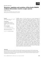
Fig. A. Major differences between Apoptotic and Necrotic types of Cell Death.
PS, Phosphotidylserine; Chr. Cond., Chromatin Condensation; IAP, Inhibitory
Apoptotic Protein.
3
other essential cellular functions. Some of the molecular components of apoptotic
machinery have been conserved through evolution (Steller H, 1995). An intact death
pathway is required for successful organogenesis in embryonic life and maintenance
of normal tissue homeostasis in adult organisms. As opposed to necrosis, apoptosis
minimizes the leakage of cell contents into extracellular space, which in turn results in
a minimal inflammatory response and tissue damage. Apoptosis has been studied
most extensively in the worm, C elegans. Genetic studies have identified 14 genes in
C. elegans that affect programmed cell death (Steller H, 1995), homologues of some
of these genes have been identified in mammals. For example, two of C. elegans’
genes, ced-9 and ced-3 (ced stands for cell death defective), are homologous to
mammalian genes: the proto-oncogene bcl-2 and ice (interleukin-1-β-converting
enzyme), respectively
Deregulation of apoptosis can be very detrimental to the organism. Excessive cell
death can lead to a number of diseases, for example, AIDS, neurodegenerative
disorders and ischemic injury (Thompson CB, 1995). In contrast, impaired apoptosis
is a significant factor in the etiology of diseases like cancer, autoimmune disorders
and viral infections.
I.1.c. Apoptotic Machinery
Apoptosis is a complex phenomenon of morphological and biochemical processes.
The field of apoptosis has witnessed an explosion of information over the past two
decades. The C. elegans hermaphrodite undergoes a distinct programmed cell death
pattern in which the same 131 cells out of 1090 cells die during the development of
this worm (Brenner et al., 1974; Sulston et al., 1976). In more complex organisms,
4
like mammals the regulation of apoptosis and its mechanism is far more intricate and
complex.
Apoptotic cell death occurs in two phases: first a commitment to cell death, followed
by an execution phase characterized by specific morphological changes in cell
structure. Classically, Apoptosis can be initiated with or without the involvement of
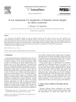
mitochodria. In cell-surface receptor induced apoptosis, activation of Fas or TNF
receptor leads to the activation of initiator caspase 8, followed by the activation of
downstream effector caspases (Fig B). In mitochondrial or intrinsic pathway, upon
apoptotic triggers there is a release of mitochondrial contents, most notably
cytochrome C, into the cytosol. This leads to the formation of a complex between
cytochrome C, Apaf1 and pro-Caspase 9, known as “Apoptosome”. Bcl-2 and Bcl-xL
block death by preventing the release of mitochondrial contents into cytosol. On the
other hand, pro-apoptotic members of Bcl-2 family like Bad and Bax, play an
important role in facilitating apoptosis (Fig C).
Executioners of apoptosis include a cascade of proteases termed caspases. Currently
11 human caspases has been identified. Initiator caspases including caspase-1, 2, 4, 5,
9, 11 and 12 interact with upstream adapter molecules and once activated lead to
downstream activation of executioner caspases (caspase-3, 6 and 7). A striking feature
of these enzymes is their specificity of substrate cleavage after an Asp residue
(Degterev A et al, 2003). Caspase activation leads to the cleavage/degradation of a
number of cellular proteins like PARP, Lamin A.
To make the picture more complex, other families of proteins have been identified
recently which are involved in the regulation of apoptosis. IAPs (Inhibitor of
apoptosis proteins) bind to caspases and inhibit their activity. Another player with the
5
Fig. B. Death-Receptor mediated Apoptosis.
Fig. C. Mitochondial pathway of Apoptosis.
6
dual name of Smac/DIABLO has been identified which promotes caspase activation
and inhibits xIAP (Douglas RG, 2000). In summary, apoptotic machinery consists of
a host of proteins interacting with each other in a complex and intricate manner, one
group of proteins favoring apoptosis and the other opposing it. Thus, the decision to
die is a matter of balance amongst anti- and pro-apoptotic proteins.
To our interest, numerous studies have demonstrated redox-regulated functional
modifications of many of the proteins involved in apoptotic machinery (Dechao L et
al. 2004; John JH, 2004; Irani K and Pascal JGC, 1998).
I.2. Reactive Oxygen Species and Apoptosis
Cells generate reactive oxygen species (ROS) during aerobic metabolism. Higher
levels of ROS are detrimental to cell’s functions, thus each cell has an extensive
antioxidant defense system to scavenge excessive amounts of ROS. The intracellular
redox state is controlled by the thioredoxin and glutathione systems (Mates JM and
Sanchez-Jimenez F, 1999). Mitochondria are the major source of ROS, where
electrons carried by the electron transport chain may leak out of the pathway and react
with oxygen to form superoxide (O2.-). Other sources of O2.- include enzymes such as
NADPH oxidase (NOX), lipoxygenases, cyclooxygenases, xanthine oxidase, and
cytochrome P450. Once O2.- is generated it is rapidly converted to hydrogen peroxide
(H2O2) by superoxide dismutase. Hydrogen peroxide can then react with Fe2+ to form
hydroxyl radicals via the Fenton reaction.
I.2.a. Pro and Anti-Apoptotic functions of ROS
Over the past decade or so there has been a paradigm shift in the understanding of
ROS and the functions they can play in the cell. The intracellular concentration of
7
these reactive molecules is kept under tight regulation by cells’ anti-oxidant systems.
The anti-oxidant defense mechanisms include scavenger enzymes superoxide
dismutase (SOD), catalase and glutathione peroxidase (Halliwell B and Gutteridge
JMC, 1999). Therefore, accumulation of ROS in the cell is a function of the overall
production and the efficiency of the anti-oxidant defences which could be cell
specific. Several reports have suggested that phorbol esters stimulate the production
of O2.- not only in phagocytic cells but in other cultured cells as well (Bonser et al,
1986; Fischer et al, 1986). This small amount of O2.- produced in non-phagocytic cells
in response to mitogenic stimuli may play some physiological role in the signal
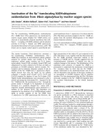
transduction. Reactive oxygen species (ROS) and Reactive nitrogen species (RNS)
can affect a wide variety of cellular functions (Droge W, 2001; John JH, 2004; Finkel
T, 2003). There is a growing consensus that redox status of a cell plays a regulatory
role on a wide range of cellular functions: gene transcription, cell proliferation,
differentiation and adaptation on one end, and apoptosis and necrosis on the other end
of the spectrum (see Fig. D).
Whereas the role of ROS in inducing necrotic cell death is well established, the role of
ROS in apoptosis is more controversial. However, there is increasing body of
evidence to support the role of ROS in apoptosis.
The activity of caspase proteases has been shown to be influenced by the redox status
of these enzymes (Hampton MB et al, 1998). Apoptosis in neutrophils and their
clearance by macrophages has also been shown to be ROS dependent (Fadeel B and
Kagan VE, 2003)
Fas receptor activation is a major trigger for apoptosis, and it has been shown that O2.can act as a natural inhibitor of Fas-induced cell death in tumor cells (Clement MV
8
Fig. D. Wide array of functions attributed to Reactive Oxygen Species (ROS).
John J. Haddad (2004)
9
and Stamenkovic I, 1996). Mitochondria can play an important role in modulating
apoptosis through generation of ROS. It has been postulated that reversal of
mitochondrial F0F1-ATPase in the inner membrane would lead to an increased
concentration of H2O2 in the cytosol which in turn, would lead to PARP activation
and ATP depletion. This depletion in ATP levels could lead to increase acid load in
the cell either by production of H+ or inhibition of H+ transporters (Gossmann DL et
al, 2004).
Interestingly, cell surface receptor and mitochondrial pathways cross talk with each
other through Bid that is a pro-apoptotic member of Bcl-2 family. Pro- and antiapoptotic proteins of this group form heterodimers and block each other’s activity.
Expression levels of these proteins can be controlled at multiple levels: transcription,
heterodimer formation and ubiquitination. Anti-apoptotic Bcl-2 family proteins, when
phosphorylated, fail to bind to each other. Thus, it has been suggested that the
phosphorylation status of Bcl-2 family proteins might affect their ability to regulate
apoptosis (Ruvolo PP et al, 2001). ROS have been shown to induce apoptosis by
regulating the phosphorylation and ubiquitination of Bcl-2 family proteins (Dechao L
et al, 2004).
The tumor suppressor p53, nick named Guardian of the Genome, plays an important
role in the regulation of cellular response to DNA damage. p53 has been shown to
participate in sensing oxidative DNA damage and modulates BER (base excision
repair) function in response to persistent ROS stress (Achanta G and Huang P, 2004).
In a recent study, stress-induced p53 activation showed strong ROS sensitivity both in
leukemic and normal lymphocytes. These observations identified mitochondrial
activity and ROS levels, as a critical intracellular determinant of the p53 stress
10
sensitivity and suggest potential implications of this linkage in the mechanisms of
chemoresistance of acute leukemia cells (Karawajew L et al, 2005).
Thus, it can be concluded that ROS can modulate or alter the activity of a number of
very important proteins involved in cell death. Process of apoptosis can be divided
into three distinct phases: initiation, effector and degradation. ROS can be involved in
all three of these phases.
Reactive Nitrogen Species (RNS) have a more established role in modulation of cell
death. Nitric oxide (NO) is an important bioregulatory molecule in the nervous,
immune and cardiovascular systems. NO participates in the regulation of many
cellular functions as well as in cytotoxic events. It possesses a controversial effect on
cell viability by acting both as a protection against apoptotic stimuli or by inducing
apoptosis when produced at elevated concentrations (Blaise GA et al, 2005).
The role of ROS will be discussed in more detail as the work presented in this
manuscript was undertaken to study the role of ROS in apoptosis.
I.2.b. Rac1 and Superoxide anion production
Rac1 is a ubiquitously expressed small GTP-binding protein, that functions
downstream of oncogene Ras. p21ras (c-Ras) has many functions in the cell, including
proliferation, differentiation, apoptosis and cytoskeletal organization. Mutations in a
ras allele that make it constitutively active have been found in 30% of all human
tumors, making it the most widely mutated human proto-oncogene. Multiple
pathways exist downstream of Ras, including activation of Rac1.
Activated Rac1 leads to the generation of ROS, including O2.- (Irani K and Pascal
JGC, 1998). Activation of Rac is classically known to trigger clustering of an enzyme
complex, NADPH oxidase (NOX) in phagocytic cells. Activation of NADPH in these
11
cells catalyses the generation of O2.- (also known as “respiratory burst”) which in turn
kills the ingested bacteria. Until recently, the single example of ‘deliberate’
generation of ROS in mammalian cells was the NOX of phagocytes (Phox). This
enzyme is inactive in resting neutrophils, but is activated by exposure to
microorganisms or inflammatory mediators, resulting in the rhobust production of
ROS. Although the exact structure and localization of NADPH-like enzyme system
has not been identified in non-phagocytic cells, Mox1 (mitogenic oxidase 1) has been
cloned and characterized as a homologue to neutrophil gp91phox, which participates
in ROS production (Suh YA, 1999). In contrast to a robust production of ROS in
phagocytic cells, lower levels of ROS produced in non-phagocytic cells appear to act
as secondary messengers or signaling molecules. Recent data suggests ROS produced
downstream of Rac might play a role in the regulation of growth, transformation and
apoptosis (Finkel T et al, 1999; Irani K et al, 1997). Rac isoform 1 has been identified
in many non-phagocytic cells and is responsible for production of intracellular O2.-, as
opposed to isoform 2 that has been described in phagocytic cells. The expression of
these enzymes in various tissues provides evidence that generation of ROS is a
general feature of many and perhaps all cells. Many cell types express NOX enzymes,
probably accounting for the diverse cellular ROS generation seen in many of the
earlier studies. Examples of non-phagocytic cells where NOX enzymes or its
components have been identified include osteoclasts, fibroblasts, glomerular
mesangial cells, chondrocytes, endothelial cells and keratinocytes (Bunn and Poyton,
1996; Suh YA, 1999).
As described earlier, O2.- is the primary ROS generated by normal cellular
metabolism, whereas H2O2 is a catalytically derived intermediate in the conversion of
O2.- to O2 (Fridovich I, 1976; Halliwell B and Gutteridge JMC, 1989). In phagocytic
12
cells, large scale production of Nitric Oxide (NO) by macrophages, or O2.- by
neutrophils, provides the host with defense function against invading pathogens, while
when produced in smaller amounts in non-phagocytic cells, these same reactive
molecules instead of causing damage to the cell function as signaling molecules
(Finkel T, 2001).
I.2.c. Superoxide anion and inhibition of apoptosis
Mammalian cells possess multiple sources of ROS generation; most evidence
suggests that plasma-membrane associated oxidases may provide one source of ROS
associated with resistance to apoptotic triggers. O2.- has been shown to contribute to
the unchecked proliferation in Ras-transformed fibroblasts (Irani K and Pascal JGC,
1998). Although the exact source of O2.- in non-phagocytic cells is still under
investigation, Rac has been implicated as a major component of O2.--generating
system, and presence of NOX enzymes in a variety of cells suggests a role for ROS in
various cellular functions.
In addition, plasma membrane of many cells has another ROS-generating enzyme
utilizing NADH as an electron donor. Both of these flavin-containing oxidases are
inhibited by diphenylene iodonium (DPI). Taken together, NADH and NADPH
oxidases may provide candidate sources of O2.- production associated with cells’
resistance to apoptotic cell death. As described in the previous section, O2.- can block
the Fas-induced cell death in tumor cells.
The regulation of tumor cells’ sensitivity to death stimuli has been shown to be linked
to the intracellular levels of O2.- and H2O2 (Pervaiz S and Clement MV, 2002 and
2004; Clement MV and Pervaiz S, 1999 and 2001). Interestingly, an increase in
intracellular O2.- concentration achieved by either its direct overproduction (Clement
13
MV and Stamenkovic I, 1996), drug-induced (Pervaiz S et al, 1999; Ahmad KA et al,
2004), activation of the small GTPase Rac1 (Pervaiz S et al, 2001), or as a result of an
inhibition of the O2.- scavenger Cu/Zn SOD (Pervaiz S et al, 1999), inhibits tumor cell
apoptosis triggered by either the CD95 receptor or anticancer drugs. In contrast, H2O2
is a widely accepted trigger of apoptotic cell death (Hirpara JL et al, 2001) and nontoxic levels of H2O2 sensitize cells to death triggers (Clement MV et al., 1998).
Earlier reports have highlighted the regulatory role of intracellular redox status on
death signaling by demonstrating an effect on caspase family protease, the central
executioners of apoptotic signals (Hampton M and Orrenius S, 1998; Chandra J et al,
2000).
I.3. Intracellular milieu
Various mechanisms of how ROS can lead to cell transformation have been proposed.
O2.- or other “oxidants” may induce targeted damage to chromosomal DNA, leading
to enhanced rate of oncogenic mutations or they can directly regulate the signaling
cascade that underlies malignant transformation (Irani K et al, 1997). ROS have been
shown to activate NF-κB, a transcription factor whose activation has been linked to
apoptosis. Evidence has grown in this poorly understood field and many signaltransducing proteins and transcription factors have been added to the list of “redoxsensitive” proteins (Sundaresan M et al, 1996).
Apoptosis is a tightly regulated chain of reactions that involves many enzymatic
reactions and proper functioning of all of its components is essential to execute a cell
in a predetermined fashion. The executioners of apoptosis, especially caspases are
very sensitive to redox alterations and require a reducing environment to be
functional. All caspases contain an active site thiol group necessary to perform their
14