Specificity and diversity in the vertebrate nervous system an analysis of two genes
Bạn đang xem bản rút gọn của tài liệu. Xem và tải ngay bản đầy đủ của tài liệu tại đây (20.76 MB, 146 trang )
i
SPECIFICITY AND DIVERSITY IN THE VERTEBRATE NERVOUS
SYSTEM : AN ANALYSIS OF TWO GENES
MAHENDRA D. WAGLE
(M.Sc., University of Mumbai-India)
A THESIS SUBMITTED
FOR THE DEGREE OF DOCTOR OF PHILOSOPHY
TEMASEK LIFESCIENCES LABORATORY
NATIONAL UNIVERSITY OF SINGAPORE
2005
ii
ACKNOWLEDGEMENT
I am thankful to Dr. Suresh Jesuthasan for having me introduced to developmental
neurobiology and for the supervision of my project. It was wonderful experience
working in his lab and I thank him for his guidance and support.
I am grateful to Dr. Karuna Sampath, Dr. Wen Zilong and Dr. Edward Manser for
being on my thesis advisory committee. I am also thankful to Dr. Naweed Naqvi, Dr.
Suniti Naqvi and Dr. Mohan Balasubramanian, for showing keen interest in my
projects and valuable suggestion. I am thankful to Dr. Amita Joshi for valuable
suggestion in ChIP experiments.
I would like to acknowledge following people for sharing the reagents. Suzanne
Lang, for providing the silicon stamp and the method for stamping. John Ngai for
unc76-GEP, Chi-Bin Chien for pESG, Ajay Chitnis and Motoyuki Ito for the
HuC∆Eco promoter, Joanne Chan for EphrinB2a, and Mary Hallaran for the Hsp70
promoter.
I would like to thank all the members of Dr. Jesuthasan’s lab for their cooperation, in
particularly Dr. Subbu Sivan and Cristiana, for the technical assistance. I am thankful
to Aniket for scientific discussions, Ventris and Bindu for proof reading of thesis.
Also thanks to all collogues, DNA sequencing and support facility as well as
administration staff at TLL for the help and support.
Last but not least I am thankful to my parents, family member and my wife Meghana
for great support and encouragement.
iii
Table of Contents
ACKNOWLEDGEMENT ii
Table of Contents iii
Abstract vi
Summary vii
List of Figures x
Abbreviation xi
Publications xi
Chapter-I : Introduction 1
1.1 Central Nervous System (CNS) development 1
1.1.1. Neural differentiation 1
1.1.2. Neuralation and patterning of neural tube 2
1.2 Neuronal diversity 5
1.3 Axon guidance –mechanism 8
1.4 Model systems and methods to study axon guidance 10
1.5 Principles of axon guidance 13
1.5.1 Netrins: 13
1.5.2 Semaphorins: 14
1.5.3 Slit-Robo 15
1.5.4 Eph-Ephrins 16
1.5.5 Secreted molecules: Shh, BMP and Wnt 18
1.5.6 Other signaling molecules 19
1.5.7 Interpretation of guidance cues (effect of Calcium and cyclic
nucleotides) 20
1.6 Aim of the thesis 21
1.6.1 Study of EphrinB2a in zebrafish visual system 21
1.6.2 Study of Rag1(Recombination activating gene-1) in neurons 22
Chapter II : Development of a baculovirus mediated misexpression system and its
application to the study of EphrinB2a function in Zebrafish visual system 25
2.1 Introduction 25
2.1.1 Vertebrate visual system: 25
2.1.2 Eph-Ephrins 26
2.1.3 Neuronal roles of Ephrin 28
2.1.4 Ephrins in Retinotectal projection and topographic mapping 30
2.1.5 The zebrafish visual system 32
iv
2.2 Aim of the project 34
2.3 Methods 36
2.3.1 Chemicals and general protocols 36
2.3.2 Zebrafish Adults and Embryos 36
2.3.3 Constructs 36
2.3.4 Virus production and injection 37
2.3.5 X-gal staining 38
2.3.6 DiI labeling 38
2.3.7 In-situ hybridization 39
2.3.8 Microscopy 39
2.3.9 Stripe assay 39
2.3.10 Ligand binding assay 40
2.4 Results 40
2.4.1 Baculovirus can drive gene expression in zebrafish 40
2.4.2 Baculovirus-mediated EphrinB2a misexpression affects segmentation. 42
2.4.3 EphrinB2a expression in the optic tectum 46
2.4.4 Retinal ganglion cell axon behaviour in a mutant with ectopic tectal
neurons 48
2.4.5 Baculovirus-mediated ephrinB2a misexpression affects RGC axon
migration 52
2.4.6 Effect of EphrinB2a on RGC axons in vitro 54
2.5 Discussion 56
Chapter III : Studying The Role of Rag1 (recombination activating gene-1) in
neurons 60
3.1 Introduction: 60
3.1.1 Similarities between the vertebrate adaptive immune system and the
CNS : Molecular link 60
3.1.2 Development of the adaptive immune system 62
3.1.2.1 B-cell and T-cell development : Immunoglobulin and T-cell receptor
structure 62
3.1.2.2 Genomic locus of immunoglobulins, TCR and V(D)J rearrangement:
Role of Rag1 67
3.1.2.3 Rag1 structure, function and regulation 68
3.1.3 Rag-1: role in neurons – facts and hypothesis. 70
3.2 Aim of the project 71
3.3 Materials and Methods: 72
3.3.1 Antibody, enzymes, chemicals and general protocols: 72
3.3.2 Oligonucleotide primers : 72
3.3.3 Buffers and solutions: 73
3.3.4 Mice and tissue collection : 74
3.3.5 P19 cells and differentiation into neurons: 75
v
3.3.6 Antibody staining 76
3.3.7 Imaging: 77
3.3.8 Construction of artificial recombination substrate 77
3.3.9 Chromatin immunoprecipitation 78
3.3.9.1 Tissue preparation: 78
3.3.9.2 Crosslinking: 78
3.3.9.3 Cell Lysis and preparation of soluble chromatin: 79
3.3.9.4 Incubation with antibodies and pull-down with beads: 79
3.3.9.5 Second round of antibody incubation and pull-down 80
3.6.9.6 Purifying double ChIP-DNA.: 80
3.3.10 ChIP-DNA analysis by specific PCR: 81
3.3.11 End-repair and adaptor ligation 82
3.3.12 LMPCR and DIG-labeled probe synthesis 82
3.3.13 Screening YAC and BAC library macroarrays 82
3.3.14 End sequencing of YACs 83
3.3.15 BACs southern hybridization 83
3.3.16 Screening BAC subclone 83
3.4 Results : 84
3.4.1 Detection of RAG1 protein in thymocytes and neurons: 84
3.4.2 Checking the V(D)J like recombination in RAG1 expressing neuronally
differentiated P19 embryonic carcinoma cells 89
3.4.3 Testing the possibility (standardization) of ChIP (chromatin
immunoprecipitation) assay: 92
3.4.4 Chromatin immunoprecipitation and Screening YAC library
macroarray : 96
3.4.5 Mapping of YACs to their genomic locus 101
3.4.6 Analysis of the putative RAG1 binding site 101
3.4.7 BAC macroarray hybridization 106
3.5 Discussion : 109
Appendix……………………………………………………………………………113
Refrences 114
vi
Abstract
This thesis describes two genes that may establish different identities in neurons and
thus mediate the formation of synaptic connections. The first gene, ephrinB2a, is
expressed strongly in posterior zebrafish tectal neurons that are contacted by retinal
axons. Ectopic expression of ephrinB2a in the anterior midbrain, with the aid of
baculovirus, causes stalling of retinal axons. EphrinB2a may thus signal some retinal
axons that they have reached their target neurons. The second gene, Rag1
(recombination activation gene-1), which mediates diversity in the immune system, is
surprisingly also expressed in the vertebrate nervous system. Here, RAG1 protein is
shown to be nuclear localized in a subset of differentiated mouse neurons. Chromatin
immunoprecipitation, coupled with macroarray screening, identified a 5’ repeat
region in a LINE-1 retrotransposon, as a potential target of RAG1 in neurons. This
raises the possibility that Rag1 may have a function in neurons by regulating a mobile
element.
Keywords: vertebrate, zebrafish, ephrinb2, baculovirus, Rag1, chromatin
immunoprecipitation, L1 retrotransposon.
vii
Summary
Neuronal networks are built up through the connections of neuronal processes
– axons and dendrites. Cues from surrounding tissues guide axons towards their
targets during development of the nervous system. Once an axon reaches its target it
needs to find a partner to make synaptic connections. Signals from the target itself
could help the axon to make necessary modifications for synapse formation. To make
precise connections it is also important that each neuron exhibit a unique identity.
This thesis describes the study of two molecules that are expressed in the
nervous system. EphrinB2 a signal from target cells that could induce presynaptic
modification and RAG1, a molecule that generates diversity in immune system,
which is also present in specific subsets of neurons.
In this study, the role of EphrinB2 in the zebrafish visual system is examined.
EphrinB2 belongs to a family of ligands for Eph receptor tyrosine kinases. It is B-
type Ephrins which are transmembrane molecules. Ephrins are known for their role in
topographic mapping of retinal ganglion cell axons on the optic tectum (O'Leary and
Wilkinson, 1999; Wilkinson, 2000). EphrinB2 is known as a repellant cue for axon
guidance and also has been found in a retinorecipient layer of chick tectum where
RGC axons make synapses (Braisted et al., 1997).
With RNA-in-situ hybridization I found that zebrafish EphrinB2 is expressed
in tectal neurons in the posterior part of the tectum when RGC axons enter the
neuropil. Receptors for EphrinB2 on zebrafish RGC axons were detected by in-vitro
receptor-ligand binding assays. As reported earlier in other systems, zebrafish RGC
axons showed repulsive response to EphrinB2 in stripe assays. Studies with the
viii
retinotectal projection mutant “gnarled” pointed out that the expression of ephrinB2
in ectopic cells in the anterior tectum of mutants could cause a premature stopping of
RGC axons (Wagle et al., 2004). To verify this observation, a baculovirus-based gene
expression system was developed which allowed temporal-spatial control over gene
misexpression in zebrafish (Wagle and Jesuthasan, 2003). Ectopic expression of
ephrinB2a in the anterior midbrain of wildtype embryos, with the aid of baculovirus,
was found to inhibit RGC axon entry into the tectum. It is thus proposed that
ephrinB2 may signal a subpopulation of RGC axons that they have reached their
target neurons in the tectum.
The Recombination activating gene-1 (RAG1) is expressed in the vertebrate
immune system and in the nervous system, including the zebrafish visual system
(Chun et al., 1991; Frippiat et al., 2001; Jessen et al., 2001). RAG1 is well
characterized for its role in generating diversity in immune system by V(D)J
recombination (Schatz et al., 1989). Rag1 plays a key role in the initiation of this
process of genomic rearrangement by recognizing and cutting recombination signal
sequences (RSS) (Schatz et al., 1992). Detection of Rag1 transcripts in the mouse
nervous system led to the idea that the genome may rearranged in neurons, but there
has been no conclusive experimental evidence. In spite of the studies done over the
last decade, the presence of RAG1 protein in neurons has not been demonstrated and
its functions are questionable.
RAG1 protein was detected in specific neurons from the mouse brain at P10-
14 and in neuronally differentiated P19 embryonic carcinoma cells. To identify
potential RAG1 binding sites in neurons, chromatin immunoprecipitation (ChIP)
ix
coupled with macroarray screening of a genomic YAC library was carried out. As a
positive control, ChIP- DNA pulled down from thymocytes with anti-RAG1 antibody
was used to generate probe. Signals obtained in this experiment partially overlapped
those obtained from a T-cell receptor locus probe, showing the feasibility of this
approach. ChIP-DNA from brain and neuronally differentiated P19 cells were then
used to generate probes. A YAC clone that showed signal with both probes was
analyzed further. Fine mapping by Southern analysis of BAC clones covering the
YAC locus narrowed the potential target to a region which harbors a retrotransposon
element. Binding of RAG1 to this region was further confirmed by analyzing ChIP-
DNA from brain with the specific PCR. Analysis of the target sequence indicated the
presence of a conserved heptamer found in the RSS. Although the YAC clone
mapped to chromosome-9, PCR analysis and BAC macroarray screening with brain
ChIP-DNA showed that the repeat region identified here as a potential target may not
be specific to chromosome-9.
Identifying a retrotransposon as a potential target of RAG1 in neurons does
not immediately answer the question of whether RAG1 could generate diversity in
neurons as it does in the immune system. Nevertheless this finding indicates that
RAG1 has distinct binding activity in neurons and puts us one step further in
understanding the role of RAG1 in neurons.
x
List of Figures
1.1 Primary neuralation and neural tube patterning 3
1.2 Growth cone structure and guidance 9
1.3 Schematic of CNS axon guidance 12
2.1 Ephrin/Eph classification and receptor ligand binding 29
2.2 Zebrafish visual system 35
2.3 Baculovirus mediated gene expression in zebrafish 43
2.4 Baculovirus mediated independent expression of two reporter genes 44
2.5 Effect of baculovirus mediated misexpression of ephrinB2a in somites 45
2.6 EphrinB2a expression in zebrafish tectum and transneuronal labeling 47
2.7 Retinotectal projection defects in gnarled 49
2.8 Midbrain morphology of gnarled 50
2.9 Neurogenesis and gene expression in gnarled 51
2.10 Baculovirus mediated misexpression of ephrinB2a in tectum and its effect on RGC
axons
53
2.11 In vitro assay with zebrafish RGC axons 55
3.1 Schematic of IgG structure, V(D)J recombination and RAG1 protein 65
3.2 Mechanism of V(D)J recombination : Role of RAG1 66
3.3 Detection of RAG1 protein in mouse thymocytes 85
3.4 Detection of RAG1 protein in mouse brain 86
3.5 P19 embryonic carcinoma cells differentiation and detection of RAG1 protein 88
3.6 Design of artificial recombination substrate 91
3.7 Flowchart of ChIP protocol and schematic representation of principle of ChIP 93
3.8 Analysis of ChIP-DNA from thymocyte 94
3.9 Mouse genomic YAC library macorarray hybridization with thymocyte ChIP-DNA
probe
98
3.10 Mouse genomic YAC library macorarray hybridization with brain ChIP-DNA probe 99
3.11 Mouse genomic YAC library macorarray hybridization with P19 ChIP-DNA probe 100
3.12 Mapping of potential Rag1 target in neurons 103
3.13 Sequence and features of potential target region 105
Table 3.1 BAC macroarray analysis……………………………………………………… ……107
xi
Abbreviation
A-P – anterior – posterior
BAC –Bacterial artificial chromosome
BDNF - Brain derived trophic factor
BMP – Bone morphogenic protein
ChIP – Chromatin Immunoprecipitation
CNS – Central nervous system
DAB - Diaminobenzidine
DAPI – 4,6-Diamidino-2-phenyindole, dilactate
DOPA - Dihydroxyphenylalanine
DSCAM - Down syndrome cell adhesion molecule
DRG – Dorsal root ganglion
D-V – Dorso-Ventral
FAK – Focal adhesion kinase
FGF – Fibroblast growth factor
GABA -gamma-aminobutyric acid.
LGN – Lateral geniculate nucleus
LINE – Long interspersed element
LMPCR – Ligation mediated PCR (polymerase chain reaction).
LTR – Long terminal repeat
MAPK – Mitogen activated kinase
NGF – Nerve growth factor
NMDA – N-methyl D-aspartate
PAK – p21 associated kinase
pcdh – Protocadherin
PCR – Polymerase chain reaction.
PSF - Pre m-RNA splicing factor
RAG –Recombination activating gene
R-C –Rostro-Caudal
RGC – Retinal Ganglion Cell
RPE – Retinal pigmented epithelium
RSS –Recombination signal sequence
SC- Superior colliculus
TGF – Transforming growth factor
YAC –Yeast artificial chromosome
xii
Publications
1. Wagle M, Jesuthasan S. 2003. Baculovirus-Mediated Gene Expression in
Zebrafish. Marine Biotechnology 5:58-63.
2. Wagle M, Grunewald B, Subburaju S, Barzaghi C, Le Guyader S, Chan J,
Jesuthasan S. 2004. EphrinB2a in the zebrafish retinotectal system. J Neurobiol
59:57-65.
3. Wagle M, Jesuthasan S. 2005 A Specific Target For The Recombination
Activation Gene-1 (Rag1) In Mouse Neurons. (Manuscript in preparation).
1
Chapter-I
Introduction
During embryonic development, the nervous system develops from a mass of
neuroblasts (neuronal precursor cells). These cells divide and build a network of
interconnected neurons. This is a crucial step in embryonic development, as a precise
neuronal network is eventually responsible for most of the activities of an organism.
1.1 Central Nervous System (CNS) development
Various model organisms ranging from worms and insects to mammals have
been used to understand neural development. With the help of mutants and other tools
of genetic manipulation, the mechanisms of neural differentiation and nervous system
patterning have been elucidated. As proposed by Goodman and Doe, the whole of
neurogenesis can be viewed in eight steps (1) Induction and patterning of neuron
forming regions, (2) birth and migration of neurons and glia, (3) generation of
specific cell fates, (4) guidance of axonal growth cones to specific targets, (5)
formation of specific synaptic connection, (6) binding of specific trophic factors for
survival and differentiation, (7) competitive rearrangement of functional synapses and
(8) continued synaptic plasticity during the life of an organism (Goodman and Doe,
1993). The first three steps are part of neural development and differentiation whereas
the last three steps are activity-dependent. In this chapter, I will briefly describe the
first steps of neural differentiation with a focus on axon guidance.
1.1.1. Neural differentiation
Soon after the embryo starts developing into a mulitcellular mass of cells from
a single cell stage, it begins gastrulation. During this stage cells proliferate and
2
migrate. Involution of these cells converts the embryo into a multi-layered structure.
Three germ layers – ectoderm, mesoderm and endoderm are formed. Specialized cell
movements known as convergent-extension transform the embryo into a primitive
body plan. Maternally deposited factors along with zygotically expressed genes
pattern the embryo along the dorso-ventral (D-V) and anterior-posterior (A-P) axes.
During gastrulation, a group of cells from the dorsal ectoderm is assigned a neuronal
fate. These neuronal precursor cells migrate to their appropriate position to form the
preliminary central nervous system in the form of a neural tube.
1.1.2. Neuralation and patterning of neural tube
In most vertebrate species, the anterior neural tube is formed by primary
neuralation involving cell proliferation, invagination and pinching off from the rest of
the cells, whereas the posterior part of the neural tube is formed by secondary
neuralation in which the neural tube arises from a solid chord of cells which
subsequently hollows out (Figure: 1.1A). The neural tube is patterned in A-P and D-V
axis by the action of several genes. The anterior neural tube folds into forebrain
(prosencephelon), midbrain (mesencephalon) and hindbrain (rhombencephalon)
(Figure 1.1B). During the formation of preliminary brain structure from the anterior
neural tube, optic vesicles are derived from the forebrain. Other sensory organs
develop while the neural tube is transforming into the CNS. The posterior neural tube
forms the spinal cord. Within the neural tube, neurons are specified to carry out
different roles.
3
z1
4
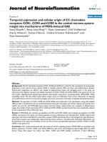
Figure 1.1: Schematic – Primary neuralation and patterning of neural tube
(Adapted & modified from Developmental Biology – 5
th
Edition: Scott F. Gilbert)
(A) The ectodermal plate consists of the neural tube in the middle and presumptive
epidermis on either side separated by neural crest cells. The presumptive epidermis
moves towards the center pushing the neural tube below it. This results in formation
of an outer epidermis and neural tube contacted by neural crest cells that eventually
migrate away from the neural tube to form peripheral neurons, glia and skin pigment
cells.
(B) In the anterior region, the neural tube folds into three major structures
Prosencephalon (Fore brain): Æ Telencephalon and Diencephalon
Mesencephalon (Mid brain)
Rhombencephalon (Hind brain) Æ Metencephalon and Myelencephalon
Structures in the adult brain such as the olfactory lobe, hippocampus and thalamus are
derived from these structures.
(C) the neural tube is patterned along the dorsal-ventral axis by signals from the
ventral floor-pate and dorsal epidermis that specify different types of motor neurons
and interneurons by activating transcription of specific genes.
5
1.2 Neuronal diversity
The nervous system of vertebrates comprises many types of neurons. In the
human brain, there are approximately 10
12
neurons of various types, for example there
are about two dozen types of inhibitory neurons in the hippocampus alone (Parra et
al., 1998). There is diversity in anatomy, gene expression and physiological
properties. Morphologically, there are four different types of neurons, i.e. axonal,
monopolar, bipolar, and multipolar. Based on their function in the nervous system,
neurons are classified as sensory neurons, interneurons and motor neurons. Different
types of sensory neurons are found within each sensory organ, depending on the
stimulus they respond to. In the retina there are at least one dozen different types of
ganglion cells (Devries and Baylor, 1997). Similarly in the olfactory epithelium each
neuron has its own identity based on odorant receptor expression (Mombaerts et al.,
1996). Motor neurons have distinct anatomical connectivities and gene expression
properties. Neurons within the CNS have differences in neurotransmitter identities;
they may be DOPAergic or GABAergic for example. They also differ by expression
of surface molecules such as protocadherins.
This diversity is created by the action of several signaling molecules that act
during the development of the nervous system. Two mechanisms have been described
for neuronal fate specification: lineage dependency and extrinsic signal/morphogen
dependency. Proneural genes belonging to bHLH family initiate neural fate and
generate progenitor cells that are committed to differentiate (Bertrand et al., 2002).
Studies in Drosophila have shown that lateral inhibition involving Notch-Delta
signaling plays a crucial role in specification of neuronal fate in neuroblasts. A
6
similar mechanism exists in vertebrates as well (Lewis, 1998). Asymmetric cell
division of neuronal progenitors allows the inheritance of cell fate determining factors
to one daughter cell, thus resulting cells may be specified as neuronal or glial (Chia
and Yang, 2002). Neuronal specification and diversity has been well studied in the
CNS with respect to patterning of hindbrain along rostro-caudal axis and D-V axis in
the neural tube. During development, FGF and several Hox genes pattern different
regions of the brain to specify neurons within these structures (Dasen et al., 2003;
Salie et al., 2005). The neural tube is patterned along the D-V axis by the action of
TGF-β from dorsal and Sonic hedgehog from the ventral floorplate or notochord
(Echelard et al., 1993; Roelink et al., 1994; Liem et al., 1995; Liem et al., 2000;
Nguyen et al., 2000). Motor neurons and interneurons are specified along the D-V
axis within the neural tube by the combinatorial effect of these factors (Figure 1.1C).
These factors induce expression of transcription factors and genes which govern
various properties of the neuron such as expression cell surface molecule/receptors,
production and response to neurotransmitters. Thus various neurons are specified
during the development of the nervous system. This allows neurons to carry out their
specialized functions as well as to connect with their synaptic partner.
Sperry’s chemoaffinity theory postulates a cytochemical specificity to
individual neurons (Sperry, 1963). Cell surface molecules are the best candidate to
satisfy this assumption. Indeed, in Drosophila, Down syndrome cell adhesion
molecule (DSCAM) could generate diversity in neurons (Schmucker and Flanagan,
2004). The Dscam locus contains three arrays of alternative exons that are combined
with 20 constant exons and two alternative transmembrane domain by alternative
7
RNA-splicing (Wojtowicz et al., 2004). This generates a huge repertoire of DSCAM
molecules containing different extracellular domains. These molecules show
homophilic interaction and are involved in axon guidance (Schmucker et al., 2000;
Wojtowicz et al., 2004). The diversity in neuronally expressed DSCAM provides a
mechanism for selective axon fasciculation and recognition of synaptic targets.
Although vertebrate orthologs of Dscam do not show this diversity, other cell surface
molecules such as protocadherins (Pcdh) exist and these are good candidates for
generating diversity in the vertebrate nervous system (Serafini, 1999). The Pcdh
genes are clustered in the genome and show similar organization as that of
immunoglobulins or T-cell receptors (Wu and Maniatis, 1999; Wu et al., 2001). Like
Dscam, individual Pcdh mRNA are generated by splicing of variable exons to the
constant 3’end (Wu and Maniatis, 1999). Pcdh are localized in synapses, and have
been proposed to offer synaptic specificity along with other cadherins (Kohmura et
al., 1998; Serafini, 1999). Other molecules such as cochlear potassium channels and
synaptic neurexins also show various isoforms through alternative RNA splicing
mechanism and may further contribute to neuronal diversity (Black, 1998; Missler
and Sudhof, 1998).
Thus neuronal diversity is achieved by the expression of various genes. In spite
of this diversity and large number of neurons in the vertebrate nervous system,
neurons are connected precisely to their targets. In fact this diversity is an essential
criteria for building a complex neuronal network and its functionality.
8
1.3 Axon guidance –mechanism
Apart from differentiation and migration of neurons to their appropriate
position in the embryo, it is also important that these neurons are connected to each
other in a specific manner to build a functional neuronal network. Once neurons are
specified, they send out processes called axons and dendrites to connect with each
other. An axon can extend many cell diameters to connect to other neurons. Axons
grow in a stepwise manner and surrounding tissue along the axon path may act as
guide posts. Examples of such cells are those at the midline for peripheral axons
(reviewed in Tessier-Lavigne and Goodman, 1996). The tip of the axon is called the
growth cone. It has microtubules at the base and dynamic actin filaments that form
finger like protrusions (filopodia) and web-like lamellipodia (Figure 1.2A). It also
bears receptors at the surface that sense cues from surrounding tissues (reviewed in
Tessier-Lavigne and Goodman, 1996)
During embryonic development, axon guidance is mainly independent of
neuronal activity and relies on surrounding cues. These cues could be in the form of
secreted molecules or cell surface molecules that either attract or repel the growth
cone (Figure 1.2). In the case of secreted signaling molecules, axon behavior is
termed as chemoattraction or chemorepulsion, whereas in the case of guidance
molecules bound to cell surface the phenomenon is known as contact mediated
attraction or repulsion (Figure 1.2 B). Receptor ligand interactions at the growth cone
lead to changes in the axon cytoskeleton. Signaling molecules may trigger different
types of signaling pathways that eventually result in cytoskeletal rearrangements.
9
z2
10
1.4 Model systems and methods to study axon guidance
Several model organisms have been used over the last few decades to study axon
guidance. Studies in invertebrates, mainly in C. elegans and Drosophila, have been
successful in identifying many axon guidance molecules. The simple nervous system
architecture, for example 302 total neurons in C. elegans and segmental arrangement in
Drosophila allowed connection of individual neurons with their targets to be studied
during development. Moreover these systems are easily amenable to genetic
manipulation. Through the study of such simple systems, well-conserved mechanisms
were elucidated. One example is the crossing of axons at the midline (Figure 1.3). Axons
from peripheral neurons are attracted towards the midline and once they cross the midline
they are kept away from the midline. The change in axon response to the same guidepost
has been studied in depth in Drosophila. Molecules such as Roundabout,
Commissureless, Netrins were identified by genetics and characterized extensively
(Kaprielian et al., 2001). Thus genetics in invertebrates has been a powerful tool to
identify guidance cues. Many of these genes have orthologs in vertebrates where they
also function at midline crossing.
Biochemical approaches have also identified several cell adhesion and signaling
molecules. A key requirement is an assay system to test effects of these molecules.
Conventionally neuronal explants and co-cultures were used for axon guidance studies.
Neuronal extensions (neurite growth) can be studied in response to secreted molecules by
placing neurons in proximity to cells expressing those molecules (Kennedy et al., 1994;
Serafini et al., 1994). Two assays that are widely used studying effects of various
molecules on growth cone behavior are “stripe assays” for analyzing membrane
11
associated molecules, and the “pipette assay” or “growth cone turning assay” for soluble
molecules. Initial stripes assays developed by Bonhoeffer’s group used stripes of
membrane preparations from tectal cells to study the growth of retinal axons from
anterior or posterior retina (Walter et al., 1987). In a modification of this assay, stripes of
purified proteins have been used to examine the response of retinal axons (Drescher et al.,
1995). Chemo- attraction or repulsion could be better studied in pipette assays. A glass
capillary pipette holding a solution of the molecule to be tested is positioned close to
growth cone. Pulses of these molecules create a concentration gradient between the
pipette tip and growth cone. Growth cone response to this gradient could be studied by
time-lapse microscopy (Lohof et al., 1992).
12
z3
(Adapted and modified from : Developmental Biology – 5
th
Edition: Scott F. Gilebert)
D
rosophila
D
rosophila
13
1.5 Principles of axon guidance
In 1892 Ramón y Cajal proposed that chemotactic cues guide axons in the nervous
system (Ramón, 1892). About hundred years later the molecular nature of these cues
became clear when several axon guidance molecules were identified based on approaches
described above. Below is the brief summary of these molecules which describe the
principles of axon guidance.
1.5.1 Netrins:
The idea of chemoattractant-mediated axon guidance was supported when Netrins
were isolated from chick brain, based on their ability to promote outgrowth and reorient
comissural axons in an in vitro assay system. (Kennedy et al., 1994; Serafini et al., 1994).
Netrins form a small family of secreted proteins similar to laminin, an extracellular
matrix protein. In mice, loss of Netrin-1 leads to abnormal commissural axons projection
(Serafini et al., 1996). Similarly C.elegans mutant for UNC-5, a homolog of Netrin-1,
show a defect in circumferential axon guidance. Also Netrin-A and Netrin-B are
expressed in the Drosophila ventral nerve cord during commissure formation, and
deletion of both these genes leads to formation of thinner than normal commissures
(Harris et al., 1996; Mitchell et al., 1996). Thus the role of Netrin as a chemoattractant at
the midline remains evolutionarily conserved.
Netrins act through their receptors known as Deleted in Colorectal Cancer (DCC)
and UNC5H in mammals. Mice mutant for DCC shows similar defects in commissural
axon projection as that of the Netrin-1 mutant (Fazeli et al., 1997). Surprisingly in C.
elegans, the Netrin ortholog UNC-6 acts as an attractant for some axons and a repellant


![vmware esx and esxi in the enterprise [electronic resource] planning deployment of virtualization servers](https://media.store123doc.com/images/document/14/y/mx/medium_mxh1401475429.jpg)