A multiplex comparative proteomic analysis of hypoxia influence in the presence and absence of p53 in HCT116 cells
Bạn đang xem bản rút gọn của tài liệu. Xem và tải ngay bản đầy đủ của tài liệu tại đây (5.82 MB, 134 trang )
A MULTIPLEX COMPARATIVE PROTEOMIC
ANALYSIS OF HYPOXIA INFLUENCE IN THE
PRESENCE AND ABSENCE OF p53 IN HCT116 CELLS
TAN WEE WEE
(MSc.), NUS
A THESIS SUBMITTED FOR THE DEGREE OF
MASTER OF SCIENCE
DEPARTMENT OF BIOLOGICAL SCIENCES
NATIONAL UNIVERSITY OF SINGAPORE
2008
ii
ACKNOWLEDGEMENTS
This thesis is dedicated to all who make it possible. Without them, this thesis
would not be available today. Therefore, I would like to sincerely thank both my
supervisors, Professor Hew Choy Leong and Dr. Liou Yih-Cherng, for giving me this
invaluable opportunity to work on this project and their constant guidance. I would
like to thank Dr. Liou for his confidence in me as well. I would like to thank Dr. Lin
Qingsong for sharing his knowledge, time, and encouragements during my MSc.
project. Furthermore, I would like to thank the members of Dr. Liou’s laboratory and
the staffs of Protein and Proteomic Centre for their assistance and friendship. Last, but
not the least, I would like to thank my parents and my girlfriend, Weng Ruifen, for the
tolerance and understanding during my course of study. For others whom I have failed
to mention, please accept my apologies and my gratitude for the contributions that
you have given to me.
~ Tan W W, August 2007~
iii
TABLE OF CONTENTS
Content Page
TITLE PAGE i
ACKNOWLEDGEMENTS ii
TABLE OF CONTENTS iii
SUMMARY 1
LIST OF FIGURES 3
LIST OF TABLES 5
LIST OF ABBREVIATIONS 6
CHAPTER 1: INTRODUCTION 7
1.1 Cancer 9
1.1.1 Cancer development 9
1.1.2 Colorectal cancer 13
1.1.3 Diagnosis & treatment 13
1.1.4 Hypoxic effects on diagnosis, treatments and
prognosis
15
1.2 Hypoxia 18
1.2.1 The nature of hypoxia 18
1.2.2 The flipside of hypoxia 19
1.3 Hypoxia-inducible factor-1 21
1.3.1 The structure of HIF-1 21
1.3.2 HIF-1α & β subunits
24
1.3.3 The regulation of HIF-1 26
1.3.4 Target gene s of HIF-1 28
1.3.5 HIF-1α & cancer
31
1.4 TP53: Tumor Protein 53 32
1.4.1 Tumor suppressor p53 32
1.4.2 The structure of p53 35
1.4.3 The regulation of p53 37
1.4.4 Target genes of p53 39
1.4.5 p53, hypoxia and HIF-1α
40
iv
1.5 Proteomics 41
1.5.1 Proteomics versus genomics 43
1.5.2 Proteomic techniques 46
1.5.2.1 Two-dimensional gel electrophoresis
& two-dimensional difference gel
electrophoresis
46
1.5.2.2 Cleavable isotope-coded affinity tags &
isobaric tags for relative and absolute
quantification
49
1.5.2.3 Stable isotope labeling with amino acids in
cell culture
49
CHAPTER 2 – OBJECTIVES 51
CHAPTER 3 – MATERIALS & METHODS 52
3.1 Antibodies 52
3.2 Primers 52
3.3 Cell culture 52
3.4 Normoxia, hypoxia and hypoxia-mimetic drugs treatments 53
3.5 Protein extraction 53
3.6 Protein quantification 54
3.7 Sodium dodecyl sulphate polyacrylamide gel
electrophoresis (SDS-PAGE)
55
3.8 Staining and destaining of SDS-PAGE gels 55
3.9 Immunoblot assay 55
3.10 iTRAQ 57
3.10.1 iTRAQ – Protein extraction 58
3.10.2 iTRAQ – Reduction & cysteine blocking 58
3.10.3 iTRAQ – Trypsin digestion 59
3.10.4 iTRAQ – Sample labeling 59
3.10.5 iTRAQ – Sample clean-up prior to LC/MS/MS
analysis
60
3.10.6 iTRAQ – Two-dimensional liquid chromatography
(LC) separation & MS/MS
61
3.10.7 iTRAQ – MS data analysis and protein 63
v
identification
3.10.8 iTRAQ – Protein quantification and statistical
analysis
64
3.11 RNA purification 65
3.12 cDNA synthesis 65
3.13 Quantitative real-time polymerase chain reaction (RT-PCR) 66
CHAPTER 4 – RESULTS 68
4.1 HIF-1α protein stabilizes and accumulates in cells under
artificially-induced hypoxia
68
4.2 iTRAQ data analysis 68
4.2.1 Effects of hypoxia on protein profiles in the
presence/absence of p53
68
4.2.2 Gene ontology and protein-protein interaction
analysis using Ingenuity Pathway Analysis (IPA)
tool
73
4.3 Downstream validations using a subset of iTRAQ results 77
4.3.1 Real-time PCR analysis 82
4.3.2 Immunoblotting 89
CHAPTER 5 – DISCUSSION 91
5.1 Increased accumulation of HIF-1α in HCT116 cells in the
presence of p53
92
5.2 p53 protein does not accumulate under hypoxia 93
5.3 A multiplex comparative proteomic analysis using iTRAQ
and mass spectrometry
94
5.3.1 Gene ontology – potential p53 and hypoxia affected
targets
94
5.3.2 Downstream validations of iTRAQ results 95
5.3.3 Proposed targets influenced by p53 97
5.3.3.1 Annexin A2 97
5.3.3.2 Pterin-4 alpha-carbinolamine dehydratase 99
5.3.4 Proposed targets influenced under hypoxia
treatment
101
5.3.4.1 Cyclin-dependent kinase subunit-2 101
vi
5.3.4.2 EF-hand domain family, member D2 103
5.5 General comments on application of iTRAQ and mass
spectrometry to multiplex comparative proteomic studies
104
CHAPTER 6 – CONCLUSION AND FUTURE PERSPECTIVES 106
REFERENCES 108
APPENDICES 124
1
SUMMARY
Cells are constantly maintained and renewed in our body under a stringent
homeostatic regulation. In the event when cellular damages are beyond repairs, these
cells will be destroyed via the programmed cell death (PCD) pathway. In cancer, the
PCD pathway becomes dysfunctional due to genetic mutations. Consequently, cells
proliferate uncontrollably and lead to disruption of the vascular network. This results
in the formation of hypoxic microenvironments within the tumor due to insufficient
oxygen supply to the cells and the presence of hypoxic regions has been shown to
correlate with poor prognosis and therapeutic resistance. Cellular activities of cancer
cells undergo changes to cope with the oxygen-deprived (hypoxia) condition and
these changes are achieved mainly by the action of hypoxia-inducible factor-1 (HIF-
1), a transcription factor. In the presence of hypoxia, apoptotic-resistant tumor cells
are selected, such as through the attenuation of p53 apoptotic response. However,
attempts to confirm the relationship between p53 and hypoxia/HIF-1 have met with
conflicting results. In this study, we investigate the differential gene expression in
cultured human colorectal cancer cells, HCT116, subjected to hypoxic condition using
isobaric tags (iTRAQ) and mass spectrometry. Using p53 knockout (KO) cells, we
also examine the elusive relationship between hypoxia and p53 by analyzing their
protein profiles. At 95% C.I., a total of 217 proteins were identified in our iTRAQ
experiments and of which, the expression levels of 54 proteins were found
significantly altered with at least 30% fold change in terms of protein abundance.
Among the significantly affected proteins, 14 were potentially regulated by hypoxia
and this includes the known hypoxia affected proteins, PGK1, LDHA, and FAS.
Fifteen proteins were found potentially regulated by p53 and the remaining 25
2
proteins were affected by both hypoxia treatment and the presence of p53. An
ontology analysis of these 54 proteins revealed that they were mainly involved in the
regulation of cellular growth and proliferation. Downstream validation analysis using
RT-PCR and immunoblotting assays further confirmed the observations in our
iTRAQ results. Both RT-PCR and immunoblotting results strongly indicate that
ANXA2 and PCBD1 may be novel interacting targets of p53 while the regulation of
EFHD2 and CKS2 may be influenced by hypoxia (1% O
2
) treatment. Therefore, we
proposed that these distinct differentially expressed proteins may be used as potential
biomarkers and/or therapeutic targets in colorectal cancer.
3
LIST OF FIGURES
Figure Title Page
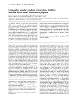
1.1 Singapore mortality rates for all causes from 1990 to 2001 12
1.2 Effects of tumor blood flow and oxygen-carrying capacity of blood
in tumor tissue
20
1.3 A flow diagram showing how hypoxia leads to therapy resistance
and the development of a more aggressive tumor phenotype
22
1.4 HIF-1 structure and its regulation 23
1.5 Genes that are transcriptionally activated by HIF-1 29
1.6 Activation and functions of p53 34
1.7 A schematic diagram illustrating the domains of p53 36
1.8 Proposed model showing different levels of HIF-1-p53 interactions
in the presence of hypoxia and anoxia
42
1.9 Different p53 isoforms and their mechanisms of production 45
1.10 Numbers of publications in proteomics and genomics each year
from 1995 to 2006 according to PudMed database
47
4.1
Stabilization and accumulation of HIF-1α under hypoxia
69
4.2 A representation of a MS/MS spectrum used to determine protein
abundance ratio in iTRAQ-labeled samples
71
4.3 Gene ontology analysis of potential iTRAQ targets affected by p53
and hypoxia according to their biological functions using IPA tool
75
4.4 A graphical display of a merged top 3 protein-protein interaction
network generated by IPA tool from the 54 iTRAQ target proteins
with at least 30% abundance change in protein expression level
78
4.5 A protein expression and interaction network of proteins, involved
in cellular growth, proliferation and cell cycle, under hypoxia in
the presence and absence of p53
79
4.6 A protein expression and interaction network of proteins, involved
in cellular growth, proliferation and cell cycle, in the absence of
p53 under normoxia and hypoxia
80
4.7 A subset of iTRAQ targets chosen for downstream validation 83
4.8 Representative graphs of real-time PCR results for targets selected
from iTRAQ results
87
4
4.9 Downstream validations of iTRAQ results by immunoblotting 90
Supplementary figure
1 Dissociation curve and amplification plot of PGK1 primer set 124
5
LIST OF TABLES
Table Title Page
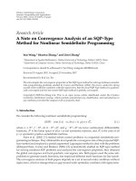
1.1 Colorectal cancer staging, stage distribution, and survival 12
1.2 Genes upregulated by HIF-1 classified into four main
categories based on their biological involvements
30
3.1 Preparation for different percentages of SDS-PAGE gels 56
4.1 Number of proteins identified by LC/MS/MS through
iTRAQ-based quantitation strategy
72
4.2 Number of potential protein targets influenced by p53
and/or hypoxia satisfying the given criteria
72
4.3 Top 5 funcitons and diseases identified by IPA 76
4.4 Tabulation of common proteins regulated in cells under
hypoxia in the presence and absence of p53 as well as in the
absence of p53 under hypoxia and normoxia
81
4.5 List of selected targets based on iTRAQ result and selection
criteria for downstream validations
86
Supplementary table
1
List of primers used for RT-PCR. All primers are arranged
in the order of 5’ to 3’
125
2 Tabulation of the 54 targets selected from iTRAQ analysis 127
6
LIST OF ABBREVIATIONS
WT-N Wildtype treated under normoxia
WT-H Wildtype treated under hypoxia
KO-N p53 knockout treated under normoxia
KO-H p53 knockout treated under hypoxia
SDS-PAGE Sodium dodecyl sulphate polyacrylamide gel electrophoresis
iTRAQ Isobaric Tags for Relative and Absolute Quantification
MS Mass spectrometry
MS/MS Tandem mass spectrometry
LC Liquid chromatography
RP Reverse phase
TOF Time of flight
MALDI Matrix-assisted laser desorption/ionization
RPA Relative peak area
m/z mass to charge ratio
RT-PCR Real-time polymerase chain reaction
PCD Programmed Cell Death
PTM Post-translational modification
PMSF Phenylmethylsulfonyl fluoride
DEPC DiethylenePyrocarbonate
7
CHAPTER 1 – INTRODUCTION
Cells are constantly kept in a dynamic equilibrium of proliferation and cell
death. At any given time point in their life spans, the number of cells in each organism
is kept relatively constant, with each individual cell being highly regulated by
transcription factors that control the expression of genes to synthesize the necessary
proteins for carrying out all cellular functions in order to maintain this homeostatic
condition and viability in response to extracellular biological (e.g. hormones and
neurotransmitters) and non-biological (e.g. temperature and oxygen fluctuations)
signals. In the presence of cellular dysfunction, genes regulating cell cycle (e.g. p16,
p21
WAF1/CIP1
, p53, cyclins, and CDKs) will be activated to arrest the cell for repair
(Brugarolas et al., 1995; Gartel and Radhakrishnan, 2005; Zhang et al., 1994). If the
damage is extensive and beyond repair, the dysfunction cells will be directed for
programmed cell death (PCD) by activation of pro-apoptotic genes such as p53, BID,
BAX and caspases. Hence, a constant homeostatic condition is maintained in the body.
In cancer, this dynamic equilibrium does not exist or is being disrupted. Thus,
cells proliferate uncontrollably and are more resilient to cell death. This phenomenon
is mainly due to multiple genetic alterations or mutations in the genome that impaired
the cell’s ability to regulate its cellular activities normally (Calabretta et al., 1985;
Renan, 1993). As a result, in the presence of cellular dysfunction, the cell is not
arrested and bypasses PCD, leading to tumor formation and cancer development.
Therefore, tumors are characterized by cells which have the ability to escape the
natural cell death program that maintain cellular homeostasis. Newly developed tumor
can be benign initially and are non-cancerous. However, they can develop, gain
8
malignancy and become capable of invading into surrounding tissues or metastasize –
a key characteristic of cancer cells (Hanahan and Weinberg, 2000). Other hallmarks
of cancer include the ability to evade apoptosis, self-sufficiency in growth signals,
insensitive to anti-growth signals, sustained angiogenesis and unlimited replicative
potential.
Cancer develops as a result of a series of genetic mutations, which gives rise
to the 6 key characteristics of cancer. Among the many genes that are affected, the
gene that encodes for a transcription factor known as p53 is frequently found mutated
in cancer. The p53 protein is also a well known tumor suppressor protein that plays
important roles in cell cycle arrest and apoptosis in the event of cellular dysfunctions
(Yu et al., 1999). However, these functions of p53 can be abolished when mutations
occur in the p53 gene or its upstream/downstream regulating genes. This results in
cellular dysfunction and cells containing genetic defects get propagated, leading to the
development of cancer.
In cancer, the microenvironment plays an important part in affecting cancer
progression as well as cancer treatment. The presence of hypoxic microenvironment is
a common phenomenon observed in many cancer tumors. Rapid cell growth during
cancer development results in the disruption of vascular network within the cancerous
tissue/tumor. As a consequence, the supply of oxygen and nutrients supplied to the
cells becomes inadequate and certain regions in the tumor become hypoxic (Semenza,
2000b). Hypoxic cells in tumors undergo a series of biological changes in order to
survive since hypoxia is an unfavorable condition for cell growth. These biological
changes are controlled by a major transcription factor, called hypoxia inducible
9
factor-1 (HIF-1), that acts as a chief regulator of oxygen homeostasis. The activation
of HIF-1 downstream target genes promotes the survival of hypoxic cells as well as
selection of apoptosis-resistant cells in the tumor and hence, promoting a more
malignant cancer phenotype (Giaccia et al., 2004).
Interestingly, although hypoxia positively correlates with tumor malignancy,
several contrasting reports have indicated that hypoxia can cause accumulation of p53
in a HIF-1 dependent manner as well as inducing cell death via the p53-dependent
pathway (An et al., 1998; Graeber et al., 1994; Yao et al., 1995). These conflicting
findings question the intriguing, yet elusive, relationship between hypoxia, HIF-1 and
p53. Thus, it is critical to elucidate this complex relationship to better understand the
hypoxic effects in cancer progression.
In the following chapters, I aim to review the intertwining relationship shared
between cancer, p53 and HIF-1 under hypoxic condition. This chapter may also
provide the updated background of my thesis studies.
1.1 CANCER
1.1.1 Cancer Development
Cancer is a disease of genes and it involves dynamic genetic alterations or
mutations in the genome that produce over-active oncogenes (gain-of-function) and
inactivated/attenuated tumor suppressor genes (loss-of-function) (Bishop and
Weinberg, 1996). The former promotes the abnormal rapid proliferation and survival
of cells under unfavorable condition while the latter allows cells to evade cell cycle
arrest/checkpoints and thus, apoptosis too. Although most types of cancers have been
10
reported to be sporadic, some are recognized as hereditary due to the inheritance of a
mutated allele, often a tumor suppressor. A classic example is familial adenomatous
polyposis (FAP) which is an autosomal dominant inherited colorectal cancer
syndrome. The cause of this disorder has been attributed to germline mutations in the
adenomatous polyposis coli (APC) gene inherited from the parents (Lamlum et al.,
2000; Miyoshi et al., 1992). APC gene is a tumor suppressor gene that promotes
apoptosis in colonic cells and is also involved in the sequestration of β-catenin, which
leads to an inhibitory effect on the β-catenin’s stimulatory effects on the cells
(Neufeld et al., 2000). Mutations in the APC gene result in a truncated/non-functional
protein that does not trigger apoptosis and instead allows β-catenin to accumulate in
the cell, promoting abnormal cell proliferation. Thus, FAP patients are characterized
by multiple non-cancerous polyps growing in the colon and the number of polyps will
increase with age. If these benign polyps are not removed, they will eventually
become malignant and develop into colorectal cancer.
On the other hand, patients with sporadic cancers do not inherit cancer-causing
mutated alleles/genes from their parents. Instead, the spontaneous mutations are
usually the results of DNA damage that can be caused by exposures to carcinogens
and/or mutagens. Carcinogens are cancer-causing agents (e.g. asbestos, cigarette
smokes, acrylamide, etc.) and typically, mutagens are carcinogenic as multiple
mutations will lead to development of cancers. Mutagens are any substance that
causes genetic mutations; for example, ethidium bromide (EtBr), nitrous acid (HNO
2
),
sodium azide (NaN
3
) and radiations (ultraviolet and gamma). Some carcinogens do
not cause mutations but affect the level of transcriptions of certain genes that are
critical to cell regulation instead. Furthermore, not all genetic mutations are caused by
11
mutagens. Some are due to errors in DNA replication (e.g. base pair substitution,
frame-shifts), repair (e.g. mismatch repair) and recombination of DNA sequences.
Normally, these genetic errors would be repaired or the cells would be destroyed if
the genetic damage is irreparable; but due to the multiple mutations, the genetic
defects get retained and are propagated to future generations which can lead to cancer
development. Typically, highly proliferating tissues such as liver and bone marrow,
which divide more frequently, will have a higher risk of developing cancer.
Cancer can occur in any person, regardless of races, genders and ages.
Generally, the risk increases with age too (Jemal et al., 2006) and there are many
types of cancers (e.g. breast, colorectal, skin, prostate, cervical, etc.), with occurrences
reported in most, if not all, tissues in human. Currently, it is reported that there are
more than 11 million people diagnosed with cancer each year and the number of new
cases reported will soar to a predictive number of 16 million every year by 2020 (Cho,
2007). Cancer is also one of the leading causes of death in the world, accounting for
7.6 million (13%) of the global mortality in 2005 alone (Cho, 2007). In Singapore,
death by cancer is the 2
nd
highest mortality rate listed (Figure 1.1) according to a
report released by National Cancer Center Singapore (NCCS), with colorectal cancer
(CRC) as the commonest cancer diagnosed – with 1 in 4 cancer patients detected
(Seow et al., 2004). The average 1- and 5-year survival rates
1
for CRC are 83% and
62% respectively (Kauh and Umbreit, 2004). However, if CRC is detected at an early
stage (modified Dukes’ stage A and B), the 5-year survival percentage has been
shown to be higher compared to detection at a later stage (modified Dukes’ stage C
and D) (Table 1.1). Yet, only a low percentage of CRC patients are typically detected
1
Cancer survival rates or survival statistics indicates the percentage of people who survive a certain type of cancer
for a specific amount of time and they are based on research that comes from information gathered on a big
population of cancer patients.
12
Figure 1.1: Singapore mortality rates for all causes from 1990 to 2001. Death caused
by cancer was maintained constantly at 2
nd
place with no sign of decrease. Extracted
from NCCS Singapore Cancer Registry report volume 6, pp 9 (Seow et al., 2004).
Table 1.1: Colorectal cancer staging, stage distribution, and survival. Data obtained
is just a representation as actual percentage might vary among different surveys.
(Extracted from Melville et al., 1998)
13
at the early stages (Kauh and Umbreit, 2004; Melville et al., 1998). Therefore, it is
critical to have more sensitive and accurate cancer diagnostic methods such that
patients suffering from cancer can be diagnosed at an earlier stage. This leads to a
strong urgency and requirement for the development of key biomarkers.
1.1.2 Colorectal Cancer
Colorectal cancer refers to the cancer of the colon and rectum. The colon is the
longest portion of the large intestine, measuring about 5 to 6 feet in length. The main
function is to convert liquid stool into solid stool by absorbing excess water into the
body. This process can take several hours to several days. On the other hand, the
rectum, which is located at the end of the colon, is about 5 inches in length and is
usually empty except prior to excretion of stool. CRC normally develops initially in
the colon and spread to the rectum in most cases, thus leading to the commonly use of
the combined name. It may be hereditary or spontaneous. However, only about
5~10% of CRC are linked to inherited genes, e.g. APC, MYH. There are many causes
that have been proposed to influence CRC development and some examples are
family history, diet, environment, gender, lifestyle, and the number of existing polyps.
1.1.3 Diagnosis & Treatment
Diagnosis of cancer is an attempt to accurately identify the origin and
malignancy of the disease, as well as the type of cells involved. The effectiveness of
treatment and prospects for survival depend critically on early detection of cancer.
Currently, diagnostic methods in practice include the use of ultrasound equipment to
detect lumps, blood tests, computed tomography (CT) scan, and tumor biopsy. An
example of cancer markers is carcinoembryonic antigen (CEA) used for detecting
14
several types of cancer (such as gastrointestinal, lung and breast cancers). However,
the sensitivity and specificity of these diagnostic methods are often insufficient and
inaccurate. Moreover, early detection of cancer is made more difficult due to the lack
of specific symptoms in the early stage (before invasion – Dukes’ stage A) as well as
limited understanding of etiology and oncogenesis. For example, the use of CA 15-3,
a blood tumor marker for breast cancer, is useless for early detection as it has low
sensitivity (41.9%) (Lumachi et al., 2000). Thus, there is a critical need for an
expedited development of biomarkers with greater specificity and accuracy and the
use of proteomic technique is a common approach used for identification of novel
potential biomarkers that can be used for cancer diagnosis and even cancer therapies.
Conventionally, cancer patients undergo a combination series of therapeutic
treatments involving surgery (excision of tumors), radiotherapy, and chemotherapy to
control and eradicate the cancer cells from their bodies. Radiotherapy involves the use
of ionizing radiations, usually X-rays, to damage DNA and kill the cancer cells while
chemotherapy utilizes chemical substances, called anticancer chemo-drugs, to treat
cancer. Adriamycin
®
, Platinol
®
(cisplatin), 5-fluorouracil and hydroxyurea are some
common examples of chemo-drugs used in chemotherapy to slow and hopefully halt
the growth and spread of a cancer. These chemodrugs are developed to (i) damage
DNA in cells (induce apoptosis), (ii) inhibit new DNA strands synthesis (inhibits
repair), and/or (iii) stop mitosis/cytokinesis (inhibits cell multiplications). Nonetheless,
like radiotherapy, a majority of these drugs are not specific, i.e. they target normal
cells too, and often many common side effects (e.g. hair loss, weight loss, edema, etc.)
arise when used in cancer therapy. Furthermore, the administration of cancer
treatments and their efficiencies are often influenced or hindered by various biological
15
and non-biological factors, including tumors’ location(s), the stage of cancer
development, presence of drug resistance transporters, altered drug metabolism,
altered DNA repair, over-expression of anti-apoptotic genes, inactivity of pro-
apoptotic genes, and non-autonomous features of tumor growth in vivo, such as the
presence of hypoxic microenvironments in solid tumors (Albiero and Pozzi, 1994).
1.1.4 Hypoxic Effects on Diagnosis, Treatments and Prognosis
The effects of oxygen are of interest in cancer treatment because high levels of
hypoxia in tumors have been shown positively to be correlated with treatment failure
or relapse for many cancers, independently of treatment (Brizel et al., 1997; Fyles et
al., 1998; Sundfor et al., 2000). Solid tumors are often in a low-oxygen state known as
hypoxia due to the existence of limited arteriolar supply and arteriolar deoxygenation
(Dewhirst et al., 1996), low vascular density and disrupted vascular architecture
(Secomb et al., 1993), insufficient oxygen supply (Secomb et al., 1995), and an
unstable blood supply to the tumor cells (Kimura et al., 1996). Although angiogenesis
and neovascularisation do occur in these tumors, the newly formed blood vessels are
often inadequate, disorganized and prone to collapse (Helmlinger et al., 1997).
Together, these physiological factors contribute to the formation of hypoxic
microenvironments/regions heterogeneously distributed within the solid tumors
(Padhani et al., 2007; Semenza, 2003).
The presence of hypoxic regions poses a huge obstacle for effective cancer
therapies as cells in hypoxic regions are less sensitive to the effects of radiotherapy
and chemotherapeutic drugs than their normal counterparts (Erler et al., 2004; Teicher,
1994; Vaupel, 2004). In radiotherapy, oxygen are essential to make DNA damage
16
permanent and it is known that DNA damage can be chemically restored in hypoxia
(Alper and Howard-Flanders, 1956; Harrison et al., 2002). According to the oxygen
fixation hypothesis (OFH), developed based on the works of Alexander and Charlesby
on polymer chemistry in the 1950s, oxygen is a radiation sensitizer. DNA radicals
produced by radiation will react with oxygen to form organic peroxides that in turn
“protects” radiation-damaged DNA from restoring to an undamaged state. Stable
DNA damage accumulates and leads to an increased lethality from a given dose of
radiation in cells, inducing apoptosis eventually. Therefore, in a hypoxic condition,
DNA damage is not accumulated as much as under normal condition and tumors
become more resistant to the effects of radiation. The solid mass of tumor also makes
it difficult for radiation to penetrate into the tumor core. It has been reported that for a
similar biological effect in hypoxic tissues as in normoxic tissues, a higher therapeutic
dose of 2.5- to 3-fold of radiation (e.g. x-rays and gamma rays) is required (Teicher,
1995; Wachsberger et al., 2003) or only about one third lethal DNA lesions reported
in hypoxic cells compared with aerobic cells when subjected to the same amount of
irradiation (Koch, 1982). Much research has been done to improve radiotherapy
efficiency on solid tumors and one such promising method is the use of metal-based
small molecules, such as
64
Cu-ATSM, as agents for higher effective cancer
radiotherapy (Lewis et al., 2001; Obata et al., 2005).
The disordered tumor cell profusion and constricted blood vessels that
contributes to hypoxia also leads to an inefficient delivery of some chemotherapeutic
drugs to the site of action as the delivery relies on the tumor vasculature. Studies on
solid tumor cells have also further suggested that through induction of apoptosis by
cytotoxic chemotherapeutic drugs, hypoxia may select for cells with defective
17
apoptotic regulators such as p53 and thus, gaining a more malignant phenotype in the
end (Graeber et al., 1996). Furthermore, the presence of multiple hypoxic regions
within a tumor may confer tumor inhomogeneity, resulting variations towards
treatment sensitivities (Vaupel et al., 2002). Inevitably, this may contribute to relapses
even after years of remission as not all cancer cells were eradicated by the treatment.
In cancer treatment, the level of hypoxia in a tumor may also be used to help
predict the response of the tumor to the treatment. The poor prognosis association
with tumor hypoxia has stimulated the development of equipment for measuring
oxygen concentrations of tumors in vivo. Such tools can be used to evaluate patient-
specific distributions of hypoxia within a tumor so that more effective treatment can
be administered. An example is the use of polarographic electrodes, commonly
known as the Eppendorf electrode, to measure partial pressure of oxygen (pO
2
) of
tumor in vivo (Fyles et al., 1998; Movsas et al., 2002; Parker et al., 2004). The
downside is that this method is invasive and it is restricted to only superficial tumors.
On the other hand, techniques such as positron emission tomography (PET) and the
use of endogenous hypoxia-induced proteins can allow the potential for non-invasive
assessment of a tumor’s hypoxic condition. Hence, there is a paramount importance
for a deeper understanding of the biological mechanism behind hypoxia in tumors in
order for the discovery of endogenous protein markers as well as the development of
more effective and sensitive cancer treatments.
18
1.2 HYPOXIA
1.2.1 The Nature of Hypoxia
Hypoxia is a condition in which the level of oxygen supplied to the
body/tissue becomes inadequate, i.e. much lesser than the norm. It is also a hallmark
characteristic of most tumors and tumor hypoxia results from an imbalance between
the cellular oxygen consumption rate and the oxygen supply to the cells (Semenza,
2003; Vaupel and Harrison, 2004). During tumor expansion, growing cells rapidly
outstrip the supply of oxygen and nutrients while the growing cell mass also limits the
availability of oxygen and nutrients to each individual cell by existing blood vessels.
Formation of new blood microvessels within the tumor (i.e. tumor neovascularisation)
would be required for growth beyond 2 mm in order to supply adequate oxygen and
nutrients to the cells. Although many factors can contribute to tumor hypoxia, they
can be classified generally into 3 types – perfusion-, diffusion-, or anemia-related
(Hockel and Vaupel, 2001; Padhani et al., 2007; Vaupel et al., 2002). Perfusion-
related hypoxia is an acute type of hypoxia and it is often temporary. It arises as a
result of inadequate blood flow (ischemic) in the tissues due to severe structural and
functional abnormalities of tumor neovascularisation, such as disorganized vascular
network, dilations, lack of functional receptors, incomplete endothelial lining, absence
of flow regulation, and an elongated tortuous shape. Diffusion-related hypoxia, on the
other hand, is a chronic type of hypoxia that results as a consequence of tumor
expansion which increases the oxygen diffusion distance. Tumor cells that are distant
(greater than 70 µm) from the microvessels receive inadequate oxygen supply.
Anaemic hypoxia results from reduced oxygen-carrying capacity of the blood which
may be due to factors relating to treatments or tumor-associated. Furthermore, it has
been shown experimentally that the combined effects of low blood perfusion rate to
19
tumors and low oxygen-carrying capacity of blood amplifies hypoxia due to lowered
oxygen supply to the tumors (Figure 1.2) (Vaupel et al., 2001).
1.2.2 The Flipside of Hypoxia
Interestingly, despite conferring resistance to cancer treatment, hypoxia can
also have a direct toxic effect as a form of stress on many cell types. Numerous
reports have shown that hypoxia can induce necrosis and apoptosis in normal cells
(Yamaguchi et al., 2001; Zhu et al., 2002) as well as in tumor cells with cell death
observed most notably in the zones furthest from the tumor vasculature (Shimizu et al.,
1995; Yao et al., 1995). Therefore, this illustrates hypoxia with two seemingly
opposing effects on tumor biology – one protective and the other toxic. The toxic
effect of hypoxia is exhibited by its ability to arrest cell at G
0
/G
1
checkpoint and
induce p53 accumulation, which can lead to p53-dependent PCD (Graeber et al.,
1994). Although, p53 is known to be involved in cell cycle regulation, many reports
indicated that hypoxia-induced p53 is transcriptionally inactive but serves more as a
transcription repressor in tumor cells (Koumenis et al., 2001). Further evidences have
also indicated that p53 accumulation induced by hypoxia did not induce p21
WAF1/CIP1
,
a well-established p53 downstream gene involved in cell cycle G
1
arrest (Gartel and
Radhakrishnan, 2005; Koumenis et al., 2001). Therefore, the accumulation of p53
during hypoxia does not play a role in cell cycle arrest. Hypoxia has also been
implicated with the development of a more malignant cancer phenotype and
metastases through functioning as a selection pressure for p53-deficient tumor cells
with reduced apoptotic potential to hypoxic areas within the tumors (Graeber et al.,
1996). While it has been widely known that hypoxia can protect tumors by increasing
their resistance to radiotherapy and chemotherapy, there is a possibility that these