Identification and characterization of protein kinase CK2 as a novel interacting protein of neuronal CDK5 kinase and its functional role in microtubule dynamics
Bạn đang xem bản rút gọn của tài liệu. Xem và tải ngay bản đầy đủ của tài liệu tại đây (2.96 MB, 182 trang )
I
I
D
D
E
E
N
N
T
T
I
I
F
F
I
I
C
C
A
A
T
T
I
I
O
O
N
N
A
A
N
N
D
D
C
C
H
H
A
A
R
R
A
A
C
C
T
T
E
E
R
R
I
I
Z
Z
A
A
T
T
I
I
O
O
N
N
O
O
F
F
P
P
R
R
O
O
T
T
E
E
I
I
N
N
K
K
I
I
N
N
A
A
S
S
E
E
C
C
K
K
2
2
A
A
S
S
A
A
N
N
O
O
V
V
E
E
L
L
I
I
N
N
T
T
E
E
R
R
A
A
C
C
T
T
I
I
N
N
G
G
P
P
R
R
O
O
T
T
E
E
I
I
N
N
O
O
F
F
N
N
E
E
U
U
R
R
O
O
N
N
A
A
L
L
C
C
D
D
K
K
5
5
K
K
I
I
N
N
A
A
S
S
E
E
A
A
N
N
D
D
I
I
T
T
S
S
F
F
U
U
N
N
C
C
T
T
I
I
O
O
N
N
A
A
L
L
R
R
O
O
L
L
E
E
I
I
N
N
M
M
I
I
C
C
R
R
O
O
T
T
U
U
B
B
U
U
L
L
E
E
D
D
Y
Y
N
N
A
A
M
M
I
I
C
C
S
S
LIM CHEE BENG
(B.Sc. (Hons), University of Melbourne)
A THESIS SUBMITTED
FOR THE DEGREE OF DOCTOR OF PHILOSOPHY
INSTITUTE OF MOLECULAR AND CELL BIOLOGY
THE NATIONAL UNIVERSITY OF SINGAPORE
2004
Acknowledgements
Robert Qi, my supervisor, for guidance, constant support and encouragement, his
critical reading of all my manuscripts. Also for his constant inspiration throughout all
the years.
Walter Hunziker, my co-superviser, for his support, guidance, encouragement and
critical reading of all my manuscripts.
Edward Manser and Cao Xinmin, my supervisory committee members, for useful
advices and critical comments on my work.
Alice Tay for her administrative support, Tang Bor Luen for sharing his materials and
helpful discussions and Wong Boon Seng for helpful advices and critical comments of
my manuscript. All the past and present members of IMCB and our laboratory for
making it a great place to work in, as well as for all the help, advices and scientific
discussions
My parents and siblings for their constant moral support.
Finally, the biggest Thank-you to my wife Liting, for her love, encouragement and
support throughout all the years.
II
Table of Contents
Title page I
Acknowledgements II
Table of Contents III
Abbreviations VII
Abstract XI
1 Introduction
1
1.1 Protein Kinase CK2:Composition and Structure 3
1.1.1 Tissue-specific distribution and subcellular localization 7
1.1.2 Regulation of CK2 9
1.1.3 Biological effects of CK2 13
1.1.3.1 Regulation of adhesive proteins 13
1.1.3.2 Regulation of cytoskeletal elements 14
1.1.3.3 Regulation of substrates involved in signal transduction 15
1.1.3.4 Regulation of proteins associated with synaptic vesicle recycling 16
1.1.3.5 Regulation of transcription factors 17
1.2 Cyclin-Dependent Protein Kinase Family 19
1.3 Neuronal Cdk5 kinase 23
1.3.1 Regulation of Cdk5 27
1.3.2 Physiological roles of Cdk5 and its mediated functions 30
1.3.2.1 Cdk5 in cytoskeletal dynamics and microtubule-based transport 30
1.3.2.2 Cdk5 in synapses and focal adhesion sites 31
1.3.2.3 Cdk5 in neurosignaling 33
III
1.3.2.4 Cdk5 in transcriptional machineries 34
1.3.3 Molecular organization of Cdk5 complexes 36
1.3.3.1 Methods used in isolating protein-interacting partners 38
1.4 Microtubule Dynamics 40
2 Materials and Methods 46
2.1 Materials 47
2.1.1 Chemicals and reagents 47
2.1.2 Cell lines 48
2.1.3 Antibodies 48
2.2 Experimental Procedures 50
2.2.1 Plasmid constructions 50
2.2.2 Recombinant protein preparation 50
2.2.3 Isolation of p35-binding proteins 52
2.2.4 Mass spectrometry 53
2.2.5 Biochemical binding assays 53
2.2.6 Protein size exclusion chromatography 54
2.2.7 Cdk5 In vitro kinase assay 55
2.2.8 Transient transfections 55
2.2.9 Immunoprecipitation 55
2.2.10 Microtubule assembly 56
2.2.11 Differential tubulin extraction from intact cells 56
2.2.12 RNAi 57
2.2.13 Immunofluorescence microscopy 57
2.2.14 Miscellaneous techniques 58
IV
2.2.15 Statistical analysis and presentation of data 58
3 Results and Discussion 60
3.1 Isolation of p35-associated proteins and identification of
protein kinase CK2 as an inhibitor of neuronal Cdk5 kinase 61
3.1.1 Introduction 62
3.1.2 Results 64
3.1.2.1 Isolation of p35-binding proteins by affinity purification and co-
immunoprecipitation 64
3.1.2.2 Identification of CK2
α
as a p35-binding protein 67
3.1.2.3 CK2α associates with p35 and Cdk5 in vivo 69
3.1.2.4 Direct association of CK2 with p35 and Cdk5 71
3.1.2.5 CK2 inhibits Cdk5 activation 74
3.1.2.6 CK2 inhibits Cdk5 in a phosphorylation-independent manner 78
3.1.2.7 CK2 blocks complex formation between Cdk5 and p35 80
3.1.3 Discussion 83
3.2 Direct regulation of Microtubule Dynamics by Protein
Kinase CK2 87
3.2.1 Introduction 88
3.2.2 Results 90
3.2.2.1 CK2 forms a direct complex with microtubules 90
3.2.2.2 CK2 induces microtubule polymerization 94
3.2.2.3 CK2 stabilizes microtubule in vivo 101
3.2.3 Discussion 105
V
4 Summary and Perspectives
108
5 References
116
6 Appendix
139
6.1 Tables 140
6.2 Publications 145
VI
Abbreviations
a.a. amino acid
AD Alzheimer’s disease
APP amyloid precursor protein
ATP adenosine triphosphate
c-abl c-Abelson
CAK Cdk activating kinase
CaMKII Ca
2+
/calmodulin-dependent protein kinase II
cAMP cyclic adenosine monophosphate
Cdk cyclin-dependent kinase
Cdk5 cyclin-dependent kinase 5
cDNA complementary deoxyribonucleic acid
CK1 casein kinase 1
CK2 casein kinase 2
CKI Cdk inhibitor
CNS central nervous system
cpm counts per minute
DARPP-32 dopamine- and cAMP-regulated phosphoprotein, M
r
32 kDa
dATP deoxy-adenosine triphosphate
dbpA DNA-binding protein A
DEAE diethylamino-ethylcellulose
DMEM Dulbecco’s modified Eagle’s medium
DNA deoxyribonucleic acid
DTT 1,4-dithiothreitol
VII
ECL enhanced chemiluminescence
EDTA ethylenediaminetetra acetic acid
EGTA ethyleneglycoltetra acetic acid
ER endoplasmic reticulum
FAK focal adhesion kinase
FCS fetal calf serum
FPLC fast protein liquid chromatography
GSH glutathione
GSK-3 glycogen synthase kinase-3
GST glutathione-S-transferase
GTP guanosine 5’-triphosphate
HA hemagglutinin
Hepes N-(2-hydroxyethyl)piperazine-N’-(1-ethane sulphonic acid)
hr hour
HSP-70 heat shock protein 70
INK4 inhibitor of Cdk4
IPTG isopropyl-1-thio-β-galactopyranoside
JNK c-Jun N-terminal kinase
kDa kilo Dalton
KIP kinase inhibitor protein
LB Luria-Bertani medium
M molar
MAP microtubule-associated protein
MAPK mitogen-activated protein kinase
MEF2 myocyte enhancer factor 2
VIII
MEK mitogen-activated protein kinase kinase
min minute
ml milliliter
MOPS 4-morpholinepropanesulfonic acid
M
r
molecular mass
NFH heavy chain of neurofilament protein
NFM intermediate chain of neurofilament protein
NGF nerve growth factor
nickel-NTA nickel nitrilotriacetic acid
NLS nuclear localization signal
NMDA N-methyl-
D-aspartate
NP-40 nonidet P-40
PAGE polyacrylamide gel electrophoresis
PAK p21-activated protein kinase
PBS phosphate buffered saline
PCR polymerase chain reaction
PI3-K phosphatidylinositol 3’-kinase
PIPES piperazine-N,N’-bis-(2-ethanesulfonic acid)
PKA cAMP-dependent protein kinase
PKC protein kinase C
PMSF phenylmethylsulphonyl fluoride
PP1 protein phosphatase 1
PP2A protein phosphatase 2A
pRb retinoblastoma protein
PVDF polyvinylidene difluoride
IX
rpm revolutions per minute
RNAi ribonucleic acid interference
RT reverse transcription
SDS sodium dodecyl sulfate
siRNA small interfering ribonucleic acid
sec second
Tris 2-amino-2(hydroxymethy)-1-3-propanediol
UV ultraviolet
X
Abstract
Neuronal cyclin-dependent kinase 5 (Cdk5) has been shown to play an
important role in a variety of cellular processes, including neuronal cell
differentiation, apoptosis, neuron migration and synaptic plasticity (Dhavan and Tsai,
2001; Lim et al., 2003). The active kinase consists of a catalytic subunit, Cdk5, and a
regulatory subunit, p35 or p25, which are expressed primarily in neurons. Little is
known about the regulation of Cdk5/p35 kinase turnover/activity, apart from the fact
that it is degraded through the ubiquitin proteosome pathway (Patrick et al., 1998).
We were interested to explore the regulation of neuronal Cdk5 activity. To achieve
this, p35 and its fragments were made as GST-tagged fusion proteins and utilized in
biochemical affinity purification attempts to isolate novel p35-binding proteins. We
have also employed a co-immunoprecipitation approach in our search for novel
interacting proteins. Using the former approach, the catalytic α subunit of protein
kinase CK2 (formerly known as casein kinase 2) was isolated from rat brain extracts.
The direct associations of CK2 with p35, as well as with Cdk5, were demonstrated
and the CK2-binding sites of p35 were delineated. We showed that CK2 exhibited a
strong inhibition on Cdk5 activation by p35 in vitro and in vivo. Cdk5 inhibition
however is not associated with CK2 kinase function, since a kinase-dead CK2 mutant
displayed a similar level of Cdk5 inhibitory activity as the wild-type protein.
Interestingly, further analysis revealed that CK2 acts by blocking the formation of a
complex between Cdk5 and p35. Hence, CK2 exerts a direct negative effect on Cdk5
activation by p35 through its physical interaction with p35.
Regulation of microtubule dynamics is essential for many vital cellular
processes such as morphogenesis and motility and Cdk5-p35 complex co-exists with
XI
microtubules in the brain (Sobue et al., 2000; Paudel et al., 1993). Since we have
identified CK2 as a p35-interacting protein and previous studies have implied that
CK2, a ubiquitously expressed protein kinase involved in diverse cellular functions
(Litchfield, 2003; Meggio and Pinna, 2003), may be involved in regulating
cytoskeleton reorganization (Serrano et al., 1987; Diaz-Nido et al., 1988; Serrano et
al., 1989), we therefore investigated if CK2 is also involved in mediating microtubule
dynamics. The CK2 holoenzyme is composed of two catalytic α or α’ subunits and
two regulatory β subunits. We showed that the α subunit of CK2 binds directly to
both microtubules and tubulin heterodimers. The CK2 holoenzyme (but not its
individual subunits) exhibited a potent effect in inducing microtubule assembly and
bundling. Moreover, polymerized microtubules were strongly stabilized by CK2
against cold-induced depolymerization. In addition, the kinase activity of CK2 is not
required for its microtubule-assembly and stabilizing function since a kinase-inactive
mutant of CK2 displayed similar microtubule-assembly activity as the wild-type.
Knockdown of CK2α/α' in cultured cells by RNA interference dramatically
destabilized their microtubule networks. The destabilized microtubules were thus
readily disrupted by colchicine at a very low concentration. Further, over-expression
of chicken CK2α or its kinase-inactive mutant in CK2α/α'-depleted cells fully
restored microtubule resistance to low doses of colchicine. To our knowledge, these
findings demonstrate for the first time that CK2 is a microtubule-associated protein
that confers microtubule stability in a phosphorylation-independent manner.
XII
Section 1
Introduction
1
1. Introduction
The ability of cells to function and proliferate depends largely on their
response to the immediate intracellular environments as well as the external stimuli.
These cellular signals trigger a specific set of mechanisms within the cells to bring
about a change in cell function. These mechanisms are highly regulated to control
cellular functions, commonly by means of changes in protein conformation. A process
of signal transduction conveys the external message to the internal cellular organelles.
Protein phosphorylation is one such mechanism, and is catalyzed by enzymes
known as protein kinases, while protein phosphatases catalyze the reverse process,
dephosphorylation (Cohen, 2002; Hunter, 2000). Protein kinases are classified into
the serine/threonine-specific, tyrosine-specific, histidine-specific or the dual
specificity (Ser/Thr and Tyr) class of kinases, depending on the residue being targeted
for phosphorylation. All known protein kinases of this class share a related catalytic
domain and are distinguished by their unique regulatory domains.
Mammalian brains are highly compartmentalized into groups of functionally
specialized neurons. Cell migration and neurite outgrowth must be tightly
orchestrated to achieve this level of organization. Likewise, cellular processes such as
DNA replication and cell division must also be tightly regulated during
embryogenesis to produce a viable organism. Many protein kinases have emerged to
be important regulators of these processes. Together, they play distinct roles in
coordinating the transition of different cellular functions.
2
1.1 Protein Kinase CK2: Composition and Structure
Casein kinases are multifunctional, highly conserved, serine/threonine protein
phosphotransferase that are ubiquitous in yeast and higher eukaryotes (Pinna, 1990).
They are cyclic-nucleotide-independent protein kinases that preferentially
phosphorylate acidic proteins (Hathaway and Traugh, 1982). Two distinct casein
kinases have been found in many different cell types. They have been designated
casein kinase 1 (CK1) and casein kinase 2 (CK2) according to the elution profile
obtained by diethylamino-ethylcellulose (DEAE-cellulose) chromatography
(Hathaway and Traugh, 1979).
Protein kinase CK2 is an oligomeric enzyme with molecular mass (M
r
) of 130-
150 kDa, as determined by sedimentation velocity and equilibrium analysis
(Hathaway and Traugh, 1979; Pinna, 1990), with the exception of a porcine liver CK2
of 210 kDa (Baydoun et al., 1986) and a monomeric human spleen CK2 of 43 kDa
(Gounaris et al., 1987). The holoenzyme of CK2 consists of subunits α, α’ and β
which associate to form several distinct heterotetramers, namely α2β2, α’2β2 and
αα’β2. A number of studies have reported that these subunits may also exist
individually in the cells which are devoid of their counterparts (Stigare et al., 1993;
Heriche et al., 1997; Kusk et al., 1999). The reported Mr for the β subunit, as
determined by gel electrophoresis in sodium dodecyl sulfate (SDS), is usually 24-26
kDa, while the values for the α and α’ subunits ranges from 35 to 44 kDa (Hathaway
and Traugh, 1982; Litchfield et al., 1990).
3
The α, α’ and β subunits are the products of three distinct genes (Allende and
Allende, 1995). All subunits have an extraordinarily high degree of evolutionary
conservation. Firstly, the sequence of the α subunit is largely conserved across
mammalians and other species (Drosophila, chicken, mouse, rat, pig, bovine, human)
where sequence identity ranges from 67 to 90%. Secondly, the α and α’ subunits are
structurally very homologous, despite the differences in their C-terminal regions
(Lozeman et al., 1990). Thirdly, the cDNA sequences of the β subunit of Drosophila,
mouse, rat, pig, bovine and human are also highly homologous (Pinna, 1990).
The α and α’ subunits are catalytically active, whereas the β subunit is
inactive. Identification of catalytic subunits CK2α and CK2α’ was based on their
enzymatic activity in the absence of the β subunit. The functions of the β subunit are
to confer stability, regulate the enzymatic activity of the holoenzyme and the
specificity of the catalytic subunit (Faust and Montenarh, 2000). The catalytic domain
of the α subunit is homologous to the catalytic domains of other protein kinases.
There is a short N-terminal segment, termed “glycine-loop on phosphate anchor”,
which makes contact with the β-phosphate of the bound ATP and is involved in the
recognition of peptide substrates (Fig. 1). A stretch of basic residues, which is
probably the most striking hallmark of CK2, is located just downstream from the
invariant lysine (Lys-68) and is recognized as an essential residue involved in ATP
binding in all protein residues. This high concentration of adjacent basic residues is
unique among protein kinases in this region. This is the region which interacts with
the β subunit and is involved in the down-regulation by the β subunit towards some
substrates (Marin et al., 1997; Sarno et al., 1998). In addition, it is also implicated in
the inhibition of CK2 by heparin (Vaglio et al
., 1996). Another intriguing function for
4
this basic sequence is based on the fact that it falls into the description of a nuclear
localization signal (NLS) that are known to mediate the attachment of molecules to
transporter proteins for their regulated nuclear import (Boulikas, 1996). It is
interesting to note that none of the other protein kinases possess a NLS motif with
more than three basic residues. Although it cannot be fully excluded that the NLS
sequences of α and α’ subunits have little to do with nuclear transport since these
subunits lack an acidic stretch close to the NLS which enhances binding with
transporter proteins (Boulikas, 1996), it is nevertheless possible that these strong
NLS-like motifs allow targeting of the kinase to the nucleus ascribed to this kinase
(Rihs et al., 1991). The next segment contains the functional elements termed the
‘activation loop’, followed by a C-terminal tail that has been shown to be
phosphorylated by the protein kinase p34
cdc2
(Litchfield et al., 1992).
Within the N-terminal of the β subunit of CK2 lies an autophosphorylation
site, which has been shown to be phosphorylated readily in vitro (Meggio et al.,
1989). A cyclin-like ‘destruction box’ also lies within this region of the protein, which
is followed by an acidic region known to be responsible for the intrinsic down-
regulation of CK2 (Meggio et al., 1994). The C-terminal segment is responsible for β-
β dimerization, association with the α subunit, protection against denaturation and
proteolysis, and up-regulation of activity (Boldyreff et al., 1993; Boldyreff et al.,
1996; Marin et al., 1997; Krehan et al., 1996). In addition, this region possess a
phosphorylatable Ser209 which has been shown to be phosphorylated by p34
cdc2
,
although no physiological role has been implicated for this modification (Litchfield et
al., 1992).
5
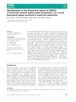
FIG. 1. Schematic diagram depicting various motifs of the CK2 proteins. Within the α subunit of
human CK2, the domain in grey includes the invariant Lys-68 which is involved in ATP binding,
followed by a cluster of basic residues which associates with the β subunit, and a strong karyophilic
putative NLS signal. The domain in pink comprises a series of six basic residues regularly spaced,
which appear to be involved in the recognition of peptide substrates, and the catalytic loop. The domain
in blue contains the functional elements termed ‘activation loop’. There are four phosphorylatable
residues (Thr-344, Thr-360, Ser-362 and Ser-370) which are identified as potential targets for Cdc2
(Litchfield et al., 1992). Within the β subunit of human CK2, the domain in yellow is highly conserved
between organism, and it includes seven acidic residues and is responsible for negative regulation of
CK2 and its association with plasma membrane. The domain in purple is mainly responsible for β-β
dimerization and α-β structural interaction. There is a phosphorylatable serine residue (Ser-209) at the
C-terminal end (Litchfield et al., 1992).
6
1.1.1 Tissue-specific distribution and subcellular localization
CK2 is expressed by various species at different stages of development. In
fact, almost all tissues of all higher organisms express CK2. By in situ hybridization
and CK2 transcripts, Mestres and co-workers showed that both CK2 protein subunits
were detected in nearly all organs of the mouse embryo, suggesting a general role
during embryonic development (Mestres et al., 1994). In general, the level of CK2
transcripts correlates with protein expression. A survey was also made of the activity
of CK2 in extracts made from various tissues of adult rat. The highest activity was
found in brain, testis and liver, whereas CK2 activity in kidney and spleen is low
(Singh and Huang, 1985; Nakajo et al., 1986; Guerra et al., 1999). CK2 activity, as
well as its immunoreactivity, were also present in all brain regions studied (Girault et
al., 1990). CK2 activity was also studied in mouse cortex and caudate-putamen during
development (De Camilli and Greengard, 1986). Its levels was found to be high at
embryonic day 16 and during the early post-natal period, and appeared to decrease
slightly in the adult.
Numerous workers have investigated the subcellular localization of CK2
though many of the initial results were somewhat confusing. However, it turned out
that CK2 was present not only in the nucleus and the cytoplasm (as reported initially)
but nearly everywhere in the cell. Oligomeric forms of CK2 were observed to be
closely associated with the plasma membranes prepared from A431 cells and from
SF9 insect cells expressing the catalytic and regulatory subunits of CK2 (Sarrouilhe et
al., 1998). The holoenzyme seems to be targeted to the plasma membrane by the β-
subunit of CK2. On the other hand, others have reported that CK2 activity and its
immunoreactivity as predominantly cytoplasmic (Singh and Huang, 1985; Girault et
7
al., 1990). Using immunofluorescence and immunoelectron microscopy, Yu and
coworkers showed that CK2α and CK2β are localized to the cytoplasm during
interphase and are distributed throughout the cell during mitosis (Yu et al., 1991). In
contrast, CK2α’ is localized in the nucleus during G1 phase and in the cytoplasm
during the S phase (Yu et al., 1991). Likewise, CK2 immunoreactivity had been
reported to be either associated with the nucleus or distributed between the nucleus
and cytoplasm (Schneider and Issinger, 1989). It is apparent that the variety of nuclear
substrates found for CK2 makes a nuclear/nucleolar function for this protein kinase
likely (Meggio and Pinna, 2003).
A closer look at the subcytosolic localization of CK2 reveals that this enzyme
is also associated with cytosolic organelles. CK2 has been purified from bovine
kidney mitochondria (Damuni and Reed, 1988). An in vivo localization of CK2 at the
membrane of the mitochondria seems to be reasonable since some potential substrates
of CK2 are localized in the matrix of mitochondria (Meggio and Pinna, 2003). CK2
has been identified as an endoplasmic reticulum (ER)-associated kinase responsible
for the in vitro phosphorylation of calnexin and signal sequence receptor-α (Ou et al.,
1992). These proteins are implicated to function as chaperones in the ER. Moreover,
the cytosolic domain of calnexin was found to be phosphorylated in vivo at CK2 sites
Ser534 and Ser 544 and these modifications play a role in targeting calnexin to the
ribosomes (Chevet et al., 1999).
8
1.1.2 Regulation of CK2
CK2 was initially isolated as a cyclic nucleotide-independent protein kinase
that preferentially phosphorylates acidic proteins (Hathaway and Traugh, 1982),
which led to much debate and controversy over its regulation in cells (Litchfield et al.,
1994). The fact that CK2 activity is generally detected in cell or tissue extract even in
the absence of any stimulation or addition of cofactors, or when it is expressed in
bacteria, lends itself to the conclusion that CK2 is constitutively active or unregulated.
Till now, studies reporting on the activation of CK2 in response to a diverse array of
stimuli have not yielded any consistent insights into the mechanisms responsible for
CK2 regulation in cells. Some of the mechanisms that contribute to the regulation of
CK2 in cells include regulated expression and assembly, modulation by covalent
modification and regulatory interactions with protein and/or non-protein molecules.
In the case of the Cdks, it is evident that their kinase activity is absolutely
dependent on the presence of regulatory cyclin subunit (Pines, 1995). In this respect,
CK2β is analogous to the cyclins that it modulates the catalytic activity and substrate
specificity of CK2 as well as the assembly of CK2 complexes. The existence of a
putative destruction box within the sequence of CK2β and the demonstration that
CK2β is ubiquitinated and degraded through a proteasomal pathway further
emphasizes its potential similarities with the cyclins (Zhang et al., 2002a).
Furthermore, it has been reported that CK2 activity oscillates during the cell cycle,
analogous to the Cdks (Carroll and Marshak, 1989; Bosc et al., 1999). Generally, it
appears that CK2 levels correlate to proliferation rate, as cells with higher
proliferation rates generally exhibit higher levels of CK2 (Munstermann et al., 1990).
9
As noted above, CK2 has traditionally been considered a tetrameric enzyme,
with CK2β exerting control over the catalytic activity of CK2 at a number of possible
levels. However, there is mounting evidence to suggest that the catalytic subunits of
CK2 exist outside the tetrameric CK2 holoenzyme. It is intriguing that there are
substrates which can be phosphorylated by CK2α or by CK2α’ but not the tetrameric
CK2 (Marin et al., 1999; Litchfield, 2003). There is a possibility that tetrameric CK2
complexes undergo regulated disassembly in cells. This is supported by recent studies
on the dynamic localization of individual CK2 subunits showing independent
movements of CK2α and CK2β within cells (Martel et al., 2001; Filhol et al., 2003).
Furthermore, recent crystal structure of tetrameric CK2 revealed that the surface
contact between the catalytic and regulatory subunits were considerably fewer than
those typically observed in stable protein complexes (Niefind et al., 2001). In this
respect, CK2 may indeed undergo regulated disassembly and reassembly in cells
(Allende and Allende, 1998).
For many protein kinases, it is apparent that stimulus-dependent
phosphorylation of sites within an activation loop is required for their activation. By
comparison, the catalytic subunit of CK2 exhibit robust activity when expressed in
bacteria in either presence or absence of CK2β (Grankowski et al., 1991; Hinrichs et
al., 1993). Similarly, there has been limited support for the suggestion that
phosphorylation regulates the activity of CK2 in response to cellular stimulation
(Agostinis et al., 1987; Ackerman et al., 1990; Mulner-Lorillon et al., 1990; Palen
and Traugh, 1991; Litchfield et al., 1991). Taken together, these data indicate that
phosphorylation is not absolutely required to activate CK2. On the other hand, both
CK2α and CK2β are phosphorylated in a cell cycle-dependent manner (Litchfield
et
10
al., 1992; Litchfield et al., 1991). Though these sites do not appear to directly effect a
dramatic change in the catalytic activity of CK2, they may, by controlling the stability
of CK2β autophosphorylation, indirectly regulate cellular CK2 activity (Zhang et al.,
2002a). The C-terminal phosphorylation of CK2α may also regulate CK2 indirectly
through interaction of phosphorylated CK2α with the peptidyl-prolyl isomerase Pin1
(Messenger et al., 2002). Interactions between Pin1 and CK2 do not appear generally
to influence CK2 activity, but do inhibit the CK2-catalyzed phosphorylation of
topoisomerase IIα in vitro.
CK2 is typically known to be independent of those small molecules that are
involved in the activation of second messenger-dependent kinases. However, it has
been established that CK2 is inhibited by negatively-charged compounds such as
heparin and activated by positively-charged compounds such as polyamines (Tuazon
and Traugh, 1991). Further finding that CK2 level and activity were elevated in mice
with enhanced polyamine levels, resulting from forced overexpression of ornithine
decarboxylase, supports the possibility that CK2 levels can indeed be modulated by
polyamines in vivo (Leroy et al., 1997).
A large body of evidence indicates that protein-protein interactions represent a
major mechanism for the regulation of specific protein kinases (Pawson and Nash,
2000). The identification of several proteins that interact with CK2 is consistent with
this conjecture that CK2 may be directly, or indirectly, regulated by interacting
proteins. CK2 interacts with proteins such as fibroblast growth factor 1 and HSP-90
that may directly alter or stabilize its catalytic activity (Skjerpen et al., 2002; Miyata
and Yahara, 1995). Studies have demonstrated that CK2 also interacts with other
11
proteins, such as tubulin and FAS-associated factor 1, that may be involved in the
targeting of CK2 to specific sites or structures within the cells (Faust et al., 1999;
Jensen et al., 2001). Overall, it is evident that many distinct mechanisms may
contribute to the regulation of CK2 in the cells. In this respect, it is conceivable that
many distinct, independently regulated subpopulation of CK2 exist in cells in order to
carry out its myriad of cellular functions.
12
1.1.3 Biological effects of CK2
In the last few decades, a great deal of research has been devoted in the study
of CK2 and its cellular implications (Litchfield, 2003; Meggio and Pinna, 2003). By
its interaction with more than 300 binding partners and substrates, CK2 modulates the
action of proteins that are involved in cell signaling and adhesion, cytoskeletal
structure, synaptic-vesicle recycling, as well as transcriptional machineries. Moreover,
CK2 is instrumental and necessary for promoting cell survival (Litchfield, 2003;
Ahmed et al., 2002), which further substantiates the mandatory roles of CK2 in the
cells.
1.1.3.1 Regulation of adhesive proteins
Studies have shown that phosphorylation might function as a regulatory
mechanism for adhesive components of the cell (Stepanova et al., 2002; Serres et al.,
2000; Seger et al., 1998). Till now, the phosphorylation by CK2 has been linked to
the functions of several cell adhesion molecules, including vitronectin and E-cadherin.
Vitronectin, a secretory product of the astrocytes, is known to be an important
adhesive glycoprotein. It participates in the regulation of the complement function and
promotes cell attachment spreading and migration through an Arg-Gly-Asp (RGD)
sequence that is known to be recognized by integrins, one type of the adhesive
transmembrane receptors present in focal adhesions (Hynes, 2002). Interestingly,
vitronectin was unearthed to be a substrate of CK2. The phosphorylation by CK2 on
vitronectin is selectively targeted to two threonine residues that are vicinal to the
RGD sequence, resulting in a significant modulation of cell adhesion (Seger et al.,
1998). Hence, CK2 phosphorylation converts vitronectin from cellular ‘glue’ to a
13