A proteomics study of chemically induced cirrhosis in rat liver revealed the mechanism of thioacelamide hepatotoxicity 1
Bạn đang xem bản rút gọn của tài liệu. Xem và tải ngay bản đầy đủ của tài liệu tại đây (266.15 KB, 56 trang )
Chapter 1
Introduction
Chapter 1
1
1.1 Objective of Our Study
In this project, we are interested in studying the changes in protein expression not
only from a global but also from a temporal point of view. A conventional 2DE-MS
proteomics approach was applied to analyze the global protein profiles of livers of rats
administered with 300 mg/kg of TAA for 3, 6 and 10 weeks respectively. This is
followed by 2-DE-MS and identification of differentially regulated proteins. This effort
will give us an idea of the biochemical changes resulting from the induction of liver
cirrhosis upon TAA administration. In turn, this will provide a framework for
understanding the mechanism of TAA-induced liver cirrhosis.
The objective of our study is twofold. Firstly, the diagnosis of fibrotic liver has
been hampered by the unavailability of specific and early biomarkers. By using the
proteomics approach, we hope to identify as many disease-related proteins as possible
and these proteins can serve as potential biomarkers if proved to be disease-specific and
found in body fluids.
Secondly, we wish to tap into the differential display capabilities of the 2-DE-MS
approach to help us identify proteins that are differentially expressed in normal compared
to the control male Wistar-Furth rat livers. Although TAA is an established approach of
inducing liver cirrhosis, very little is known about the underlying mechanisms of toxicity
and fibrogenesis. Therefore, it is illuminating to find proteins or pathways that are
Chapter 1
2
associated with the disease onset process. Due to the unbiased, discovery nature of
proteomics approach, unexpected or novel findings can be obtained.
Table 1-1. Outline of our strategy in the study of global protein profiles of TAA
administered Wister-Furth rat livers.
Experiments
Results
3 groups of 6 male Wistar-Furth were treated with
TAA for 3, 6 and 10 weeks respectively. The paired
controls were left untreated. As a result, liver fibrosis
and cirrhosis of varying intensity were observed in the
three groups of rats. These samples could, not only
provide us data about the protein expression differences
but also temporal protein expression.
2DE
Samples were prepared in buffer compatible with 2-
DE. Triplicates gels were run in 2-DE for each sample
to ensure spot reproducibility.
Image Analysis
2-D gel images were analysed both manually and using
PDQuest software to identify spots that were
consistently and differentially present between the
control and experimental gels.
Mass Spectrometry
Spots of interest were excised from gels, trypsinized
and the peptides were analysed on a MALDI-TOF MS
for peptide mass fingerprint and the protein identified.
Analysis of Results
Once differentially proteins have been identified,
proteins or pathways that are involved are deduced.
TAA
3 week
6 week
10 week
Chapter 1
3
1.2 Liver - the metabolic center
The liver is the largest organ in the body and is essential in keeping the body
functioning properly. It removes germs and bacteria as well as neutralizes poisons in the
blood besides produces immune agents to fight infection. It also synthesizes proteins that
regulate blood clotting and produces bile to help absorb fats and fat-soluble vitamins. In
short, it is the metabolic center of our body.
1.3 Structural and functional organization of the liver
The liver tissue consists of lobules of muralium made of liver cell plates, which
extend linearly from the portal tract to the central vein, and the transmural spaces that
contain hepatic sinusoids. In the liver cell plate, 15 to 25 hepatocytes extend as a
monolayer, which is located between the portal tract and the terminal hepatic venule (Fig.
1-1A). Within the portal tract, there is a portal venule and a hepatic arteriole, in addition
to bile ducts. These vessels channel blood into the hepatic sinusoids so that it can perfuse
the liver cell plates before finally reaching the hepatic venules where the blood exits to
the systemic circulation. In contrast to the direction of blood flow, bile flows in the
opposite direction, from hepatocytes near the hepatic venules to portal tract bile ducts.
Chapter 1
4
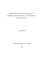
Figure 1-1
A.
The basic structural elements of the liver cell plate.
Only hepatocytes are shown.
B. A
traverse view of the liver cell plate. The microvilli
of hepatocytes extend into the perisinusoidal
space of
Disse for exchange of solute escape
from the sinusoids
through the fenestrations. Hepatic stellate cells (HSC)
are located in the perisinusoidal
space. Kupffer cells are
intra-
sinusoidal, participating in the removal of large
particles.
A
B
Hepatocytes
Sinusoids
Hepatic Venules
Portal Tract Blood
Bile
Portal
venule
Hepatic
artery
HSC with
vitamin A
granules
Kupffer
Cells
Endothelial Cells
Space of
Disse
Sinusoids
Fenestrations
Microvilli
Biliary canaliculi
Hepatocytes
Chapter 1
5
1.4 Compositions of the liver
The liver consists of two main components; the liver cells and the extracellular
space. Table 1-1 discloses the relative volumes of different cellular and inter-celullar
compartments in a typical liver of a rat, the animal model adopted in our current project.
These data was derived from stereological morphometric investigations (Blouin et al.,
1977). As shown in Table 1-1, hepatocytes, sinusoids, perisinusoidal space of Disse and
billiary canaliculi (collectively named lobular parenchyma) make up 96% by volume of a
healthy rat liver. The remaining 4% are portal triads, the hepatic and central veins
(Weibel et al., 1969).
Liver cells consist of parenchymal cells (hepatocytes) and non-parenchymal cells
(Fig. 1-2A). The latter includes endothelial cells, Kupffer cells, hepatic stellate cells
(HSCs) (also called Ito cell, fat storing cells or lipocytes), bile duct cells, and pit cells.
Although non-parenchymal cells do not contribute much to the total volume of the liver,
they occupy (Blouin et al., 1977) 26.5% of the total cell membrane surface and 55% of
the total fat droplet volume.
On the other hand, the extracellular space is made up of the perisinusoidal space
of Disse, the sinusoidal lumen, billiary canaliculi and extracellular matrix (ECM)
proteins. Hepatic sinusoids, where Kupffer cells are located, lead the mixed blood of the
terminal branches of the hepatic artery and portal vein to the central vein. Billiary
canaliculi, where bile is accumulated and drained, is formed by the invaginations of the
adjacent plasma membranes of two hepatocytes (Fig 1-1B). The space of Disse contains
many cytoplasmic dendritic projections and cell bodies of hepatic stellate cells (HSCs),
Chapter 1
6
abundant microvilli and hepatocytes, nerve endings and a non-electron dense complex
ECMs.
Table 1-2 Composition of a healthy rat liver (from Blouin et al., 1977)
Relative
Volume
Relative number of
cells
Extracellular space compartment
Sinusoidal lumen 10.6%
Disse Space 4.9%
Billiary canaculi 0.4%
Relative part of cells in total liver
volume
Hepatocytes 78.0% 60%
Non-hepatocytes
6.3%
Sinusoidal endothelial cells 2.8% 19%
Kupffer cells 2.1% 15%
Hepatic stellate cells 1.4% 6%
Chapter 1
7
LIVER CELLS
Parenchymal
i.) Hepatocytes
Nonparenchymal
i.) Endothelial Cells
ii.) Kupffer Cells
iii.) Stellate Cells
iv.) Pit Cells
v.) Bile Duct cells
Presumed embryological origin
Epithelial
i.) Hepatocytes
ii.) Bile Duct Cells
Mesenchymal
i.) Endothelial Cells
ii.) Hepatic Stellate Cells
Hematopoieti
c
i.) Kupffer Cells
ii.) Pit Cells
A.
B.
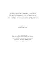
Figure 1
-
2A
. The different cell types in the liver.
Figure 1
-
2B
.
The presumed
embryological origins of different cell types in the liver. Upon activation
, hepatic stellate
cells (HSCs) can
actually undergo transition from mesenchymal cells to more contractile
myofibroblast-like cells. This is an important feature of liver fibrosis. (
Adapted from
Kaplowitz, 1996)
Chapter 1
8
1.4.1 Liver cells
1.4.1.1 Parenchymal cells
1.4.1.1.1 Hepatocytes
Hepatocytes are important effector cells that play key roles in the majority of liver
functions. Among others, they are responsible for glucogenolysis and gluconeogenesis of
the liver, besides being involved in the inactivation of toxic ammonia in the urea cycle.
They also synthesize serum proteins such as albumins, components of the complement
system and acute-phase proteins. The hepatocytes are involved in the synthesis of many
classes of lipoproteins as well as the catabolism of blood-derived cholesterol-enriched
proteins. Therefore, they also contribute to the metabolism of exogenous and
endogenous lipids. They are well-equipped for oxidative stress too and are thus capable
of detoxification of some endo- and exogenous substances. Besides, the production of
bile components such as bile acids, cholesterol, phospholipids and conjugated bilirubin
takes place in the hepatocytes.
After liver resection or severe liver injury, the parenchymal cells have
phenomenal capacity for proliferation to restore organ mass. The process of liver
regeneration was reported to be regulated by extracellular factors as well as by many
substances released by neighbouring nonparenchymal cells (Fausto, 2000).
Chapter 1
9
1.4.1.2 Nonparenchymal cells
1.4.1.2.1 Sinusoidal endothelial cells
Endothelial cells of the liver sinusoids (SECs) are structurally and functionally
unique from other endothelial cells. Not only do they lack basement membrane, they are
also enclosed by the cytoplasmic processes of the hepatic stellate cells (HSCs). The
SECs form a filtration barrier between the sinusoidal lumen and the hepatocytes (Fig. 1-
1B), controlling the exchange of various substances between the two compartments via
the dynamic changes of the fenestrae size.
Due to the presence of numerous plasma membrane receptors, SECs possess a
large pinocytotic and endocytic capacity (Wisse, 1972). In fact, a key physiological
function of SEC is the receptor-mediated endocytosis of circulating collagen, hyaluronic
acid, fibronectin, laminin, nidogen and chondroitin sulphate proteoglycan (Smedsrod et
al., 1994). They also perform immunological functions by acting as antigen-presenting
cells (APCs) as they constitutively express MHC class I and II (Rubinstein et al., 1986),
CD4, CD11, CD54 and CD106 necessary for the presentations of antigens to T cells.
1.4.1.2.2 Kupffer cells
Kupffer cells are located within the lumen of the sinusoids, whose double lining
is contributed by the cellular extensions of the Kupffer cells themselves (Motta, 1984).
Being the resident macrophages of the liver, Kupffer cells constitute the largest
population of macrophages in the mammalian body (Bouwens et al., 1986). They are
Chapter 1
10
involved in the removal of gut-derived bacteria and potent bacterial toxins such as
endotoxins or peptidoglycans from the post-mesentric blood. Besides having high
phagocytic and endocytic activity, Kupffer cells secrete a host of mediators which
interact in a paracrine manner with neighbouring cells, mainly the hapatocytes and the
HSCs.
1.4.1.2.3 Hepatic stellate cells
The HSCs are stellate-shaped cells that are located in the perisinusoidal space of
Disse, sandwiched between the sinusoidal capillaries and the parenchymal cells (Wake,
1980). The HSCs are involved in retinoid metabolism and are responsible for the storage
of 80-90% of the retinoids in the liver in the form of lipid droplets (Hendriks et al.,
1985). They also modulate sinusoidal blood flow through the contraction and dilation of
the sinusoidal lumen (Bauer et al, 1994), besides being the main producers of extra-
cellular matrix (ECM) macromolecules and ECM-degrading enzymes (Arthur, 2000;
Friedman et al., 1993). Upon liver injury, the HSCs can interact with other cell types and
the ECM components to contribute to liver tissue repair. HSCs are involved in
intercellular communication through the synthesis of cytokines and growth factors. The
HSCs express hepatocyte growth factors, transforming growth factor-β (TGF-β) and
insulin growth factor (IGF)-binding proteins and synthesize other cytokines that interact
with all types of neighbouring cells, including hepatocytes.
Chapter 1
11
The HSCs can exhibit dual phenotypes. In a normal liver, HSCs exhibit a
quiescent phenotype whereby the cells contain abundant lipid droplets, have low
proliferative rate and synthetic activity. In contrast, chronically diseased liver; especially
in liver fibrosis contains activated HSCs that have a myofibroblast-like phenotype. This
phenotype is characterized by the loss of lipid vacuoles, increased cell proliferation and
enhanced synthesis of ECM components (Friedman et al., 1993).
1.4.2 Extracellular space
1.4.2.1 Space of Disse
The perisinusoidal space of Disse is located in between the sinusoidal domain of
the hepatocyte plasma membrane and the endothelial cells that form the walls of the
hepatic sinusoids (Fig. 1-1B). This is the location where substances released from
sinusoidal blood and hepatocytes mix. This contact is made possible by the extension of
the microvilli of the hepatocytes into the space of Disse to mix with solutes released from
the sinusoidal space through the sinusoidal fenestrations. This contributes to the removal
of the solute. Excess solutes and water in this space is believed to contribute to hepatic
lymph formation.
Chapter 1
12
1.4.2.2 Hepatic Sinusoids
Unlike other capillaries, hepatic sinusoids lack basement membrane and the wall
of the sinusoids is interrupted by endothelial cell pores or fenestrations (Fig. 1-1B).
These pores can undergo dynamic changes in size so that most solutes, except for large
complexes, have free access to the space of Disse. The hepatic sinusoids also serve as an
intermediate connecting the terminal portal venule and the terminal hepatic arteriole with
the hepatic venules.
1.4.2.3 Biliary canaculi
The bile canaliculi are formed by two hemicanaliculi, each contributed by one
hepatocyte (Fig. 1-1B). Bile accumulates in canaliculi between hepatocytes and drains
via collecting ducts in portal triads, small liver ducts and finally to common hepatic duct
which meets the cystic duct.
1.4.2.4 Extracellular matrix
1.4.2.4.1 Collagens
There are two types of collagens in the liver: the fibrillar (interstitial) collagens
and the basement membrane-like collagens. The fibrous external capsule, septa,
periductal and perivascular areas and portal tracts of the liver are particularly rich in
interstitial collagens such as collagen type I, III, V and fibronectin (Schuppan, 1990)
Chapter 1
13
while collagen IV and VI (basement membrane-like collagens) together with laminin and
fibronectin are the major matrix protein in the space of Disse. It has been reported that
the composition of the ECM connecting sinusoidal space with the brush-border of
hepatocytes resembles that of basement membrane (Hahn et al., 1980; Martinez-
Hernandez, 1984; Arenson et al., 1988).
1.4.2.4.3 Glycoproteins
The collagens do not function alone but are complexed with proteoglycans and
glycoproteins such as laminin, fibronectin, entactin and elastin. Laminin, being produced
by HSCs and the endothelial cells, is a glycoprotein that promotes cell adhesion,
migration, differentiation, and growth (Castronovo et al., 1991; Herbst et al., 1988;
Kleinman et al., 1986; Carley et al., 1988; McGuire et al., 1992). It also assists the
endothelial cells in forming the capillary (Kubota et al., 1988; Grant et al., 1989, 1991).
Meanwhile, fibronectins, large but thin filaments associated with collagen fibres, can
exist in plasma and cellular forms. Entactin, also called nidogen, is a highly sulphated,
glycoprotein restricted to basement membranes. Though normally scattered throughout
the portal tracts, elastin fibres are deposited in fibrous septa of the cirrhotic liver over
time.
Chapter 1
14
1.4.2.4.3 Proteoglycans
Collectively, this group of proteins is made up of chondroitin sulphate, heparan
sulphate, hyaluronic acid and dermatan sulphate. Generally, these proteins consist of a
core protein decorated with un-branched side chains of repeating sulphated disaccharide
units. The proteoglycans can serve as adhesion molecules (Elenius et al., 1990) and
receptor molecules on cell surfaces (Andres et al., 1989). Therefore, they can be an
important reservoir for cytokines and growth factors (Fausto, 2000).
Table 1-3 Extracellular matrix components (based on Martinez-Hernadez, 1984).
Component Normal Distribution
Collagens
Type I
Portal tract matrix, hepatic veins, points of inflection
in hepatic cords
Type III Portal tract matrix, space of Disse
Type IV Portal tract basement membranes, space of Disse
Type V Portal tract matrix, space of Disse
Type VI Portal tract matrix, space of Disse
Glycoproteins
Laminin Portal tract basement membranes, space of Disse
Fibronectin Portal tract matrix, space of Disse
Entactin (nidogen) Portal tract basement membranes
Elastin Portal tract matrix
Proteoglycans
Heparan sulphate Portal tract basement membranes
Chapter 1
15
1.5 Liver fibrosis and cirrhosis
Liver cirrhosis accounts for more than 27 000 deaths in the USA per year,
making it the ninth leading cause of death (Grant et al., 1991). In Singapore, chronic
liver diseases and cirrhosis account for 0.8% of the total deaths reported in 2001,
according to the figure published by the Ministry of Health in Singapore. This translates
to over 120 deaths on an annual basis. It is also consistently the eighth principal cause of
deaths in Singapore from 1999 to 2001. (Table 1-3).
1.5.1 Definition of fibrosis and cirrhosis
The term “fibrosis” refers to the excessive accumulation of connective tissues in
parenchymal organs. Liver becomes fibrotic following a chronic insult of sufficient
intensity to trigger a “wound-healing”-like reaction (Poli, 2000). These insults are
usually initiated by hepatocyte damage that leads to the recruitment of inflammatory cells
and platelets, activation of Kupffer cells and the subsequent release of cytokines and
growth factors. Subsequently, hepatic stellate cells (HSC) become activated and secrete
extra-cellular matrix proteins (ECM) to form a fibrous scar. In spite of the variable
etiology, there are common features (Table 1-4) in any fibrosis events. These common
features encompass the mechanisms and cell types involved in the overproduction of
ECM proteins and the induction of fibrosis.
Chapter 1
16
Table 1-4 A figure published online by the Ministry of Health in Singapore
indicates that chronic liver diseases and cirrhosis are the 8
th
principal cause of
death in Singapore, consistently from year 1999 to year 2001.
(
Principal Causes of Deaths in Singapore
1999
2000
2001
Total Number of Deaths
15 516
15 693
15 367
% of Total Deaths
1. Cancer
26.6 27.0 28.2
2. Ischaemic and other heart diseases
25.7 25.1 26.3
3. Pneumonia
10.6 11.4 10.0
4. Cerebrovascular disease
10.5 10.4 9.2
5. Injuries
6.9 7.2 6.7
6. Diabetes Mellitus
2.3 2.3 3.3
7.
Nephretis, Nephrotic Syndrome &
Nephrosis
1.2 1.3 1.7
8. Chronic liver disease and cirrhosis
1.0 0.7 0.8
9. Bronchitis, emphysema and
asthma
0.9 0.7 0.7
10. Tuberculosis
0.7 0.6 0.7
Chapter 1
17
On the other hand, “cirrhosis” is a morphological term which refers to “a diffuse
process characterized by fibrosis and the conversion of the normal liver architecture into
structurally abnormal nodules” (Anthony et al., 1977). This widely-accepted definition
was coined in 1977 by the World Health Organization (WHO). Liver cirrhosis is the end-
stage consequence of liver fibrosis. Unlike fibrosis, once a liver becomes cirrhotic, it is
irreversible (Popper et al, 1975). In cirrhotic livers, delicate bands, broad scars with
nodules are observed in hepatic parenchyma. These nodules can vary form less that 3
mm in diameter (micronodules) to 3 mm to several centimeters in diameter
(macronodules). As a result, the parenchymal structure of the entire liver is disrupted.
Chapter 1
18
Table 1-5 Main biochemical and histological features of fibrosis, sclerosis and
cirrhosis of the liver (Poli, 2000)
Fibrosis
Increase of connective tissue within parenchymal organs
Main Features:
o Accumulation of both fibrillar and basement membrane like
collagens
o Increase in laminin and fibrobectin
o Thickening of connective tissue septae
o Capillarization of the sinusoids
Sclerosis
Ageing of fibrotic tissue
Main Features:
o Decrease of hyaluronic acid and heparan sulphate
proteoglycans
o Increase of chondrotin sulphate proteoglycans (decorin,
biglycan)
o Progressive fragmentation and disappearance of elastic
fibers
o Distortion of sinusoidal architecture and parenchymal
damage
Cirrhosis
End-stage process of liver fibrotic degeneration
Main Features:
o Whole anatomy heavily distorted by thick bands of collagen
surrounding nodules of hepatocytes with regenerative foci
Chapter 1
19
1.5.2 Etiology
Liver fibrosis is initiated by a variety of causative agents. These etiological
agents can be broadly categorized into six classes as shown in Table 1-5.
Table 1-6 Causes of liver cirrhosis
Causes of Cirrhosis
Drugs and toxins
Alcohol, Methotrexate, Isonazid, Methyldopa, Amiodarone
Infections
Hepatitis B, Hepatitis C, Schistomiasis
Autoimmune
Autoimmune hepatitis, Primary biliary cirrhosis, Primary sclerosing
cholangitis
Inherited Metabolic
Defects
Haemochromatosis, Wilson’s disease, α
1
-Antitrypsin deficiency,
Galactosemia, Tyrosinaemia, Type IV glycogen storage disease,
Hereditary fructose intolerance, Urea cycle disorders,
Abetalipoproteinaemia, Progressive familial intrahepatic cholestasis,
Cystic fibrosis
Acquired bile duct
disease
Biliary atresia, Gallstone obstruction, Common bile duct stricture
Vascular
Budd-Chiari syndrome, Veno-occlusive disease, Chronic right heart
failure, Hereditary haemorrhagic telangiectasia
Miscellaneous
Non-alcoholic steatohepatitis, Indian childhood cirrhosis, Intestinal
by-pass surgery, Hypervitaminosis A, Sarcoidosis
Cryptogenic
Chapter 1
20
1.5.3 Features of fibrotic livers
When the liver becomes increasingly fibrotic due to chronic injury, significant
changes occur. These changes include increases in the total content of collagens and
non-collagenous components of the ECM from three to eight fold (Anthony, 1988).
Besides, excessive type I and III collagen are laid down in both the portal tracts and the
lobule, resulting in delicate or broad septal tracts. This is then accompanied by a shift in
the type of ECM in the sub-endothelial space from the normal low-density basement
membrane like matrix to the interstitial type.
As a result, the sinusoids are converted into capillaries with a basement
membrane, impairing the blood-hepatocyte solute exchange despite maintenance of
absolute hepatocyte volume (Ohara et al., 1993). This “capillarization” process leads to
the loss of hepatocyte microvilli and the disappearance of endothelial fenestrations.
Central to this fibrogenesis process is the HSCs which are the major source of ECM
proteins (Arenson et al., 1988; Maher et al., 1988).
1.5.3.1 Activation of hepatic stellate cells
HSCs have been established as the unequivocal source of extracellular matrix
proteins and thus HSC activation is a key event in the fibrosis process. HSC activation
involves the transition of HSCs from mesenchymal cells to contractile myofibroblast-like
(MFB) cells. This process is mediated by cytokines released by resident and non-resident
cells in the diseased liver (Gressner and Bachem, 1994). Once activated, HSCs produce
Chapter 1
21
not only ECM molecules, metalloproteinases and respective inhibitors, but also a variety
of cytokines, growth factors and chemokines that act in paracrine and autocrine manners
on parenchymal cells, untransformed HSCs and MFB. Upon activation, TGF-β can
stimulate ECM synthesis, initiate the transformation of HSC and enhances ECM
production of MFB in an autocrine manner (Bachem et al., 1992).
Activation of the HSCs is arbitrarily divided into two stages. This two-stage
process consists of an “initiation” or “preinflammatory” phase (Gressner, 1996) followed
a “perpetuation” phase (Friedman, 1993).
1.5.3.1.1 Initiation
Before the liver can become fibrotic and ECM proteins accumulate, HSCs must
be made to respond to other cytokines and stimuli in the early stages of fibrogenesis.
This “initiation” step involves changes in the gene expression and phenotype of both the
parenchymal and nonparenchymal cells. For instance, hepatocytes can release mitogenic
signals, presumably insulin-like growth factor (IGF-1) and IGF-binding proteins that
induce HSCs to undergo proliferation. Besides, hepatocytes are potent source of lipid
peroxides (Gressner, 1995) which have been shown to be involved in many forms of liver
cirrhosis. The level of oxidative aldehyde adducts was also shown to correlate with
collagen gene expression by the HSCs (Paradis and Kollinger et al., 1997; Paradis and
Mathurin et al., 1997). In addition, the fact that anti-oxidant levels are severely depleted
Chapter 1
22
in cirrhotic liver (Leo et al., 1993) could also lead amplify the injurious effects of lipid
peroxides.
Meanwhile, activated Kupffer cells and macrophages (Friedman and Arthur
1989; Matsuoka and Tsukamoto 1990) enhance the ECM production by secretion of a
host of mediators. Among these, TGF-β appears to be the major profibrogenic, matrix-
production and HSC transformation-promoting cytokine of Kupffer cells and infiltrating
mononuclear cells.
Injured endothelial cells stimulate production of cellular fibronectin, which can
potentially activate stellate cells (Jarnagin et al., 1994). They are also likely to
participate in the conversion of TGF-β1 from the latent to the active, profibrogenic form
through the activation of plasmin (Gleizes et al., 1997).
Finally, platelets in injured liver region are potent source of paracrine stimuli by
generating multiple potentially important mediators including platelet derived growth
factor (PDGF), TGF-β1 and epidermal growth factor (EGF). Activated stellate cells have
also been observed in primary and metastatic human tumours (Enzan et al., 1994 & 1995)
Chapter 1
23
HSC
Kupffer
Cells
Endothelial Cells
Space of
Disse
Sinusoids
Fenestrations
Microvilli
Hepatocyte
FIBROSIS
Collagen
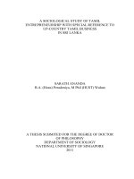
Figure 1
-
3
shows the changes of features of the liver during the fibrogenesis process. Firstly, hepatocytes in fibrotic
liver appear to lose
their microvilli. Besides, the HSCs are transformed from mesenchymal cells to contractile
myofibroblast-
like cells. This is accompanied by the heavy deposition of extracellular matrix proteins such as collagens.
The Kupffer cell also becomes activated
and starts to play an important role in enhancing the ECM production by
secretion of a host of mediators
. Finally, the fenestrations between endothelial cells are lost through this
“capillarization”.
Chapter 1
24
1.5.3.1.2 Perpetuation
Subsequently, several phenotypic changes occur in the HSCs. The combination
of these phenotypic changes contributes to the accumulation of extracellular matrix. In
this “perpetuation” phase, HSCs proliferate, as a result of up-regulation of many
mitogenic factors and their corresponding cognate receptors. Among these, PDGF is the
most potent mitogen towards the HSCs. Besides, the HSCs become more contractile as
they are transformed from mesenchymal cells to myofibroblast-like cells with increased
expression of alpha smooth actin. Loss of intracellular vitamin A is also well-
documented in the activation of the HSCs. However, it is still unclear whether retinoid
loss is a prerequisite for activation of the HSCs and which retinoid might accelerate or
prevent activation in vivo.
During this phase, increased fibrogenesis activity was detected in HSCs and this
remains the most direct proof of the role of HSCs in hepatic fibrosis. Among others, the
most potent fibrogenic factor for the HSCs was found to be TGF-β1 while other less
fibrogenic factors include interleukin -1β (IL-1β), TNF, lipid peroxides and acetaldehyde
(Li et al., 1999). Among the ECM proteins, up-regulation of collagen is among the most
obvious responses of HSCs to injury. Transriptional activation of the type 1 collagen has
been extensively studied (Racine-Samson et al., 1997; Winwood et al., 1995; Niki et al.,
1996) and it was shown that the half-life of collagen alpha1 mRNA increases 20-fold in
the activated compared with quiescent stellate cells (Ballardini et al., 1994).