Molecular modeling of localized collective motions and dynamics in proteins
Bạn đang xem bản rút gọn của tài liệu. Xem và tải ngay bản đầy đủ của tài liệu tại đây (3.06 MB, 162 trang )
PHD thesis National University of Singapore Cao zhiwei
1
Chapter 1
Introduction
1.1 Protein motion and dynamics vs. protein function
Protein motions and dynamics are involved in a variety of biological processes in living
organisms. Though in some of these processes the static structure of a protein determines
its function (e.g. collagen in tissues or α-keratin in hair), protein motions and dynamics are
crucial in others cases. Examples are metabolism, transport and synthesis of biomolecules
etc. In fact, all dynamic biological processes can find the origin in protein motions and
dynamics. Muscle contraction, for example, is based on the combined motion of actin and
myosin. Other examples are the molecular motors kinesin and F1-ATPase. Motion and
dynamics also play important roles in many other proteins whose primary function is not
mobility itself. Conformational change is actually essential to the function of many
transport proteins, enzymes, and those proteins involved in signal transduction, immune
protection or gene expression[1].
It has been noticed that the biological function of most of the globular proteins often
includes an interaction with one or more different molecules on appropriate occasions,
such as small ligand, substrate, peptide, a fragment of nucleic acids, even another protein.
In many enzymes, conformational changes serve to enclose the substrate, thereby
PHD thesis National University of Singapore Cao zhiwei
2
preventing its release from the protein and ideally positioning it for the protein to perform
its function, as in lysozyme. For example, immunoglobulins are highly flexible in order to
be able to interact with a large range of ligands. Generally, functional interactions of
flexible ligands with protein binding sites often require conformational adjustments in
both the binding ligands and the host protein. The structural changes in some proteins
regulate the interaction between ligands and protein through induced fit and allosteric
effects [2,3]
The “induced fit” theory by Koshland [4] proposed that the original structure of active
site in enzymes does not fit substrate exactly, but the presence of the substrate induces
structure changes in the active site to fit for substrate binding. It is expected that each
intermediate step of the whole cycle of enzyme catalysis requires the enzyme molecule,
especially the active site region, to be in a specific conformation different from another.
Allosteric effect is found in a special class of proteins, so-called allosteric proteins.
Substrate binding to one subunit of these multimeric proteins triggers conformational
changes in proteins which alters the substrate affinity of the other subunits, thereby
sharpening the switching response of these proteins [5,6].
Moreover, the importance of motion and dynamics to protein function has been further
confirmed by various experimental studies. Two major sources of evidence come from X-
ray crystallographic analysis and Nuclear Magnetic Resonance (NMR). One example
from X-ray analysis is myoglobin. In order to capture, bind and release oxygen (O
2
)
freely, myoglobin has been found to have more mobile character toward the periphery of
the molecule although the core surrounding of its heme group is compact [7]. A similar X-
PHD thesis National University of Singapore Cao zhiwei
3
ray analysis of lysozyme has produced the intriguing observation that the enzyme’s active
site cleft undergoes an ~1 Å closure upon substrate binding [8]. On the other hand,
NMR study revealed that several sub-states often exit for one protein or enzyme. These
sub-states, which each have slightly different atomic arrangements, randomly interconvert
at rates that increase with increasing temperature [9].
Instead of being stationary at fixed positions, the atoms in a protein molecule are rather
in a state of constant motion. The “static” view of a protein structure from X-ray analysis
is at best a representation of its average structure. The atoms in each protein molecule
exhibit sizable high-frequency fluctuations about this average. The atomic fluctuation
affects various bond interactions in proteins, especially those relatively weaker non-
covalent interactions. For example, hydrogen bonds break when the partner atoms
fluctuate out of a certain limit of distance, while alternative hydrogen bonds reform if the
new partners come closer. Hydrogen bonds keep breaking and reforming during protein
motion which gives protein molecule extra dynamic features. In summary, proteins are
constantly changing the details of their conformation. Therefore any attempt to understand
the function of proteins requires a scientific investigation of protein motion and dynamics.
1.2 Structure basis for protein motion and dynamics
The motions of the atoms in a protein tend to share certain characteristics that can be
explained in terms of the basic structure of proteins. Each protein is made up of a specific
number of small units, the amino acids. Each of the 20 different amino acids is
characterized by a side chain, a distinctive chemical group that ranges in complexity from
PHD thesis National University of Singapore Cao zhiwei
4
a hydrogen atom in the simplest amino acid, glycine, to elaborate rings of atoms in the
most complex amino acid, tryptophan. The side chain of each amino acid can vary not
only in size and shape, but also in charge, reactivity and ability of forming hydrogen
bonds.
Proteins are commonly consisting of from 50 to more than 500 amino acids, which
corresponds to some 500 to 5,000 non-hydrogen atoms. The precise sequence of amino
acids determines the average structure and other properties of the protein. In particular, the
balance of the attractive and repulsive forces between the individual atoms of which the
protein are composed causes the peptide to fold in a characteristic way essential to its
motions and its functions.
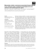
In a protein, the amino acids are linked together into a polypeptide chain by peptide
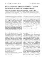
bond, as being indicated in Fig 1.1. The peptide bond itself is rigid, because it is involved
into tautomerization that gives it considerable double bond character, as Fig 1.2 shows.
However, there are many other strong bonds in the main chain of the polypeptide free to
twist. For instance, the N-C and C-C bonds relatively are free to rotate. These rotations
are represented by the torsion angles phi (φ) and psi (ψ), respectively (shown in Fig 1.3).
Phi and psi can vary to certain extent within the Ramanchandran Plots as Fig 1.4 shows
[10]. This allows the protein to potentially adopt many different conformations. Therefore,
the twisting, or bond rotation, allows one part of the polypeptide chain to move with
respect to another. As the polypeptide chain twists and turns, its various side chains move
with it. The side chains themselves have rotatable bonds which imparts additional
flexibility. Fig 1.5 shows the rotatable bonds in peptide.
PHD thesis National University of Singapore Cao zhiwei
5
The flexibility of the polypeptide backbone and of the side chains is what enables each
protein to fold into its characteristic native, or average, structure. These sites also facilitate
the fluctuations of protein atoms around their average positions. Even in the folded
protein, however, the thermal energy corresponding to the atomic velocities at room
temperature is sufficient to allow twisting motions.
Fig 1.1 Peptide bond of amino acids.
The rectangle line is the part of the peptide bond.
PHD thesis National University of Singapore Cao zhiwei
6
Fig 1.2 Tautomerization of peptide bond
Fig 1.3 Torsion angles in the main chain of protein.
φ and ψ are free to rotate.
PHD thesis National University of Singapore Cao zhiwei
7
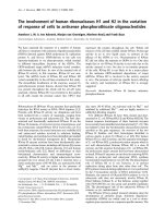
Fig 1.4: The Ramachandran plot.
In the above diagram the white areas are sterically disallowed regions for all amino
acids except glycine which lacks a side chain. The red regions correspond to
conformations where there are no steric clashes, i.e. these are the allowed regions
namely the alpha-helical and beta-sheet conformations. The yellow areas show the
allowed regions if slightly shorter van der Waals radi are used in the calculation,
i.e. the atoms are allowed to come a little closer together.
PHD thesis National University of Singapore Cao zhiwei
8
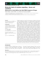
Fig 1.5 Sites of flexibility, rotatable bonds in peptide chain.
The drawing depicts only the principal atoms of a polypeptide chain. The backbone of
the chain (black bonds) consists of carbon and nitrogen atoms; the linkage called
peptide bonds is rigid, whereas the intervening bonds allow rotations (curved arrows).
The side chains shown in detail also contain rotatable bonds.
PHD thesis National University of Singapore Cao zhiwei
9
1.3 Classification of protein motion
Protein motion involves a wide variety of conformational change ranging from very
subtle, local fluctuations to large, global movements. These may be motions of individual
atoms, groups of atoms, or whole section of the molecule. Generally they can be classified
into three broad categories for convenience according to their coherence, displacement
amplitude and time-scale, as shown in Table 1A [11]. Methods used to study them are
given in the table.
Table 1A: Types of motions found in protein
Motion Spatial dis-
placement (Å)
Characteristic time
(second)
Energy
source
Method of
observation
Atomic
fluctuations
0.01 to 1 10
-15
to 10
-11
k
B
T Computer
simulation, X-ray
diffraction
Collective
motions
0.01 to >5 10
-12
to 10
-3
k
B
T NMR,
fluorescence,
hydrogen
exchange,
simulation, X-ray
Triggered
conformational
change
0.5 to >10 10
-9
to 10
3
Inter-action X-ray,
spectroscopy
The first category contains atomic fluctuations, such as individual atom vibrations.
These motions are random, very fast, but rarely cover more than 0.5
Å. The time scale of
these motions is in the order of picoseconds or less, and therefore it is usual to observe
them by vibrational spectroscopy and to model them by molecular dynamics. X-ray
diffraction can give information on the spatial distribution of atomic fluctuations though
an analysis of the spreading of atomic electron density produced by such motion. The
energy for these motions comes from the kinetic energy inherent in the proteins as a
PHD thesis National University of Singapore Cao zhiwei
10
function of temperature. They are normally driven by collisions with solvent molecules or
by random collision with neighboring atoms in the protein. Although many of them
individually may not be important for protein functions, they contain information that is of
considerable significance. There may be a correlated directional character to the active-
site fluctuations that play a role in enzyme catalysis. Furthermore, the small amplitude
fluctuations are essential to all other motions in proteins. They serve as the “lubricant”
which makes it possible for large-scale protein motions to happen on a physiological time
scale.
The second category contains collective motions, such as the movements of groups of
atoms that are covalently linked in such a way that the group moves as a unit.
Noncovalently interacting groups of atoms may also move collectively. The size of the
group ranges from a few atoms to many hundreds of atoms, or even entire structural
domains, as in the case of the flexible Fc portion of immunoglobulins [12]. There are two
types of rapid collective motion: those that occur infrequently (like internal aromatic ring-
flips), and those that occur with high probability (many collective motions of small groups
of neighboring atoms, bonded or nonbonded, are in the pico-second time regime.)
Collective motions can also be very slow, as local unfolding of a polypeptide segments.
The energy for collective motions also derives from the thermal energy inherent in a
protein as a function of temperature. The time scale of collective motions (from
picoseconds to nanoseconds or slower) allows some of them to be studied by techniques
such as NMR and fluorescence spectroscopy.
The third category contains motions which can be described as triggered
conformational changes. These are the motions of groups of atoms (i.e. individual side
PHD thesis National University of Singapore Cao zhiwei
11
chains) or whole sections of a protein (i.e. loops of chain, domains of secondary and
tertiary structure, or subunits) that occur as a response to a specific stimulus. The distance
moved can be as much as 10 Å or more. The time scale can be estimated from the rate of
binding or turnover reactions. The energy for triggered conformational changes comes
from specific interactions, such as electrostatic attractions or hydrogen bonding
interactions. The best-known example of a triggered conformational change is the
transition in tertiary and quaternary structure that occurs when ligands bind to the iron
atoms of hemoglobin [13].
Category one and two define atomic motions that, whether individual or correlated,
involve random excursions about the equilibrium conformation. In contrast, category three
is used to classify motions that involve a transition from one equilibrium conformation to
another. At any given time, a typical protein exhibits a wide variety of motions described
above. However, the effective dynamical units in proteins are those collective motions that
behave nearly as half-rigid bodies under physiological temperature. Examples include
range from small chemical groups in the side chains of residues, to a fragment of peptide
chain. Most of the time the small chemical groups display only relatively small internal
motions owning to the high energy cost associated with deformations of bond lengths,
bond angles or dihedral angles about multiple bonds. The more functionally important
collective motions, stimuli triggered or not, involve displacements of larger groups
associated with torsional motions with rotationally permissive single bonds in main chain
[11]. Comparing with the short-time relatively small amplitude motions, the substantial
displacement of this class of localized collective motions occur over longer time intervals,
PHD thesis National University of Singapore Cao zhiwei
12
and result in concomitant displacements of the different fragments of protein, like domain
or intra-domain motion in biological process.
1.4 Features of functionally important collective motions in protein
Relatively large inter- or intra-domain movements provide spectacular examples of
collective motions, and thus have been under extensive investigation because of their
important functional roles. They normally occur in proteins with half-rigid domains, or
constrained sub-domains, or functional groups, linked by short flexible linker regions,
which have fewer packing constraints and are free to undergo conformation change. An
illustrative example is opening and closing of the binding site of HIV-1 protease (HIV Pr)
that results from conformational changes in the region covering the reactive site. Based on
the different open and close even half-close states, analysis of protein crystal structures
has shown that protein inter-domain motion can take two basic forms: hinge motions that
are not constrained by tertiary packing interactions and shear motions between close-
packed segments of polypeptide[14].
PHD thesis National University of Singapore Cao zhiwei
13
The characteristic of these two forms of mechanisms of protein flexibility are
summarized in Fig 1.6 and Table 1B. According to analysis from Gerstein, motions of
close packed segments of polypeptide can be divided into those that are perpendicular to
an interface and those that are parallel. Generally, hinge motions produce a motion
perpendicular to the plane of an interface, so that the interface exists in one conformation
but not in the other, as in the opening and closing of a book or a door. Shear motion tends
to be similar to scissors’ trimming. It is parallel to the plane of the interface, which is
limited by the packing contacts involving the interdigitation of side chains. These two
basic mechanisms can be applied in a great variety of protein motions[14].
Fig 1.6 Hinged and shear mechanism
for domain closure. See table1B for a
summary of the characteristics of both
mechanisms.
PHD thesis National University of Singapore Cao zhiwei
14
Table 1B
Scheme showing the difference between shear (sliding) and hinge motions.
The essential characteristics of the various motions are summarized below.
Items
Shear mechanism Hinged mechanism
Well-packed interfaces MAINTAINED, throughout
motions
NOT MAINTAINED; rather
created, burying surface
Motion at interface Parallel to plane of interface
(shear)
Perpendicular to interface
Mainchain packing Constrained by close packing Free to kink at hinge
Mainchain torsions Small changes in many torsion
angles
Large changes in a few torsion
angles
1.4.1 Feature of hinge domain motion
The most basic motion of a protein is a few large changes in main-chain torsion angles
in the localized region, i.e., at flexible inter-domain linker regions. The deformation of an
extended strand is the simplest hinge motion because its only constraint is that the torsion
angles of the strand remain in the allowed regions of the Ramachandran diagram.
Consequently, its torsion angle changes can be very large and the resulting motion can
rotate the conjoint chain up to 60˚. For example in lactate dehydrogenase, two adjacent
torsion changes rotate a strand by ~35˚ in a direction not accessible by a single change
[15]. If the torsional changes fall in the more constrained alpha-helices, the deformation
of helices will spread over to more residues than the deformation of sheets. Because
residues in the helices are subject to more severe hydrogen-bonding and steric constraints
than those in sheets, their torsion angles are restricted to a smaller region of the
Ramachandran diagram. Thus, if residues are to remain on a helical conformation, the
possible changes in their torsion angles are correspondingly smaller than those of resides
in an expanded conformation. Such spread-out helical deformations can produce bending
PHD thesis National University of Singapore Cao zhiwei
15
motions. For instance, changes in eight torsion angles between 9˚ and 15˚ in the C-
terminus of a helix in a mutant lysozyme bend its end to produce a shift of 3.3 Å [16] .
1.4.2 Feature of shear domain motion
Large shifts of close-packed segments of polypeptide would require switching between
different interdigitating configurations. Small shear motions (Figure 1.7) that do not
involve repacking of the interface are commonly seen in domain closure. The
interdigitating side chains normally accommodate shear motions, mostly, by small (<15˚)
changes in side-chain torsion angles. The main chain of each segment in a shear motion
does not deform appreciably. The segment shift and rotate relative to each other by no
more than 2 Ǻ and 15˚, amounts likely to be the limits of low-energy conformational
adjustment. Except at very small interfaces, larger movements than these require the
combination of several shear motions.
Fig 1.7: Small shear motion in citrate synthase.
Representative shear motions between close-packed helices. Note how the mainchain
only shifts by a small amount and the sidechains stay in the same rotamer configuration.
PHD thesis National University of Singapore Cao zhiwei
16
1.4.3 Summary
In general, proteins that can undergo predominantly hinge domain motion usually have
two domains connected by flexible linker regions that are relatively unconstrained by
packing. A few large torsional changes are sufficient to produce almost the entire domain
motion. The rest of the protein rotates essentially as a rigid body, with the axis of the
overall rotation passing through the flexible linker regions.
Since an individual shear motion is small, a single one is usually not sufficient to
produce a large domain motion. Usually, a number of shear motions combine to give a
large effect. In other words, the peptides that link the shearing segments have small main-
chain torsional changes to accommodate the relative movements.
It is important to realize that hinge and shear motions are only ideal paradigms to
describe large inter-domain motions. A real protein motions often has a combination of
both motions, i.e., hinges in one part of the protein and shearing interfaces elsewhere.
Nevertheless, many protein large-scale collective motions can be described as occurring
predominantly by a hinge or a shear mechanism, or none of them.
In the smaller intra-domain protein motions, hinge and shear mechanism are also
involved in the collective motion, when individual loops or helices move relative to each
other. For example, in Trp repressor, shear mechanism between 2 helices exists to adjust
position of helix-turn-helix reading head domain to enable it to bind to DNA[17].
PHD thesis National University of Singapore Cao zhiwei
17
1.5 Computational methods to study motions and dynamics in protein
As computer hardware has become faster and computational methods become more
sophisticated, computer modeling has been developed and widely applied in studying
protein collective motions and dynamics in the past decades. Protein motion can be
derived from atomic interactions based on knowledge of the structural fluctuations that
occur as a result of thermal motion. Such fluctuations can be obtained in various ways,
such as molecular dynamics, harmonic dynamics, and stochastic dynamics, and others.
Molecular Dynamics (MD) techniques has so far provided the most detailed and direct
results on protein motions. In MD modeling, insights into molecular flexibility and
activity are sought by numerically following molecular configurations in time according to
Newton’s law of motions [18,19,20]. One of the main tasks in MD simulation is to derive
and analyze the overwhelming amount of trajectory that describes the time-dependant
changes in atomic coordinates. Several kinds of methods have been applied to facilitate
the analysis in order to reveal the concerted fluctuations with large amplitudes. Examples
are principal component analysis (PCA), Monte Carlo methods[21,22,23,24] essential
dynamics analysis (EDA) [25] or molecular optimal dynamic coordinates analysis [26].
These aspects of Molecular Dynamics modelling have been covered recently in several
reviews [27,28,29,30].
In theory, MD modeling can bridge spatial and temporal resolution and thus capture
molecular motion over a wide range of thermally accessible states. In practice, the
PHD thesis National University of Singapore Cao zhiwei
18
numerical time-step problem has limited most applications to straightforward integration
with very small time-steps compared to the motion of major interest. With current state-
of-art methods and super-computers, a microsecond protein simulation has been described
for the 36-residues subdomain of a small protein, vallin, via Cray T3E [31]. However, a
typical protein of hundreds of amino acids can only be simulated for time-scales of at most
nanoseconds [32]. Comparing to the most conformational changes taking place in proteins
(time range from 10
-12
to 10
2
s ), such simulations may not grasp the essential motions
related to biological function which occur at much longer time-scales
[33]. Thus a clear
gap exists between time scales that can currently be obtained by molecular dynamics and
the time scale required for most of biological processes.
To study the longer time-scale and more complex functionally related motions in
proteins, it is generally necessary to eye on other than the straightforward molecular
dynamics simulation method. Harmonic analysis of protein dynamics differs from straight
molecular dynamics in that they provide a concise and compact description of the
concerted motions in protein molecules. After modification and improvement, harmonic
analysis can also be applied to investigate protein dynamics of bond breaking such as
hydrogen bond disruption. In early attempts, harmonic approximation had been used to
examine dynamical properties of proteins or their fragments, and now it has developed
into one of the key computational tools to study protein motion and dynamics.
1.5.1 Normal mode analysis (NMA) for protein collective motion
Harmonic analysis was motivated by vibrational spectroscopic studies[34,35,36], in
which the calculation of normal mode frequencies from empirical potential functions has
PHD thesis National University of Singapore Cao zhiwei
19
long been a standard step in the assignment of infrared and Raman spectra [37,38]. One
assumes that the vibrational displacements of the atoms from their equilibrium positions
are small enough that the potential energy can be approximated as a sum of terms which
are quadratic in the displacements. The coefficients of these quadratic terms form a matrix
of force constants which, together with the atomic masses, can be used to set up a matrix
equation for the vibrational modes of the molecule.
A straightforward computation of normal modes in Cartesian coordinates involves a
numerical diagonalization of a matrix of size 3N*3N for a molecule with N atom. A set of
harmonic vibrational “modes” will be generated by the calculation, among which 3N-6
eigenvalues will provide the internal vibrational frequencies of the molecule. The
associated eigenvectors to respective eigenvalues give the directions and relative
amplitudes of the atomic displacements in each normal mode. There is a close connection
between mode frequency and the collective character of the protein motions. A clear drop-
off in collectivity has been found as the frequency increases, with large collectivity only
found in modes below 200 cm
-1
[39].
Each single mode comprises the concerted motions of many atoms which are useful in
characterizing fluctuations from a stable equilibrium structure. With present-day computers, it
is not difficult to study proteins up to a few hundreds of amino acids with an all-atom model
[39]. For example, computation of the normal modes for the 159 amino acid protein
dihydrofolate reductase, required about 4 hours on a Silicon Graphics R10000 workstation
[40].
PHD thesis National University of Singapore Cao zhiwei
20
The standard application of NMA to large protein molecule is computationally
expensive; however, several sophisticated numerical techniques can be applied to extract
the lowest frequency modes of large molecules [41]. A common approximation for larger
systems is that bond lengths and angles are fixed. This can reduce the size of the matrix
involved by about an order of magnitude. Calculations can be carried out by direct
construction of the potential and kinetic energy matrices in (curvilinear) internal
coordinates [42] or through matrix partitioning techniques that start from Cartesian
derivatives [43]. In general, reduction of the dimensionality of the expansion space has
noticeable but not overwhelming effects on the resulting normal mode description of the
dynamics. The directions of the lower-frequency modes are largely preserved, but
frequencies in general are higher in the lower-dimensional space [44], suggesting that
small fluctuations in bond lengths and bond angles allow the dihedral angles to become
more flexible. Many practical aspects of computing modes for large molecules are
available elsewhere [45,46].
In brief summary, Normal Mode Analysis uses harmonic approximation of the full-
atomic force-field, for which a single experimental structure suffices. Although the
harmonic model may not provide the complete description of the motional properties of a
protein because of the contribution of anharmonic terms to the potential energy, normal
mode analysis has become an increasingly active area of research on protein collective
motions over the past years [39]. For instance, domain motions within the regulatory and
catalytic chains of aspartate transcarbamylase were analyzed by NMA [47,48]. NMA has
also recently been applied to a-lytic protease in an attempt to understand the substrate
specificity [49]. Many other studies[50,51] have suggested that NMA is the most
PHD thesis National University of Singapore Cao zhiwei
21
suitable theoretical methods in determining functionally relevant motions[52]. Therefore,
NMA is adopted in this thesis as the theoretical method to study localized collective
motions in protein.
1.5.2 Modified self-consistent harmonic approach for H-bond breaking
dynamics
For protein systems that are strongly bonded and well below their melting temperature,
atom displacements are small and the simple harmonic approximation works very well in
studying the dynamic motion. However, there are often situations where the
displacements are not small enough and the anharmonic terms influence the mean-square
displacements. Examples are motions around melting temperature where the bonds
dissociate, or hydrogen bond breaking during protein fluctuation or conformational change.
In these cases, the displacements of even near neighbor atoms are necessarily large and the
simple harmonic approximation fails. When applying harmonic analysis to hydrogen
bond disruption of protein dynamics, a modified self-consistent harmonic approach
(MSHA) for the analysis of melting is necessary to be developed.
Modified self-consistent harmonic approach (MSHA), an important extension of
normal modes analysis, has been derived using quantum statistical methods [53]. It has
been applied into systems where large inter-atomic displacements arise from thermal
activation rather than quantum uncertainty [54,55]. In this approach, statistical distribution
of random thermal fluctuational motions leading to occasional disruption of individual H-
bonds is derived from that of self-consistently determined vibrational normal modes. H-
PHD thesis National University of Singapore Cao zhiwei
22
bond disruption probability due to such vibrational motions can then be derived from this
statistical distribution.
This approach and its algorithm of H-bond parameters have been used to compute
thermal fluctuational disruption probability of individual H-bonds in DNA and drug-DNA
systems in both pre-melting and melting temperature regime [56,57], with results in fair
agreement with observations. It is expected that MSHA can also be applied to protein
dynamics probe of H-bond breaking. In the second part of this thesis, protein dynamics of
hydrogen bond disruption is studied by MSHA.
1.6 Outline of this thesis
The second chapter of this thesis is concerned with the classical molecular mechanics
in relation to protein dynamics. Covalent and Non-covalent interactions are introduced
and the common potential functions for each of them are presented.
The third chapter presents an extension of theoretical methodology, harmonic
approximation in research of protein motion and dynamics. Hamonic analysis is based on
the vibrational motions of harmonic oscillator. Over the range of thermal fluctuations, the
displacements of each atom in proteins are small enough to be approximated by simple
harmonic oscillator. Thus each item of potential energy function will take the form of
harmonic potential. This chapter presents the mathematical formulation of vibrational
analysis first, followed by application issue of normal mode analysis (NMA) in study of
protein collective motion, and modified self-consistent harmonic approach (MSHA) in
study of hydrogen bond disruption dynamics. Not only the parameters used in our
PHD thesis National University of Singapore Cao zhiwei
23
calculation, but also the advantages as well as the disadvantages of the MSHA method are
discussed in this chapter.
Chapter 4 is about implementation of normal modes analysis to localized collective
motions. Protein collective motions have been the subject of extensive research because of
their fundamental implication to protein function. Especially those localized in flexible
linker region of protein structure, a few torsional change can initiate the domain or sub-
domain motions. Nevertheless, the previous NMA studies on protein collective motions so
far only focused on very low frequency modes (<20 cm
-1
) in bio-molecules. These
motions are highly anharmonic and collective that most of the residues are involved. In
order to find out those functionally import``ant motions localized in flexible linker regions,
computed normal modes are scanned for 10 structures of 5 proteins. Possible relationship
between normal modes frequency range of 20 cm
-1
to 200 cm
-1
and torsional collective
motions localized in protein flexible linker regions has been identified.
We also apply harmonic approximation into case of hydrogen bond breaking caused by
thermal fluctuation in chapter 5. Hydrogen bond is a ubiquitous feature of protein. They
are important forces in stabilizing the protein structures and were proposed as part of
“stereochemical code” for protein folding. A modified self-consistent harmonic approach
is employed to investigate how easily thermal fluctuation can lead to hydrogen bond
breaking. Based on Boltzman relationship, H-bond disruption probability can be roughly
compared to experimentally estimated free energy of H-bond deletion in proteins. As such,
proteins with available experimental data of H-bond deletion provide good examples to
test the usefulness of MSHA. Proteins with hydrogen bonds of folding code are also under
investigation. It is expected that these H-bonds will have higher level of stability.
PHD thesis National University of Singapore Cao zhiwei
24
In the end, Chapter 6 finishes the thesis with some concluding remarks on theoretical
approaches in the field of protein dynamics and an outlook to the future.
PHD thesis National University of Singapore Cao zhiwei
25
Chapter 2
Underlying Forces and Potential Energy Functions for
Protein Motion
Theoretical studies of biological molecules permit the study of the relationships
between structure, function and dynamics at the atomic level. Scientifically the most
accurate method to treat molecular system is quantum mechanics, which deals with the
electrons as well as atomic nuclei in the system. Unfortunately the protein system is too
large to be tackled by quantum mechanics because of the huge number of particles (eg.
Electrons) involved. And the calculations are too time-consuming to be feasible. Force
fields methods (also known as molecular mechanics) ignore the electronic motions and
calculate the energy of a system as a function of the nuclear positions only, which makes it
much more tractable and less computationally demanding. Molecular mechanics is based
on a rather simple model of molecular interactions within a system with contributions
from processes such as bond stretching, bond angle bending, rotation of torsion angle and
other interactions. For convenince to describe the model we first divide these underlying
forces in proteins into consideration of covalent interactions (namely bonds stretching,
angle bending and torsion) and non-bonded interactions (electrostatic interactions,
hydrogen bonding, van der waals interactions), then summarize them into a potential
energy function (PEF).