Functional studies of BPGAP1, a novel BCH domain containing RhoGAP protein
Bạn đang xem bản rút gọn của tài liệu. Xem và tải ngay bản đầy đủ của tài liệu tại đây (3.9 MB, 196 trang )
FUNCTIONAL STUDIES OF BPGAP1, A NOVEL BCH
DOMAIN-CONTAINING RHOGAP PROTEIN
SHANG XUN
NATIONAL UNIVERSITY OF SINGAPORE
2004
FUNCTIONAL STUDIES OF BPGAP1, A NOVEL BCH
DOMAIN-CONTAINING RHOGAP PROTEIN
SHANG XUN
(M.Sc., B.Sc.)
A THESIS SUBMITTED
FOR THE DEGREE OF DOCTOR OF PHILOSOPHY
DEPARTMENT OF BIOLOGICAL SCIENCES
NATIONAL UNIVERSITY OF SINGAPORE
2004
献给我最亲爱的妈妈,感谢她对我的养育和爱护。
妈妈的爱和鼓励是我的精神支柱和完成学业的最大动力。
Dedicated to my dearest mother
i
ACKNOWLEDGEMENTS
I would like to express my utmost appreciation and gratefulness to my Ph.D.
supervisor, Dr. Low Boon Chuan, who leads me into the research area of molecular
biology and cell signaling, guides me with great patience, helps me whenever I meet
problems.
I wish to thank Lim Yun Ping, for her generous assistance in the
bioinformatics including multiple alignments and genomic analysis.
I wish to thank Zhou Yi Ting, for his precious technical assistance and
discussions, for his ready-made cDNAs and a mutant construct.
I wish to thank Liu Lihui and Lua Bee Leng, who provide good suggestions
for my thesis writing.
I would like to express my sincere gratitude to all my colleagues of Dr. Low’s
lab, for their constant assistance and support through the years. They are: Zhou Yiting;
Liu Lihui; Lua Bee Leng; Soh Jim Kim Unice; Zhong Dandan; Zhu Shizhen; Jan Paul
Buschdorf; Chew Li Li; Tan Shui Shian and Soh Fu Ling.
I acknowledge the National University of Singapore for awarding me the
research scholarship.
Shang Xun
2004
ii
TABLE OF CONTENTS
Page
Acknowled
g
ements i
Table of contents ii
Summar
y
viii
List of figures xi
List of tables xiii
List of abbreviations xiv
iii
CHAPTER 1 INTRODUCTION
1.1
Rho GTPases regulate actin cytoskeleton dynamics and cell molitity 1
1.1.1
Rho GTPases 1
1.1.2
Rho GTPases regulate actin cytoskeleton organization 4
1.1.3
Rho GTPases regulate cell migration 7
1.1.3.1
Cell migration 7
1.1.3.2
Role of Rho GTPases in cell migration 9
1.1.4
Regulators of Rho GTPases 12
1.1.4.1
Guanine nucleotide exchange factors (GEFs) 13
1.1.4.2
GTPase-activating proteins (GAPs) 13
1.1.4.3
Guanine nucleotide dissociation inhibitors (GDIs) 13
1.1.5
Effectors of Rho GTPases 14
1.1.5.1
Effectors of Rho 14
1.1.5.2
Effectors of Cdc42 15
1.1.5.3
Effectors of Rac 15
1.1.6
The role of Rho GTPases in disease development 16
1.2
Definition of protein interaction domains 18
1.3
The BCH domain 21
1.3.1
BNIP-2 and Cdc42GAP 22
1.3.2
The BCH domain, a novel protein-protein interaction domain 23
1.3.3
BCH domain, a novel apoptosis-inducing sequence in BNIP-S
α
24
1.3.4
Implication of BCH domain in cytoskeletion organization by targeting Rho
GTPases 25
1.4
Rho GTPase-activating proteins (GAPs) 25
1.4.1
Overview of human RhoGAP-containing protein families 26
1.4.2
Function of Rho GTPase-activating proteins—Negative regulators of Rho
GTPases 32
1.4.2.1
Structural basis of Rho GTPase-activating reaction 33
1.4.2.2
Role of RhoGAPs in neuronal morphogenesis 34
1.4.2.3
Role of RhoGAPs in cell growth and differentiation 35
1.4.2.4
Role of RhoGAPs in tumour suppression 35
1.4.2.5
Role of RhoGAPs in endocytosis 36
1.4.3
Regulation of RhoGAPs 37
1.4.3.1
Regulation by phosphorylation 37
1.4.3.2
Regulation by lipid binding 38
1.4.3.3
Regulation by protein-protein interaction 38
1.4.4
RhoGAP: A signal convergent or divergent point 39
1.5
Proline-rich sequence, a potential target for SH3 and WW domains 39
1.5.1
Proline-rich sequences 39
1.5.2
Proline recognition domains 40
1.5.2.1
SH3 domain 41
1.5.2.2
WW domain 44
iv
1.6 Cell culture system was used to study the cellular and physiological functions
of BPGAP1………………………………………………………………………… 47
1.7 Objectives of this study…………………………………………………………48
CHAPTER 2 MATERIALS AND METHODS
2.1 Blast search for BPGAP1 50
2.2 RT-PCR cloning of BPGAP1 isoforms and plasmid constructions 50
2.2.1 RNA isolation and RT-PCR 50
2.2.2 Cloning of the BPGAP1 constructs 51
2.2.2.1 Cloning of BPGAP1 deletion fragments 51
2.2.2.2 Cloning of BPGAP1 deletion mutants by inverse-PCR 52
2.2.2.3 Point mutation by site-directed mutagenesis 52
2.2.3 Expression vectors 53
2.2.3.1 pXJ 40 FlAG-tagged and GFP-tagged expression vectors 53
2.2.3.2 pGEX4T1 53
2.2.4 Sequencing the cloned BPGAP1 constructs 54
2.3 Semi-quantitative RT-PCR for gene expression analysis 54
2.4 Cell Culture and transfection 55
2.4.1 Cell Culture 55
2.4.2 Spectrophotometric quantitation of plasmid DNA for transfection 56
2.4.3 Transfection 57
2.5 Precipitation/“pull-down” studies and Western blot analyses 58
2.5.1 Preparation of GST-fusion proteins for “Pull-down” experiments 58
2.6 Co-immunoprecipitation 59
2.7 Preparations of GST-fusion proteins for in vitro GTPase assay 60
2.7.1 Approach for the preparation of GST-fusion proteins 60
2.7.2 Bradford assay for protein concentration measurement 60
2.7.2.1 Standard curves 60
2.7.2.2 Determination of protein concentrations 61
2.8
In vitro
GTPase activity assay 61
2.9
In vivo
GTPase activity and binding assay 62
2.10 Immunofluorescence 64
2.10.1 Indirect immunofluorescence by confocal microscope 64
2.10.2 Direct fluorescence b
y
the ex
p
ression of GFP-ta
gg
ed contructs 65
v
2.11 Cell meaturement…………………………………………………………… 65
2.12 Cell migration assay………………………………………………………… 66
2.13 Ubiquitination assay………………………………………………………… 68
CHAPTER 3 RESULTS
3.1 Identifying novel GTPase-activating proteins 69
3.1.1 Bioinformatics was used to identify novel GTPase-activating proteins from
database 69
3.1.2 Cloning of BPGAP family members 71
3.1.3 Sequence comparison between BPGAP1 and Cdc42GAP 78
3.2 Expression profile of BPGAP1 83
3.3 Multiple interacting partners of BPGAP1 85
3.3.1 Protein expression of the domains of BPGAP1 in mammalian cells 85
3.3.2 BPGAP1 forms homophilic/heterophilic interactions via BCH domain 87
3.3.2.1
In vitro
“Pull Down” 87
3.3.2.2
In vivo
Co-immunoprecipitation 90
3.4 BPGAP1 targeted Cdc42, RhoA and Rac1 differentially via their BCH and
GAP domains 91
3.4.1 GAP activity in vitro and in vivo 92
3.4.1.1
In vitro
GAP activity assay 92
3.4.1.2
In vivo
GAP activity assay 93
3.4.2 Interactions between BPGAP1 with Rho GTPases 94
3.5 BPGAP1 induced pseudopodia in epithelial cells 98
3.5.1
Indirect immunofluorescence showed that expression of BPGAP1 could
induce cell protrusions 98
3.5.2 Direct fluorescence by GFP expression 99
3.5.3 BPGAP1-induced cell protrusion was NOT due to cell body retraction 101
3.6 BPGAP1-induced pseudopodia involve inactivation of RhoA but activation of
pathways downstream of Cdc42/Rac1 103
vi
CHAPTER 4 DISCUSSION
3.7 BPGAP1 promotes cell migration
via
coupling of BCH and GAP domains
with the proline-rich region. 109
3.8 Interaction of BPGAP1 with Nedd4, a ubiquitin ligase, indicates the possible
turnover of BPGAP1-induced cell signaling 110
3.8.1 BPGAP1 has multiple interacting partners via its proline-rich region 110
3.8.2 BPGAP1 interacted with Nedd4 113
3.8.3 BPGAP1 was ubiquitinated 114
4.1 Significance of multi-domain organization 117
4.2 Significance of different splicing variants of BPGAP families 118
4.3 Divergent functions of BCH domains in different proteins 119
4.4 Post-translational modification and intramolecular interaction regulate the
conformation and function of BPGAP1 120
4.5 BPGAP1 may function as an adapter protein through its interaction with
multiple interacting partners 122
4.6 GTPase activity of BPGAP1 122
4.7 Both BCH domain and GAP domain are needed for BPGAP1-induced short
and long pseudopodia 124
4.7.1 Regulation of the interaction between BPGAP1 and Rho GTPases 125
4.7.2 BPGAP1 induces short and long pseudopodia through differentially
regulating Rho GTPases 126
4.7.3 BPGAP1 induces drastic “neurite-like” structure upon Rac1 activation 128
4.8 BPGAP1-induced cell pseudopodia is not due to cell retraction 128
4.9 Roles of domains in the BPGAP1-induced cell migration 129
4.9.1 BPGAP1 facilitates cell migration through differentially regulating the Rho
GTPases activities 129
4.9.2 The contribution of proline-rich region to the BPGAP1 induced cell migration
131
4.9.3 BPGAP1-induced cell migration requires the interplay of multi-domains 132
vii
CHAPTER 5 CONCLUSIONS AND FUTURE PERSPECTIVES
5.1 Conclusions…………………………………………………………………….137
5.2 Future perspectives……………………………………………………………137
CHAPTER 6 REFERENCES………………………………………………… 141
4.10 BPGAP1 is ubiquitinated in a Nedd4-dependent manner 133
4.10.1 Binding motifs of BPGAP1 with Nedd4 133
4.10.2 Nedd4 (CS) mutant inhibits the polyubiquitination of BPGAP1 134
4.10.3 Not all the BPGAP1 expressed might be ubiquitinated 135
4.10.4 Implications of the turn-over of BPGAP1 signaling in human disease 136
viii
SUMMARY
Rho GTPases are small molecular switches of 21-25 kDa that cycle between
GTP-bound active form and GDP-bound inactive form. They control a wide variety of
signal transduction pathways that regulate cytoskeletal reorganization, leading to
changes in cell morphology and cell motility. Cdc42, RhoA and Rac1 are among the
most well-studied members of these small GTPases They are activated by guanine
nucleotide exchange factors (GEFs) which catalyze the exchange from GDP to GTP
and inactivated by GTPase-activiting proteins (GAPs) that accelerate GTP hydrolysis.
In this study, we present the cloning of a novel RhoGAP, BPGAP1 (BNIP-2 and
Cdc42GAP Homology (BCH) domain-containing, Proline-rich and Cdc42GAP-like
protein subtype-1), its expression and functional characterization in mammalian cell
signaling.
Full length BPGAP1 cDNA was isolated by reverse transcription-polymerase
chain reaction. BPGAP1 is ubiquitously expressed and shares 54% sequence identity
to Cdc42GAP/p50RhoGAP, one of the first RhoGAPs identified. GTPase assays and
protein binding assays were carried out to investigate the Rho GTPase interaction and
activities of BPGAP1 towards Cdc42, RhoA and Rac1 both in vivo and in vitro.
BPGAP1 selectively enhanced RhoA GTPase activity, but not those of Cdc42
(excepting in vitro) and Rac1, despite interacting with its GAP domain. In contrast,
the BCH domain, which is a protein-protein interaction domain, preferentially
targeted Cdc42. Pull-down and co-immunoprecipitation studies indicated that
BPGAP1 formed homophilic or heterophilic complexes with other BCH domain
ix
containing proteins such as Cdc42GAP, BNIP-2 and itself via its BCH domain and
could assume an intramolecular interaction between its BCH and GAP domain.
Furthermore, its proline-rich sequence targeted various SH3 and WW domains
including p85α, PLC-γ, c-Src and Nedd4. These protein-protein interactions imply the
involvement of BPGAP1 in multiple cell signaling pathways.
Fluorescence studies of epitope-tagged BPGAP1 revealed that it induced
pseudopodia and increased migration of human breast adenocarcinoma (MCF7) cells.
Formation of pseudopodia required its GAP and BCH domains but not its proline-rich
region, and was inhibited by co-expression of constitutive active mutant of RhoA
G14V, dominant negative mutants of Cdc42 T17N or Rac1 T17N. Interestingly, with
BPGAP1, constitutive active mutant of Cdc42 G12V caused intensed microspikes
whereas Rac1 G12V induced drastic “neurite-like” feature. However, mutant devoid
of the proline-rich region failed to confer any increase in cell migration despite the
induction of pseudopodia.
Further experiments also showed that BPGAP1 interacted with endogenous
Nedd4, a ubiquitin ligase, both in vivo and in vitro. Ubiqutination assays showed that
BPGAP1 was ubiqutinated in the Nedd4-dependent manner. These findings provided
a possible mechanism for the turn-over of BPGAP1, hence down-regulation of
signaling induced by BPGAP1.
The present study reports both the biochemical features and cellular functions
of BPGAP1, and provides evidence that cell morphology changes and migration are
coordinated via multiple domains in BPGAP1. The results present a novel mode of
x
regulation for cell dynamics by a RhoGAP protein and its possible involvement in
multiple signaling pathways.
xi
LIST OF FIGURES
Figure 1.1
Phylogenetic tree of Rho small GTPases subfamily
2
Figure 1.2
The Rho GTPase cycle.
3
Figure 1.3a
Rho, Rac, and Cdc42 control the assembly and organization of the
actin cytoskeleton. 6
Figure 1.3b
Activation of Rho, Rac, and Cdc42 by extracellular agonists and the
regulation on actin cytoskeleton. 6
Figure 1.4
A model for the steps of cell migration.
9
Figure 1.5
Rho GTPases regulate cell dynamics via their down stream effectors
during cell migration 16
Figure 1.6
Homologous domains in BNIP-2 and Cdc42GAP.
22
Figure 1.7
Summary for regulation and function of Rho GTPase-activating
proteins. 33
Figure 1.8
Protein degradation by Nedd4 dependent ubiquitination.
47
Figure 2.1
Molecular basics of GTPase activity assays that were performed by
using Enz-check
TM
Phosphate Assay Kit. 63
Figure 2.2
Cells migrate from the upper compartment to the lower compartment
through a microporous membrane. 67
Figure 3.1
Schematic representation of selected human RhoGAP
domain-containing preoteins. 70
Figure 3.2
Domain organization of Cdc42GAP-like proteins.
70
Figure 3.3
Molecular cloning of different isoforms of BPGAP family.
72
Figure 3.4
cDNA and protein sequences of BPGAP1.
73
Figure 3.5
Comparison of BPGAP1 with three other putative isoforms derived
from sequences deposited in GenBank.
75
Figure 3.6
cDNA and protein sequence of BPGAP5.
76
Figure 3.7
BPGAP1 induced cell morphogical changes while BPGAP2 could not.
78
Figure 3.8
Alignment of BPGAP1 with Cdc42GAP protein sequences reveals
regions of homology and divergence. 80
Figure 3.9
Alignment of BCH domains among BPGAP1, Cdc42GAP, BNIP-2
and BNIP-Sα.
81
Figure 3.10
Alignment of GAP domains.
82
Figure 3.11
Alignment of the proline-rich regions.
83
Figure 3.12
Expression profiles of BPGAP family cDNAs in various cell lines.
84
Figure 3.13
Expression profiles of BPGAP family cDNAs in various mouse
organs. 85
Figure 3.14
Expression constructs of BPGAP1 and its protein expression profiles
in mammalian cells. 86
Figure 3.15
In vitro “Pull-down” of BPGAP1 with other BCH domain containing
proteins. 88
Figure 3.16
In vitro “Pull-down” of BPGAP1 with other BCH domain containing
proteins. 88
xii
Figure 3.17
Intramolecular interaction of BPGAP1.
90
Figure 3.18
In vivo binding of BPGAP1 with itself and other BCH
domain-containing proteins.
91
Figure 3.19
In vitro GAP assays.
93
Figure 3.20
In vivo GTPase binding assays.
94
Figure 3.21
In vitro binding of BPGAP1 with endogenous Rho GTPases.
96
Figure 3.22
In vitro binding of BPGAP1 with overexpressed Rho GTPases.
96
Figure 3.23
In vivo binding of BPGAP1 with endogenous Rho GTPases.
97
Figure 3.24
In vivo binding of BPGAP1 with overexpressed Rho GTPases.
97
Figure 3.25
BPGAP1 induced pseudopodia.
99
Figure 3.26
BPGAP1 induced pseudopodia via BCH and GAP domains (figure).
100
Figure 3.27
BPGAP1 induced pseudopodia via BCH and GAP domains (diagram).
101
Figure 3.28
BPGAP1-induced morphological changes are protrusions/pseudopodia
and not retraction fibers. 102
Figure 3.29
BPGAP1-induced pseudopodia involve the regulation of RhoA.
104
Figure 3.30
BPGAP1-induced pseudopodia involve the regulation of Cdc42.
106
Figure 3.31
BPGAP1-induced pseudopodia involve the regulation of Rac1.
107
Figure 3.32
Coexpression of BPGAP1 with Rac1 G12V induced “neurite-like”
outgrowth of cells. 108
Figure 3.33
Effects of BPGAP1 on cell migration.
110
Figure 3.34
In vitro binding between BPGAP1 and various SH3 domains.
112
Figure 3.35
In vitro binding between BPGAP1 and various WW domains.
112
Figure 3.36
Model for the effects of BPGAP1 on cell dynamics control.
130
Figure 3.37
In vitro binding of BPGAP1 with endogenous Nedd4.
113
Figure 3.38
In vivo binding of BPGAP1 with endogenous Nedd4.
114
Figure 3.39
Nedd4-mediated ubiquitination of BPGAP1.
116
Figure 5.1
Future perspectives for the studies of BPGAP family.
140
xiii
LIST OF TABLES
Table 1.1 Selected mammalian Rho GTPase-activating proteins. 26
Table 1.2 SH3 domain-containing proteins and their ligand binding motifs. 43
Table 1.3 Classification of WW domains based on their ligand specificity 44
Table 2.1
Primers used for the cloning of BPGAP1 full length, domain and
mutant constructs. 55
Table 3.1 Structure of BPGAP1 gene locus. 77
xiv
LIST OF ABBREVIATIONS
ANOVA: Analysis of Variance
Arp2/3: Actin-Related Proteins 2 and 3
ATP: Adenosine Triphosphate
BCH domain: BNIP-2 and Cdc42GAP Homology domain
BNIP-2: BCL2/adenovirus E1B 19kD Interacting Protein 2
BNIP-S: BNIP-2 Similar
BPGAP1: BNIP-2 and Cdc42GAP homology (BCH) domain-containing, proline-rich
and Cdc42GAP-like protein subtype-1
BSA: Bovine Serum Albumin
CDART: Conserved Domain Architecture Retrieval Tool
Cdc42: Cell Division Cycle 42
EDTA: Ethylenediamine Tetraacetic Acid
GAP: GTPase-Activating Protein
GDI: Guanine Nucleotide Dissociation Inhibitor
GDP: Guanosine Diphosphate
GEF: Guanine Nucleotide Exchange Factors
GFP: Green Fluorescent Protein
GST: Glutathione S-transferase
GTP: Guanosine Triphosphate
GTPases: Guanosine Triphosphatases
xv
HEPES: 50mm 4-(2-hydroxyethyl)-1-Piperazineethanesulfonic Acid
MESG: 2-Amino-6-Mercapto-7-Methylpurine Riboside
mRNA: Messenger RNA
Nedd4: Neural precursor cell Expressed, Developmentally Down-regulated 4
PAK: p21-Activated Kinase
PBD: p21-Binding Domain of PAK1
Pi: Inorganic Phosphate
PI3K: Phosphatidylinositol 3’ Kinase
PLC-γ: Phospholipase C-γ
PtdIns-(3,4,5)P3: Phosphatidylinositol 3,4,5-Triphosphate
Rac1: Ras-related C3 Botulinum Toxin Substrate 1
Ras: Retrovirus Associated Sequence
RBD: p21-Binding Domain of Rhotekin
RhoA: Ras Homologous member A
ROK: Rho Kinase
RT-PCR: Reverse Transcription-Polymerease Chain Reaction
SDS-PAGE: Sodium Dodecyl Sulphate-Polyacrylamide Gel Electrophoresis
Ub: Ubiquitin
WASP: Wiskott-Aldrich Syndrome Protein
WAVE: WASP-like Verprolin-homologous protein
WCL: Whole Cell Lysates
Wt: Wild type
Chapter 1
Introduction
Chapter 1 Introduction
_____________________________________________________________________
1
1.1 Rho GTPases regulate actin cytoskeleton dynamics and cell molitity
Cells undergo dynamic changes as part of their adaptation and response to
extracellular stimuli. These adaptation and response include their abilities to
proliferate, differentiate, migrate or execute death (Hall, 1998). Actin cytoskeleton
reorganization plays an important role in the regulation of cell dynamics in all
eukaryotic cells. It is a major determinant of cell morphology and polarity. The
assembly and disassembly of filamentous actin structures provides a driving force for
dynamic process such as cell motility, phagocytosis, growth con guidance and
cytokinesis. Rho family of small GTPases Rho, Rac, and Cdc42 play central roles in
signal transduction pathways that link plasma membrane receptors to the organization
of the actin cytoskeleton (Hall and Nobes, 2000).
They are also the key regulators of
cell migration, cell cycle progression, vascular transportation, gene transcription, cell
polarity and microtubule dynamics (Jaffe and Hall, 2003; Moon and Zheng, 2003).
Three types of regulators have been identified to control the “on/off” switch of
GTPases, including guanine nucleotide exchange factors, GTPase-activating proteins
and guanine nucleotide dissociation inhibitors. Multiple down stream effectors of Rho
GTPases such as ROK, WASP and WAVE functions to relay signals to actin
cytoskeleton, thus to regulate cell dynamics and cell migration.
1.1.1 Rho GTPases
Rho GTPases are members of the Ras superfamily of monomeric 21-25 kDa
GTP-binding proteins. Rho is for “Ras Homology” and GTPases are for “Guanosine
triphosphatases”. So far, at least 18 different mammalian Rho GTPases have been
Chapter 1 Introduction
_____________________________________________________________________
2
identified, some with multiple isoforms. They inculde: Rho(A,B,C isoforms), Rac
(1,2,3 isoforms), Cdc42 (Cdc42Hs, G25K isoforms), Rnd1/Rho6, Rnd2/Rho7,
Rnd3/RhoE, RhoD, RhoG, TC10, TTF. They share around 50-55% identity to each
other. Phylogenetic analysis has been done to show their evolutional relationship
(Figure 1.1). The most extensively characterized members are Rho, Rac and Cdc42
(Bishop and Hall, 2000; Hall and Nobes, 2000; Wherlock and Mellor, 2002).
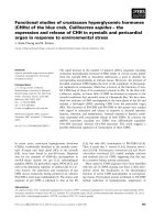
Figure 1.1 Phylogenetic tree of Rho small GTPases subfamily (adapted from
Wherlock and Mellor, 2002).
Rho GTPases are small GTP binding proteins that serve as molecular switches to
control a wide variety of signaling pathways. They are known principally for their pivotal
role in regulating the actin cytoskeleton. By switching on a single GTPase, several
distinct signaling pathways can be coordinately activated. They use a simple biochemical
Chapter 1 Introduction
_____________________________________________________________________
3
strategy to control complex cellular processes (Figure 1.2). They cycle between two
conformational states: one bound to GTP which is in the “active state”, the other bound
to GDP which is in the “inactive state”. In the active (GTP) state, GTPases recognize
target proteins and generate a response until GTP hydrolysis returns the switch to the
inactive state (Etienne-Maneville and Hall, 2002). This signaling paradigm has been
elaborated throughout evolution, which is confirmed in mammalian cells as well as in
yeast, flies, worms and plants.
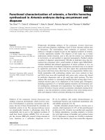
Figure 1.2 The Rho GTPase cycle. The cycle is between an active (GTP-bound) and
an inactive (GDP-bound) conformation. The cycle is highly regulated by three classes
of protein: guanine nucleotide exchange factors (GEFs), GTPase-activating proteins
(GAPs) and guanine nucleotide exchange inhibitors (GDIs) (adapted from Moon and
Zheng, 2003).
Chapter 1 Introduction
_____________________________________________________________________
4
1.1.2 Rho GTPases regulate actin cytoskeleton organization
The actin cytoskeleton regulates a variety of essential biological functions in all
eukaryotic cells. In addition to providing a structural framework around which cell
shape and polarity are formed, its dynamic properties provide the driving force for cells
to move and to divide. Understanding the biochemical mechanisms that control the
organization of actin is thus a major goal of contemporary cell biology, which also have
implications for health and disease (Hall, 1998).
The actin cytoskeleton is composed of actin filaments and many specialized
actin-binding proteins (Small et al., 1994; Stossel et al., 1993; Zigmond et al., 1996).
Filamentous actin is generally organized into a number of discrete structures
including : actin stress fibers which are bundles of actin filaments that traverse the cell
and are linked to the extracellular matrix through focal adhesions; lamellipodia which
are thin protrusive actin sheets that dominate the edges of cultured fibroblasts and
many migrating cells; membrane ruffles observed at the leading edge of the cell result
from lamellipodia that lift up off the substratum and fold backward; and filopodia
which are fingerlike protrusions that contain a tight bundle of long actin filaments in
the direction of the protrusion. They are found primarily in motile cells and neuronal
growth cones. Therefore, it is important that the polymerization and depolymerization
of cortical actin be tightly regulated. In most cases, this regulation of actin
polymerization is regulated by Rho GTPases, Rho, Cdc42 and Rac.
Members of the Rho family of small GTPases have been studied as key
regulators of the actin cytoskeleton. It is showed that in fibroblasts Rho can be
activated by the addition of extracellular stimulation such as lysophosphatidic acid
(LPA), and that activation of Rho causes the bundling of actin filaments into stress
Chapter 1 Introduction
_____________________________________________________________________
5
fibers and the clustering of integrins and associated proteins into focal adhesions
complexes (Hall, 1998; Ridley and Hall, 1992; Kozma et al., 1997). Rac can be
activated by a distinct set of agonist (for example, platelet-derived growth factor or
insulin), leading to the assembly of a meshwork of actin filaments at the cell periphery
to produce lamellipodia and membrane ruffles. And activation of Cdc42 is shown to
trigger actin polymerization to form filopodia or microspikes (Mackay and Hall, 1998;
Ridley and Hall, 1992; Ridley et al., 1992; Nobes and Hall, 1995; Kozma, 1995;
Machesky and Hall, 1997). With similar to Rho, the cytoskeletal changes induced by
Rac and Cdc42 are also associated with distinct, integrin-based adhesion complexes
(Figure 1.3a; Figure1.3b). Moreover, there is significant cross-talk between GTPases of
the Ras and Rho subfamilies: Ras can activate Rac, thus Ras induces lamellipodia;
Cdc42 can activate Rac, therefore filopodia are intimately associated with lamellipodia
(Nobes and Hall, 1995; Kozma et al., 1995); Rac1 can inactivate RhoA in NIH3T3
cells resulting in epithelioid phenotype (Sander et al., 2000; Zondag et al., 2000; Evers
et al., 2000); In contrast, in Swiss 3T3 fibroblasts, Rac1 activates RhoA instead (Ridley
et al., 1992).
From the observations above, it can be concluded that members of the Rho
GTPase family are the key regulatory molecules that link surface receptors to the
organization of the actin cytoskeleton. And this conclusion is further confirmed in a
wide variety of mammalian cell types as well as in yeast, flies and worms (Etienne-
Manneville and Hall, 2002).
Chapter 1 Introduction
_____________________________________________________________________
6
Figure 1.3a Rho, Rac, and Cdc42 control the assembly and organization of the actin
cytoskeleton. In fibroblast, activation of Rho causes the bundling of actin filaments
into stress fibers and the clustering of integrins and associated proteins into focal
adhesions complexes; activation of Rac leads to the assembly of a meshwork of actin
filaments at the cell periphery to produce lamellipodia and membrane ruffles;
activation of Cdc42 is shown to trigger actin polymerization to form filopodia or
microspikes (adapted from Hall, 1998).