Molecular basis of agrobacterium mediated gene transfer into mammalian cells 1
Bạn đang xem bản rút gọn của tài liệu. Xem và tải ngay bản đầy đủ của tài liệu tại đây (536.6 KB, 45 trang )
Chapter 1. Literature Review
Agrobacterium tumefaciens is a Gram-negative, soil-borne plant pathogen that
can cause crown gall disease, a tumorous disease at infection sites, on a wide range of
plant species (Van et al, 1974; Waston et al, 1975). Initial research in
Agrobacterium-plant interaction was intended to understand the molecular mechanism
of Agrobacterium-mediated tumor formation and to shed light on animal tumors.
Although no relationship was found between animal and plant tumors, the research
effort has introduced a possible revolution in plant genetic engineering and transgenic
technology. An overview on the mechanism of plant tumor formation is shown in Fig.
1.1. Briefly, on the wound site of the plant cell, part of the Agrobacterium DNA (T-
DNA) is processed from a large tumor-inducing (Ti) plasmid to form a T-complex
with some vir gene products. The T-complex is then transferred into the plant cell
where it will be integrated into the plant genome. In nature, the subsequent
expression of these genes carried on the T-DNA will result in the formation of
neoplastic growth, known as crown gall
tumors that serves to provide the major
sources of carbon and nitrogen for Agrobacterium (Kado, 1991; Sheng and Citovsky,
1996; Zupan and Zambryski, 1997; Stafford, 2000; Zhu et al., 2000).
Besides its natural hosts, which are dicotyledonous plants such as fruit trees and
grape vines, Agrobacterium has also been used to transform monocotyledonous plants
like rice (Komari et al, 1998; Hiei et al, 1997). Furthermore, the accumulated
knowledge of Agrobacterium has been applied to fungus, yeast and mammalian cells
as well (Bundock et al, 1995; Relic et al, 1998). Undoubtedly, the development of
Agrobacterium as a plant genetic vector has been one of the most important technical
developments in the past 25 years.
1
.
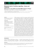
Fig. 1.1. Schematic diagram of the Agrobacterium transformation process. Critical
steps that occur to or within the bacterium (chemical signaling, vir gene induction
and T-DNA processing) and within the plant cell (bacterial attachment, T-DNA
transfer, nuclear targeting, and T-DNA integration) are highlighted, along with
genes and/or proteins known to mediate these events (Cited from Gelvin, 2000).
2
1.1. Overview of T-DNA transfer from A. tumefaciens into plant
Agrobacterium-plant interaction is the only well studied example of natural
interkingdom horizontal gene transfer system. The process of T-DNA transfer
consists of several critical steps: bacterium chemotaxis and attachment, vir gene
induction, T-DNA processing, T-DNA transfer and nuclear targeting, T-DNA
integration into the plant genome and transferred gene expression. Briefly, the T-
DNA transfer process is initialed when Agrobacterium perceives and responds to
certain
phenolic compounds, sugar, acidic pH and low phosphate level, which are
present at plant wound sites. The signal perception is mediated by the VirA/VirG
two-component transduction system. Autophosphorylation of VirA
protein and the
subsequent transphosphorylation of VirG protein result in the activation of vir gene
transcription.
Then the vir gene products are directly involved in the T-DNA
processing from the Ti plasmid and the subsequent
transfer of T-DNA from the
bacterium into the plant cell nucleus (for reviews see Tzfira et al., 2000; Kado, 2000;
Gelvin, 2000).
The T-DNA transfer process from Agrobacterium into a plant cell involves
many factors from both the bacterium and the host. There are three genetic
components of Agrobacterium that are essential for plant cell transformation. The
first component is T-DNA, the transferred segment, which is transported from the
bacterium into the plant cell (Wang et al., 1984; 1987). The T-DNA is located on the
200 kb Ti plasmid of Agrobacterium and is delimited by flanking two 25-bp imperfect
direct repeats known as the T-DNA borders. Border sequences of the T-DNA are the
only cis elements necessary for effective transformation of the plant cell (Miranda et
al., 1992). The second component is the virulence (vir) genes, which are also located
3
on the Ti plasmid. This 35-kb region of DNA, which is not transferred to the plant
cells, codes for proteins that are required for the sensing of plant wound metabolites
as well as the processing, transfer, nuclear targeting and integration of T-DNA. There
are eight major loci (virA, virB, virC, virD, virE, virG, virJ and virH) in this region.
All of the vir operons are induced as a regulon via the virA/virG two-component
system by plant phenolic compounds, such as acetosyringone (AS) and specific
monosaccharides. The third component is a set of chromosomal virulence (chv) genes,
which have been identified as necessary for tumorigenesis. Some of the chv genes are
involved in bacterial chemotaxis and attachment to wounded plant cells (Uttaro et al.,
1990; Thomashow et al., 1987; O'Connell and Handelsman, 1989; Kamoun et al.,
1989; Sheng and Citovsky, 1996), while others might be involved in the regulation of
vir gene expression
. The latter two genetic components play important roles in the
processing and transfer of the T-DNA from A. tumefaciens to the plant nucleus. In
the following subsections, the characteristics and functions of Vir proteins as well as
Chv proteins that are involved in the T-DNA transfer will be described in detail.
1.1.1. Roles of Ti-plasmid encoded virulence genes
1.1.1.1. VirA/VirG, member of highly conserved class of two-component
regulatory system
Sensing of signal molecules released by wounded plant cells is the first step of
signal transduction, which leads to vir gene expression in Agrobacterium. The vir
operons constitute a regulon which is strongly and coordinately induced in cells
growing under acidic pH conditions by two classes of plant signal molecules:
phenolic compounds, such as acetosyringone, and sugars such as glucose and
glucuronic acid. The expression of virulence genes is under the control of a two-
4
component regulatory system in A. tumefaciens, which is comprised of
VirA and VirG
(Winans, 1992; Olson, 1993).
Based
on protein sequence similarities, VirA and VirG have been assigned
to a
large group of His-Asp two-component regulatory systems,
involving a sensor and a
response regulator. VirA, an inner membrane histidine
kinase,
senses certain
phenolic
compounds released from the wounded plant cells and gets autophosphorylated
at
His-
474 (Lee et al., 1995; 1996). The phosphorylated VirA will, in
turn, transfer the
phosphate moiety to the response regulator VirG at Asp-52. Physical and genetic
evidences indicated that VirA protein exists as a homodimer in the native
conformation and the homodimer is the functional state in the plant-bacterium signal
transduction (Pan et al., 1993).
The VirA protein could be divided into four domains, which are the periplasmic,
linker, kinase and receiver domains. The periplasmic domain has been found to sense
a variety of monosaccharides
required for vir gene induction. This domain can also
interact with a periplasmic sugar-binding
protein, ChvE (Cangelosi et al., 1990; 1991).
This interaction
alone does not induce vir gene expression, but it sensitizes the
VirA
molecule to the phenolic
inducers. The VirA protein has variable efficiency in
different strains of A. tumefacines, which suggests that different chromosomal
backgrounds, especially ChvE, are not equivalent for the VirA function. The linker
domain is necessary
for perceiving phenolic compounds and acidity whereas the
kinase domain contains the conserved phosphorylatable His-474, which is required
for signal transduction in all sensor molecules. Changing this His-474 to Gln results
in a protein that can no longer be phosphorylated and a mutant carrying this
modification is avirulent and unable to induce vir gene expression in the presence of
5
plant signal molecules (Huang et al, 1990; Jin et al., 1990a; 1990b; 1990c). The
receiver domain is somewhat similar to the region of VirG, which is phosphorylated
by VirA. The function of this domain is unclear. However, it is proposed to play an
inhibitory
role in signal transduction, because once deleted,
monosaccharides alone
could induce vir gene expression in the absence
of phenolic compounds.
The VirG protein is a cytoplasmic protein. An 12-bp conserved consensus,
called vir-box, is present in the upstream region of most of the vir genes. VirG can
bind specifically to this vir box and act as a transcriptional activator of vir genes. The
C-terminus region of VirG is responsible for the DNA binding activity, while the N-
terminal is the phosphorylation domain and shows high homology to the VirA
receiver (sensor) domain. Mutants with non-phosphorylatable VirA or VirG protein
fail to induce vir gene expression (Jin et al., 1990a; 1990b; 1990c).
Both the number of copies and the types of virG gene can influence some
biological properties of A. tumefaciens. For example, multiple copies of VirG in A.
tumefaciens can greatly enhance vir gene expression and the transient transformation
frequency of some plants tissues (Liu et al., 1992). Besides, multiple copies of VirG
allow a high level of vir gene induction by acetosyringone (AS) even at alkaline pH
(Liu et al., 1993).
In addition, recent studies have revealed that
quantitative differences exist in the
interactions between VirG
and vir boxes of different Ti plasmids, suggesting that
efficient vir gene induction
in octopine and nopaline strains requires virA, virG, and
vir
boxes from the respective Ti
plasmids.
6
1.1.1.2. VirC, VirD and VirE, elements necessary for T-DNA processing
1.1.1.2.1. Roles of VirC, VirD and VirE in T-complex formation
Proteins responsible for the production of T-complex are encoded by virD and
virE operons (Grimsley et al., 1989; Toro et al., 1989; Citovsky et al., 1988; 1989;
Gietl et al., 1987; Sen et al., 1989). The T-complex consists of T-DNA, which is a
single-strand DNA segment processed from Ti plasmid, a molecule of VirD2
that is
an endonuclease covalently bound to the 5’ end of the T-DNA, and a large number of
VirE2 molecules, which is a single-strand DNA binding protein. The T-DNA is
delimited by two 25-bp direct repeats, also known as the T border, at its ends. Any
DNA between the T borders will be transferred into the plant cell as a single-strand
DNA and integrated into the plant genome. In vivo, VirD2, along with VirD1, is
sufficient for T-DNA processing in both E.coli and A. tumefaciens. virD2 encodes an
endonuclease, which cleaves the bottom strand of the T-DNA at the T-borders and
remains covalently bound to the 5’ end of the nicked DNA (Pansegrau, 1993; Jasper,
1994; Zupan et al., 2000; Gelvin, 2000). The endonuclease activity domain lies in the
N-terminal 228 aa of VirD2. This domain, along with two short regions near the C-
terminus, is the only known highly conserved domain in VirD2 protein. The possible
role of VirD1 might be its interaction with the T-borders, where ssDNA is originated.
This interaction can induce local double helix DNA destabilization and provide a
single-stranded loop substrate for VirD2. In vitro studies have shown that VirD
2
alone is enough for mediating the precise cleavage of T border sequence carried by
ssDNA templates even in absence of VirD1 protein. In contrast, VirD1 is essential
for the cleavage of supercolied strand substrate by VirD
2
.
7
Another factor, VirC1, has been found to increase the efficiency of T-strand
production when VirD1 and VirD2
proteins were limited (De Vos and Zambryski,
1989). The VirC1
protein can specifically recognize and bind to an enhancer or
overdrive sequence next to the right T-border, which is necessary for optimal T-DNA
formation.
After T strand processing, VirE2 subsequently coats ss-T-DNA along its entire
length (Citovsky et al., 1988; 1989; Gietl et al., 1987; Sen et al., 1989; Zupan et al.,
2000), forming the so-called T-complex. In this manner, VirE2 can
protect the T-
DNA from potential nucleolytic attacks. However,
recent evidences have suggested
that VirE2 protein might function primarily in the
plant cell but not necessarily in the
bacterium because plants expressing virE2 can be successfully transfected by A.
tumefaciens lacking virE2 (Citovsky et al., 1992).
Although VirE2 is associated with
the T-strand in plant cells, it is still unclear
whether this binding also occurs within the bacterial cell
or VirE2
and T-strand
molecules meet each other only inside
the host plant cell. There are two proposed
models for the VirE2 transport. On one hand, VirE2 is one of the most abundant Vir
proteins in Agrobacterium and it can bind ssDNA strongly in a cooperative way. In
addition, VirE2 and T-strand are transported
from the bacterium into
the plant cells
through the same channel. These suggest that VirE2 should bind to the T-strand in
the early
steps
of the infection process. Indeed, the T-strand and VirE2 can be
coimmunoprecipitated from the extracts of vir-induced Agrobacterium. On the other
hand, more and more evidence support that VirD2/T-DNA complexes and VirE2
might be exported into plant cells independently from the bacterium.
Complementation and co-infection studies suggest that T-strand and VirE2 are
8
exported
from the bacterial cells independently and VirE2 is not required for the
export of T-strand (Citovsky et al., 1992), while VirE2 export can be inhibited
without affecting
T-strand export. A recent biophysical report further suggested that
VirE2 itself could form channels on the artificial membranes (Dumas, 2001). Based
on the above result, Dumas et al. (2001) proposed that VirE2 is transported through
the VirB/VirD4 channel or an alternative route and subsequently inserts into
the plant
plasma membrane, allowing the transport of the ss-T-DNA-VirD2
complex.
As a specific molecular chaperone for VirE2, VirE1 is essential for the export of
VirE2 to plant cells, but not that of the T strands (McBride and Knauf, 1988; Winans
et al., 1987; Deng et al., 1999). VirE1 is a small, acidic protein with an amphipathic
-helix at its C-terminus. Yeast two-hybrid studies and extracellular complementation
suggest that VirE1 mediates T-complex formation in several
possible
ways: (i)
Although VirE1 does not
influence VirE2
transcription from the native P
virE
promoter,
virE1 indeed regulates the efficient translation of virE2; (ii)
VirE1 stabilizes VirE2 via
an interaction with the N-terminus of VirE2. VirE1-VirE2 complex is composed of
one molecule of VirE2 and two molecules of VirE1; and (iii) the formation of VirE1-
VirE2 complex, which inhibits self-interacting of VirE2 to form aggregates, might
help to maintain the VirE2
molecule in an export-competent state.
1.1.1.2.2. Roles of VirD2 and VirE2 in nuclear localization
T-complex nuclear localization is the critical step of tumorigenesis. Since T-
DNA itself
does not contain
any specific sequence, any DNA fragment located
between T-DNA borders can be transported into the plant cells and subsequently
integrated into the
plant
genome. This implies that VirD2 and VirE2,
which are
thought to associate directly with T-DNA molecule, are able to specifically mediate
9
T-complex nuclear localization instead of the nucleic
acid molecule itself. Both
proteins contain conserved bipartite nuclear localization sequence (NLS), which can
direct the T-complex into the plant nucleus through the nuclear pores (Tinland et al.,
1992; Citovsky et al., 1992; 1994). VirD2 mutants with mutations at the nuclear
localization sequence have been shown to have a reduced capability to cause
tumorigenesis, while the VirE2 mutants were completely avirulent. For the import of
short ssDNA, VirD2
alone was sufficient, but the import of long ssDNA required
VirE2
additionally (Ziemienowicz et al, 2000; 2001). These imply that the NLS of
two proteins might play different roles in nuclear localization.
The targeting of the T-DNA to nucleus is thought
to occur in a polar fashion
(Zupan and Zambryski, 1997). VirD2, which is attached to the 5' end of the T-strand,
may provide this piloting function. VirD2 molecule contains two NLS sequences, one
at each end of the molecule (Herrera-Estrella et al., 1990; Howard et al., 1992). The
N-terminal sequence possesses the monopartite type that resembles the NLS found in
the SV40 large T-antigen, whereas the C-terminal sequence belongs to the bipartite
NLS group which is characterized by two adjacent basic amino acids, a variable-
length
spacer region and a basic cluster in which any three out of
the five contiguous
amino acids must be basic (Dingwall and Laskey, 1991, Howard et al., 1992).
The N-terminal half of VirD2 required for nicking at the border sequences may
be involved in T-DNA integration in the plant nucleus, but it is not required for T-
DNA transfer because mutations in this domain could not affect T-DNA transfer
significantly (Koukolikova-Nicola et al., 1993; Shurvinton et al., 1992). It has been
reported that the N-terminal NLS of VirD2 might be occluded by the covalently
bound T-DNA because the tyrosine 29, with which
VirD2
is bound to T-DNA, is only
10
a few amino acids away from the
N-terminal NLS. The C-terminal NLS has been
found to be involved in the tumorigenesis of Agrobacterium (Rossi et al., 1993;
Narasimhulu et al., 1996). Agrobacterium mutants with genes that code for a VirD2
protein missing its C-terminal part have been found to lose their ability to induce
tumors but were efficient in the processing of T-DNA (Young and Nester, 1988).
Results from translational fusion protein and coimmunoprecipitation experiments
showed that the C-terminal of VirD2 was capable of directing a reporter gene into the
plant cell nucleus. Interestingly, the C-terminal NLS of VirD2 protein was found to
retain this function even in the mammalian cell systems. Recent evidences have
supported that VirD2 alone is sufficient to transfer short single-stranded
DNA into the
nuclei of tobacco cell and this function is strictly
dependent on the presence of the C-
terminal NLS of the VirD2
protein. A VirD2 mutant lacking its C-terminal NLS was
unable
to mediate the plant nuclear targeting of the T complexes (Rossi et al., 1993;
Ziemienowicz et al, 2000; 2001).
VirE2 protein contains two separate bipartite NLS regions (NLS1 and NLS2)
that located in the central region of the molecule in residues 212-252 and residues
288-317 respectively. Both NLSs might participate in piloting the T-DNA into plant
cell nucleus (Gietl et al., 1987; Christie et al., 1988; Citovsky et al., 1988; Das, 1988).
The relative importance of VirE2 NLSs for T-strand transfer is difficult to assess
because mutations in these sequences might also affect the binding of VirE2
proteins
to ssDNA. Analysis of VirE2 sequence have revealed that ssDNA binding domain or
the
cooperativity domain is overlapped with the NLSs of VirE2 (Citovsky et al., 1992;
Citovsky et al., 1994). Based on the results obtained from Citovsky (1992), NLS1
and NLS2 might also be involved in binding the single-strand T-DNA. Deletion of
NLS1 in VirE2 would reduce its cooperative ssDNA binding activities while deletion
11
of NLS2 or both NLS1 and NLS2 together would completely abolish ssDNA binding
and nuclear localization activities.
The contribution of VirE2 NLSs for T-complex nuclear targeting is still a
controversial issue. Some research groups have suggested that both VirD2
and VirE2
proteins play important roles in the nuclear targeting of T-complex. In one
experiment, the VirE2-GUS fusion protein was found to be localized in the plant cell
nuclei due to the nuclear targeting function of VirE2. Another experiment showed
that the fluorescently labeled single-stranded DNA together with VirE2 (lacking
VirD2), which was microinjected into plant cells, was found to have accumulated in
the plant nuclei, while naked single-stranded DNA remained in the cytoplasm. VirE2
mediated nuclear localization was found to be blocked by nuclear import
inhibitors
(Guralnick et al., 1996; Zupan et al., 1996). Unlike that in VirD2, the NLSs of VirE2
derived from the nopaline-specific Ti plasmid
are not functional in the nuclear import
of proteins in Xenopus oocytes, Drosophila
embryos (Guralnick et al., 1996) and
yeast cells (Rhee et al., 2000). However, the modified VirE2 whose NLS amino acids
was altered to resemble more
closely to animal NLS sequences could target
DNA to
animal cell nuclei (Guralnick et al, 1996), suggesting that
nuclear targeting signals in
plant and animal cells might differ
slightly (Gelvin, 2000).
On the other hand, recent studies from Ziemienowicz group showed that VirD2
alone could import a small covalently attached oligonucleotide
into the plant nucleus
and this import was absolutely dependent
on the C-terminal NLS of VirD2.
Additional evidences showed that presence of VirE2
protein could not
functionally
compensate for the deletion of the VirD2 NLS (Ziemienowicz et al., 1999; 2001).
However, when it comes to the nuclear import of big ssDNA above 250nt, VirE2
12
molecule is required even in the presence of functional VirD2 molecules.
Furthermore, it has also been found that RecA, which is a ssDNA binding protein,
could be a substitute for VirE2 in the nuclear import of T-DNA but not in the efficient
T-DNA transformation of tobacco. These results imply that (i) VirD2 might play a
role(s) in directing the T-complex to the nuclei and the NLS in VirE2 is not necessary
for the nuclear localization because RecA protein contains no motif resembling
known NLSs; (ii) VirE2 may assist nuclear uptake of the
T-complex by keeping the
T-strand in an unfolded state. In order to decipher the relative roles
of the VirD2 and
VirE2 NLSs in nuclear targeting of the T-strand, more
experiments may have to be
performed.
1.1.1.2.3. Roles of VirD2 and VirE2 in T-DNA integration
The final step of T-DNA transfer is its integration into the plant genome.
However, due to the lack of suitable systems for detailed investigation, the
mechanism of T-DNA integration into the plant genome is still unclear. It has been
proposed that this process occurs by illegitimate recombination and most of the T-
DNA transferred to the plant cell nucleus
does not integrate into the plant genome.
The integration of the
5' end of the T-strand into the plant genomic DNA is generally
precise as VirD2 is covalently linked to the 5’ end of T-strand. These facts suggest
that VirD2 might play an active role in the precise T-DNA integration into the plant
chromosome although it does not influence the efficiency of the intergration step
(Tinland et al., 1992). Shurvinton et al. (1992) demonstrated that deletion of the
conserved omega domain located near the C-terminal end of VirD2 resulted in an
approximate two orders of magnitude decrease
in tumorigenesis, while the same
mutation resulted in only a three-
to five-fold decrease in T-DNA transient expression
13
in tobacco
and Arabidopsis cells (Mysore et al., 1998; Narasimhulu et al., 1996).
These results indicated that
this mutation affected T-DNA integration
to a much
greater extent than it affected T-DNA transfer and
nuclear targeting. Mysore et al.
(1998) further proved that an Agrobacterium
strain harboring this mutation was
deficient in T-DNA integration.
The function of VirE2 protein in integration of the T-DNA into
the plant
genome is still unclear. Rossi (1996) suggested that, instead of contributing to the
efficiency
of integration, VirE2 might be involved in maintaining the integrity of the
T-DNA during the integration process.
1.1.1.3. VirB and VirD4, a type IV secretion system
The transfer of T-complex from A. tumefaciens into a plant cell relies on a type
IV secretion system (TFSS), assembled from the gene products of the ~9.5-kb
virB
operon and virD4 (Zupan et al., 1998; Deng and Nester, 1998). This apparatus
has a
pilus and may form a transmembrane channel for translocating the oncogenic T-DNA
and effector proteins from the donor to recipient cells during the process
of
A. tumefaciens infection.
As the largest operon of the vir region, the 9.5 kb virB operon encodes 11
proteins, VirB1-VirB11 (Thompson et al., 1988; Ward et al., 1988 ; 1990; Kuldau et
al., 1990; Shirasu et al., 1990). These proteins are thought to be located in or
transported to the Agrobacterium inner membrane. The proteins VirB2 through
VirB11 are absolutely required for
gene transfer and the efficient assembly of
extracellular T pili, while VirB1 is an efficiency factor for T-complex transmembrane
assembly (Berger and Christie, 1994; Fullner, 1998; Lai and Kado, 1998; Dale et al.,
1993).
14
Sequence analysis indicated that the N-terminus of VirB1 protein contains
motifs conserved among lysozymes and lytic transglycosylases, suggesting that VirB1
protein might be a putative lysozyme and locally lyse the murein cell wall to create
channels for transporter
assembly (Mushegian et al., 1996; Baron et al., 1997). This
hypothesis is supported by the findings that mutants with deletion in the putative
lysozyme homologue were attenuated in virulence (Mushegian et al., 1996).
A smaller protein, VirB1* (comprising the C-terminal 73 amino acids of VirB1
protein) is found to be secreted and loosely associated with the outer membrane.
Coimmunoprecipitation analysis showed that VirB1* and VirB9 form a large complex
(Baron et al., 1997). These findings suggest that VirB1* may mediate pilus formation
by stabilizing pilus-based contacts between Agrobacterium and plant cells (Zupan et
al ., 1998).
VirB2 and virB5
form a pilus that presumably promotes host-recipient
interaction. A processed form of VirB2
is suggested to be the major structural
component of pilus, while VirB5 could serve as essential protein stabilizers (Lai and
Kado, 1998; Shirasu and Kado, 1993). VirB3 and VirB4
might be accessory
pilus
proteins, which are required for pilus assembly
but are not the structural components
(Jones et al., 1994; Shirasu et al., 1994; Dang and Christie, 1997; Dang et al, 1999).
The other five VirB proteins (VirB6-VirB10) might form putative transmembrane
apparatus (reviewed in Kado, 2000). Most of these proteins interact with one another
and form various protein complexes. VirB6 is
firmly embedded in the inner
membrane with its five transmembrane
regions, and its presence is required for the
stability of several
other VirB proteins.
The core of the transfer apparatus is likely to
be composed
of VirB7-VirB9 heterodimers that are linked by a disulfide bridge
and
15
anchored in the outer membrane by lipid modification of VirB7. The VirB7-VirB9
heterodimer interacts,
either directly or indirectly, with VirB10 and is genetically
required for the stability of VirB4, VirB8, VirB10 and VirB11.
Purified VirB4 (Shirasu et al., 1994; Dang and Christie, 1997; Dang et al.,
1999) and VirB11 (Christie et al., 1989; Rashkova et al., 1997) were shown to
possess ATPase activity and VirB4 ATPase mutations would abolish T-pilus
biogenesis. These results clearly indicate that VirB4 promotes T-pilus
production and
configures the transfer apparatus as a dedicated
export machinery by an ATP binding-
dependent mechanism. VirB11 might also function as chaperones
to facilitate the
movement of unfolded proteins and DNA substrates
across the cytoplasmic membrane
by supplying energy for a possible gated secretion channel (Lai and Kado, 2000).
VirB11
localizes at
the inner face of the cytoplasmic membrane independently of
interactions
with other VirB proteins. Analysis of mutants with
defects in the
nucleotide triphosphate binding pocket (Walker
A motif) suggest that this membrane
interaction is modulated by
ATP binding or hydrolysis.
As the third ATPase, VirD4 is also essential for T-DNA transfer into plant cells
because the VirD4 mutants showed complete inactivity in T-DNA transfer (Zupan et
al., 1998). VirD4 is an inner membrane protein with two membrane spanning
domains near the N-terminus while both the N- and C-terminus of the protein are
cytoplsmic. The large cytoplasmic region contains a nucleotide-binding domain.
Both the periplasmic and cytoplasmic domains are essential for substrate transfer.
Although VirD4 is not required for T-pilus assembly, it is required for virulence and
thus it likely
plays a role as the coupling protein for the transfer of virulence
factors
(VirD2, VirE2, VirF and T-DNA) to the membrane-bound
components of the type IV
16
transporter by an energy dependent mechanism. It remains unclear whether VirD4
interacts physically with the T-DNA transport apparatus, and whether the interaction
would either be permanent or transient. Pantoja et al. (2002) proposed that VirD4
localizes to the cell pole and a polar VirD4 –VirB complex functions in substrate
transfer from the cytoplasm.
Proteins homologous to the VirB and VirD4 system
can also be found in many
animal pathogens, including Bartonella henselae,
Bordetella pertussis, Brucella
abortus, Brucella suis, Helicobacter
pylori, Legionella pneumophila and Rickettsia
prowazekii. In mammalian pathogens, these systems are required for
the delivery of
pathogenesis-related effector proteins and other molecules as well as for intracellular
survival. Presumably, both the T-strand and
its associated proteins are transferred
from Agrobacterium into the plant
cells through this type IV secretion system,
although the precise role of pili in
DNA transfer is still not clear in any conjugal
transfer system.
1.1.1.4. VirF
The 23-kDa VirF protein is encoded by a gene only present in the vir region of
octopine-type Ti plasmid and absent in nopaline-type Ti plasmid (Melchers et al.,
1990;
Schrammeijer et al., 1998). virF mutants were originally described as host
range mutants because the presence of virF gene on the octopine-type Ti plasmid
made Nicotiana glauca susceptible to the infection by Agrobacterium.
Besides VirD2
and VirE2, VirF is the third Vir protein that is exported to the
plant cells from Agrobacterium. The transport of VirF from Agrobacterium into the
plant cells is depended on the VirB/D4
transport system. The C-terminal amino acid
17
motif Arg-Pro-Arg, which is also present on the VirE2 molecule, is supposed to be the
export signal that can be recognized by the VirB/D4
secretion system.
VirF might function in the plant cells because virF mutant strain can be
complemented by the expression of the virF gene in the plant host cells. The results
from yeast two-hybrid experiment suggest that the VirF protein is the first prokaryotic
protein with an F box, by which it can interact with the plant homologue Skp1 protein
of the yeast. Since Skp1 proteins are part of the complexes involved in targeted
proteolysis and thus regulate the plant cell into S phase, it is suggested
that VirF might
help in stimulating the plant cells to divide and become
more susceptible to
transformation of A. tumefaciens (Schrammeijer et al., 2001).
1.1.1.5. VirJ
virJ lies between virA and virB in the vir region of an octopine-type Ti plasmid
(Pan et al., 1995; Kalogeraki and Winans, 1995). This gene is not found in the
nopaline –type Ti plasmid pTiC58. VirJ shares 50% identity at the amino acid
sequence level with a chromosome-encoded protein AcvB, which could be found in
both octopine and nopaline type strains. The homologous region lies in C-terminal
half of AcvB. The virJ gene contains a putative vir box and can be induced in a
VirA-VirG dependent fashion by the vir gene inducer acetosyringone, which,
however, has no effect on acvB.
The role of VirJ (and AcvB) in tumorigenesis is still unclear. Either VirJ or
AcvB is required for the T-DNA transfer from Agrobacterium into the plant cell (Pan
et al., 1995). The two proteins share at least some degree of functional similarity
because virJ could heterologously complement an acvB mutation in the tumorigenesis
of Agrobacterium on plant wound sites. AcvB or VirJ did not affect the attachment of
18
Agrobacterium to plant cells, but agroinfection experiments had proven that VirJ or
AcvB might be required for the T-DNA transfer (Pan et al., 1995). It has been
reported that AcvB might play a role in virulence by influencing the formation of the
pili (Parimal et al., 1999). Some researchers (Parimal et al., 1999) suggested
that
AcvB is a single-stranded DNA binding protein that could interact
with the T-strand
and assist in the export of T-DNA from the bacteria to the host cells.
If this model
were proven correct, it would explain how the T-strand
could be transferred from
Agrobacterium as a T-strand/protein
complex independent of VirE2.
1.1.1.6. Other vir genes
Using an electron microscope, an Ti plasmid conversed genetic locus was
identified at the left end of known vir gene. This locus flanks an operon designated
as virH. The virH operon contains two genes that resemble P-450-type
monooxygenases (Kalogeraki and Winans, 1998). Since VirH
1
and VirH
2
are
homologous to each other, it seems plausible that they could be functionally
redundant. The role of VirH in plant –microbe interaction requires additional studies.
1.1.1.7. Other genes on Ti plasmid
There are some other gene loci on the Ti plasmid besides vir genes. Some of
them confer ancillary functions in tumor formation, such as inter-bacterial
conjugation genes and vegetative replication genes. Inter-bacterial conjugation genes
include oriT, traAFB and trbB, which control the conjugative transfer of Ti plasmid.
Vegetative replication genes, including repAB and repC, function to control Ti
plasmid replication and partition.
19
Some of the T-DNA genes, which direct the production of plant growth
hormones, affect tumor morphology and physiology. Interestingly, the non-
transcribed regions of these genes possess many features of plant genes, including
typical eukaryotic TATA and CAAT boxes, transcriptional enhancers and poly(A)
sites. These genes include iaaM (also called aux1, tms1), iaaH (also called aux2,
tms2) and ipt (also called cyt, tmr), encode enzymes catalyzing the synthesis of auxin
and cytokinin respectively. The gene ons (or 6a) controls octopine and nopaline
export from plant cells, while tml (or 6b) increases the sensitivity of plant cells to
phytohormones (Clarence, 1991; Sheng and Citovsky, 1996; Winans et al., 1986;
1989).
1.1.1.8. Summary of the functions of Ti-plasmid encoded virulence genes
After sensing the particular plant signals such as phenolic compounds, the
VirA/VirG two-component system induces the expression of other vir genes whose
products function in processing of T-DNA and the subsequent transfer, nuclear
targeting and integration. Both VirD1 and VirD2
are responsible for producing the ss-
T-DNA (Albright et al., 1987;Wang et al., 1987). The VirD2 protein recognizes and
nicks the T-DNA borders and subsequently becomes covalently attached to the 5’ end
of ss-T-strand (Howard et al., 1989; 1992; Pansegrau et al., 1993). The 69 KDa
single-stranded DNA (ss-DNA) binding protein VirE2 coats the T-strand along its
entire length (Citovsky et al., 1989; Gietl et al., 1987; Sen et al., 1989), but it remains
unclear whether the binding of VirE2 occurs in the bacterium (Christie et al., 1988) or
in the plant cell (Binns et al., 1995; Sundberg et al., 1996). What is certain is that this
cooperative association of VirE2 and T-DNA prevents the attack of nucleases.
20
After T-DNA processing, the T-complex can travel from Agrobacterium into the
plant cells through a type IVsecretion system (TFSS, which is assembled from
11 VirB proteins and VirD4 (Dang et al., 1999; Lai and Kado, 1998; Zupan et al.,
1998). After delivered into the plant cytoplasm, the T-strand is targeted to the nucleus
and would cross the nuclear membrane before it is integrated into the host plant
genome. Both VirD2
and VirE2, which contain NLSs, play an active role in these
processes.
Mutations in the virA, B, D, E and G loci result in avirulence, whereas mutations
in virC causes attenuated virulence (Yanofsky et al., 1985; Horsch et al., 1986).
Some members of vir operon, such as virJ, F, H and E3, are required for
tumorigenesis in specific instead of all hosts or play other roles in pathogenesis.
Results of recent studies showed that VirD2, VirE2 and VirF, the three exported
virulence proteins, can also be exported from bacterial cells by a specific pathway
independent of VirB/D4
(Chen et al., 2000). Although the precise biological function
of this process is still not clearly addressed, it suggests that the transfer of the T-
complex from Agrobacterium may take place in two steps, with the first step mediated
by an unidentified pathway and the second step by the virB/D4 system (Chen et al.,
2000).
The fact that Agrobacterium possesses genes which are not only typically
eukaryotic genes with eukaryotic expression signals, but also prokaryotic genes
coding for proteins with eukaryotic features such as the nuclear localization sequences
(VirD2, VirE2 and VirE3), the F box (VirF) and eukaryotic promoter (iaaM and
iaaH) infers that some of these genetic materials were possibly introduced into
21
Agrobacterium by horizontal gene transfer from an eukaryotic organism, although no
direct evidence has been obtained so far.
1.1.2. Roles of chromosomal virulence genes of A. tumefaciens
Some Agrobacterium chromosomal virulence (chv) genes have also been shown
to play important roles in tumorigenesis (Gelvin, 2000; Zhu et al., 2000; Zupan et al.,
2000; Liu et al, 2001). In contrast to the virulence genes on the Ti plasmid, the
functions of chromosomal virulence genes have not been well
elucidated. The
pleiotropic functions of these genes make it difficult to assess their precise roles in
tumorigenesis.
The chv genes exert their functions mainly in the events of bacterial attachment
to the plant cell wall, the promotion of growth efficiency in wound site on the plant
and the regulation of virulence genes on the Ti plasmid during the early stages of
infection (Sheng and Citovsky, 1996; Zupan and Zambryski, 1997). These suggest
that, unlike those vir genes on the Ti plasmid, which are dedicated solely
to specific
steps in the interaction of Agrobacterium with the host
plant, the chromosomal
virulence genes exert their functions in
regulating the general physiology of
Agrobacterium and have been conscripted
to play ancillary but significant roles in the
interaction of
the bacterium with its
host plants.
As the best understood chromosomal virulence gene, chvE was shown to play
important roles in the sugar enhancement of vir gene induction and bacterial
chemotaxis. Mutation in this locus strongly attenuated vir gene induction and limited
the host range. chvE gene codes for a periplasmic glucose-galactose binding protein,
which is required in the VirA/VirG two component regulatory system (Winans et al.,
1994; Doty et al., 1996). This protein can sense monosaccharides in the environment
22
and then interact with the periplasmic domain of VirA, a requirement for maximal
activation of VirG and the subsequent activation of all Ti plasmid-encoded
vir genes.
Chemotaxis and attachment to plant is the beginning of infection (Vande Broek
and Vanderleyden, 1995). Genetic studies showed that non-attaching mutants had
lost the capability of tumorigenesis. Agrobacterium attachment to the plant cell is a
two-step
process. A cell-associated
acetylated and acidic capsular polysaccharide
plays an important role in the first step. The attachment in this step is reversible
because vortexing or washing with a stream of
water could dislodge the bacteria.
attR
encodes a transacetylase, which is required for the synthesis
of this polysaccharide.
Mutants of attR, which could not synthesize the acetylated polysaccharide were found
to be avirulent and could not attach to carrot suspension
cells (Matthysse and
McMahan, 1998)
The second step of attachment involves the elaboration of cellulose
fibrils by the
bacterium, which causes large number of bacteria
to colonize at the wound surface
(Matthysse and McMahan, 1998; Matthysse et al., 1995). Some chromosomal
virulence genes, chvA, chvB, and pscA (exoC), are required for this process. These
three genes were concerned with either the synthesis (chvB and pscA) or export
(chvA) of cyclic
ß-1,2-glucans and other sugars into the periplasm (Uttaro et al., 1990;
Thomashow et al., 1987; O'Connell and Handelsman, 1989; Kamoun et al., 1989) and
may be involved indirectly
in bacterial attachment by an unknown mechanism.
Mutants of chvA
or chvB could not attach to the host cells and abolished the tumor
formation ability in normal inoculation conditions (Douglas, 1982; 1985).
Interestingly, chvB mutants could partially regain virulence if
the bacteria
were grown
and inoculated at 19°C.
23
The newly characterized chromosomal gene, chvH, encodes a homologue of an
elongation factorP (efp) involved in protein synthesis (Peng et al., 2001). The chvH
gene is present as a single
copy in A. tumefaciens
and
is important but not essential for
the growth of Agrobacterium. The chvH mutant A6880 is an avirulent, pleiotropic
mutant. This strain is more sensitive to detergents such as SDS and acidic pH than its
parent strain, which suggests
that the integrity of the outer membrane is impaired.
Elongation factor P in E. coli can increase
the efficiency of formation of peptide
bonds involving aminoacyl
acceptors that bind poorly to the ribosome in its absence
(Glick 1980). Heterologous complementation of chvH mutation in Agrobacterium
could be achieved by the expression of the efp gene of
E. coli, suggesting that chvH
and efp are
functionally homologous.
As an elongation factor protein, ChvH exerts its roles at the posttranscriptional
level. The avirulence of the chvH mutant is due to the low level expression of key
proteins required for T-DNA transfer
such as VirB, VirE2 and VirG, although the
possibility that the chvH gene product
may contribute in other ways to tumorigenesis
cannot be ruled out. Further results showed that wild-type chvH locus is essential not
only for full expression
of vir genes encoded by the Ti
plasmid but also for that of
some chromosomal genes. These genes might
code for particular sequences of amino
acids, perhaps near the
start of translation, which are exceptionally dependent on
elongation
factor P for translation.
Some other genomic genes such as chvD, ros and miaA are also involved in
virulence (Gray et al., 1992; D’souza-Ault et al., 1993). chvD encoding an ABC
transporter homologue plays important roles in the virulence regulatory pathway.
Results from Liu et al. (2001) showed that ChvD controlled
virulence genes by
24
affecting virG
expression. The strains carrying a mutant chvD gene greatly attenuated
virulence and vir gene expression, while constitutive expression of virG in the same
strain
restored virulence. Using yeast two-hybrid screening, the interaction between
VirB8 and ChvD is verified. However, the biological relevance of this interaction is
still
unclear.
Another chromosomal gene ros encodes an 15.5-kDa C2H2 zinc finger protein
that represses the expression of virC and virD (Cooley et al., 1991) and a plant
oncogene ipt. ipt, whose promoter contains typical TATA boxes, is regulated by
eukaryotic transcriptional machinery in the host plant (Chou et al., 1998 ). C2H2 zinc
finger proteins are a large superfamily of eukaryotic transcription factors and are
originally thought to occur in eukaryotes. Phylogenetically, ros is distantly related to
eukaryotic zinc finger regulators, as will be described later.
Some chv genes have counterparts in other bacteria that are associated
pericelluar or intracellularly with animals and plants, either as pathogens or as
endosymbionts. One set of such genes, chvG/chvI has been under extensive studies
and are found widely in the chromosomal loci of many organisms such as
A. tumefaciens, Brucella abortus and Sinorhizobium meliloti (Sola-Landa et al., 1998;
Galibert, et al., 2001). These genes are required for establishing a successful
relationship between the bacteria
and their hosts. Sequence anaylsis of the 16S rRNA
gene showed that these genera all
belong to the same -2 subdivision of the
proteobacteria. Based on the amino acid sequence analysis, the chvG/chvI genes
encode a two-component signal transduction system. Once bacteria are internalized
into plant or animal cells, it is possible that they encounter an acidic pH environment
within the vesicles containing them. Sensing the acidity appears to be important for
25