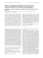
Antimicrobial activity of delaminated aminopropyl functionalize
Bạn đang xem bản rút gọn của tài liệu. Xem và tải ngay bản đầy đủ của tài liệu tại đây (1.28 MB, 8 trang )
Research Paper
Antimicrobial activity of delaminated aminopropyl functionalized
magnesium phyllosilicates
Gayathri Chandrasekaran
a
, Hyo-Kyung Han
b
, Geun-Joong Kim
c
, Hyun-Jae Shin
a,
⁎
a
Department of Chemical and Biochemical Engineering, Chosun University, Gwangju 501-759, Republic of Korea
b
College of Pharmacy, Dongguk University, Pil-dong 3-ga, Jung-gu, Seoul, Republic of Korea
c
Department of Biological Science, College of Natural Sciences, Chonnam National University, Gwangju 500-757, Republic of Korea
abstract
Keywords:
AMP clay
Antimicrobial activity
Membrane permeability
Intracellular enzymes leakage
In this study, dispersed aminopropyl functionalized magnesium phyllosilicates (AMP clay) were tested for
antimicrobial activity against various Gram negative and Gram positive bacteria including multidrug resistant
bacterial strains and fungi. The AMP clay strongly inhibits the growth of microorganisms such as Escherichia
coli, Staphylococcus aureus and Candida albicans. The inhibition of microbial growth by AMP clay is attributed
to the amino propyl groups and their charge interactions. The bactericidal effect of AMP clay occurred within
1 h, and 95% killing was observed within 2 h. In addition, AMP clay could kill microbes by disrupting
membrane integrity and thus essential components inside the cells. This effect was also evaluated through
membrane depolarization assays and measured by the release of intracellular enzymes. The assessment of cell
damage by the AMP clay was obtained by scanning electron microscopy (SEM) observation. Moreover, AMP
clay activity was stably maintained after a long storage time of up to 30 days at different temperature
conditions. The goals of this study were to synthesize the dispersed AMP clay from a commercially available
substrate, which can be applied to various biomedical applications and environmental protection.
1. Introduction
Clays are used in various scientific and industrial applications
because they are present in nature, and they can be modified both
chemi cally and physic ally (Aguzzi et al., 2007). The difference
between the natural and synthetic materials lies in their surface
areas, which are proportional to the degree of delamination or
exfoliation of the clay layers (Carrado, 2000). Generally, smectite clay
is modified organically via organo-cation exchange (Xue and
Pinnavaia, 2010). An aminopropyl-functionalized magnesium phyllo-
silicate (AMP clay) contains a trioctahedral smectite with an organic
propylamine group. There have been several recent reports on the
synthesis and characterization of organoclay-modified derivatives in
the form of nanocomposites for biomolecules encapsulation, immo-
bilization, biosensing devices, nanoreactors and enzyme reactors
(Ferreira et al., 2008; Holmström et al., 2007; Johnsy et al., 2009; Patil
et al., 2005). Therefore, the interest in the use of organically modified
clays has increased with time. In fact, they possess swelling ability,
high cation exchange capacity, high layer aspect ratio, high specific
surface area, film formation and adsorptive properties. In particular,
smectites appear to be good candidates in industry, agriculture,
environmental remediation, catalysis, coatings, ceramics, and poly-
mer materials due to their submicron size (Xue and Pinnavaia, 2010).
In addition, various applications of organoclays have been suggested
for groundwater purification, hazardous waste landfills and industrial
wastewater treatment (Pernyeszi et al., 2006). Clay materials can be
used as geotechnical barrier adsorbents for toxic organic chemicals
and heavy metals and can also serve as candidates for antibacterial
applications (Boyd et al., 1988; Oya et al., 1991). Recently, the
antimicrobial effects of nanoclay composite films, which were
prepared by organically modified nanoclays have been reported. It
has been hypothesized that these properties could result from the
quaternary ammonium groups of organically modified clays (Rhim
et al., 2009). The use of organically modified clay has greater
advantages than clay-based inorganic antibacterial materials. The
synthesis of inorganic antibacterial clays using heavy metals is
comparably difficult due to serious metal toxicity in humans and
the environment. These heavy metals also decrease the antibacterial
activity due to the formation of insoluble compounds (He et al., 2006;
Pernyeszi et al., 2006). However, the clays that have been reported to
date were insoluble, which limits their applications. As far as we
know, there has been no report on the antimicrobial activity of AMP
clay. To make dispersed clay, AMP clay was synthesized and evaluated
for antimicrobia l activity against bacteria, fungi and multidrug
resistant strains. The mechanism of antimicrobial action was
investigated by various parameters, such as membrane depolariza-
tion, release of intracellular enzymes, and scanning electron
⁎ Corresponding author. Tel.: +82 62 2307518; fax: +82 62 2307226.
E-mail address: (H J. Shin).
SCIENCEWORLDLIB.COM
microscopy. Therefore, this dispersed AMP clay may exert antimicro-
bial activity by a membrane disruption mechanism.
2. Experimental
2.1. AMP clay synthesis
The AMP clay was prepared by sol–gel synthesis (Burkett et al.,
1997; Whilton et al., 1998). In brief, magnesium chloride (1.68 g,
7.24 mM) was first dissolved in ethanol (40 g), and the solution was
stirred at room temperature. Then, 3-aminopropyltriethoxysilane
(2.6 ml, 11.7 mM) was added drop-wise under magnetic stirring. The
resulting white suspension that formed after few minutes was stirred
overnight. The resulting products were centrifuged, washed in
ethanol (3× 50 ml) and dried at 40 °C. The white solid was ground
to produce a powder (Patil et al., 2007).
2.2. AMP clay characterization
The microstructure of AMP clay was investigated with Fourier
transform infrared spectroscopy (FT-IR), silicon nuclear magnetic
resonance (
29
Si NMR), X-ray diffraction (XRD) and transmission
electron microscopy (TEM) analysis. The FT-IR spectrum of AMP clay
was obtained with KBr pellets at the ratio of 10:90 by weight
(NICOLET 6700, Thermoscientific, USA). Solid state Silicon-29-NMR
measurements were performed to determine Si associated with
different numbers of hydroxyl end groups using an Avance 400
(Bruker, Germany) at room temperature. The powder XRD pattern
was obtained with an X-ray diffractometer (XPert PRO, PANalytical
B.V., Al melo, Holland) using CuKα radiation at 20 mA and 40 kV.
Powder samples were pressed onto a glas s sample holder. S cans were
recorded between 2° and 70 ° (2θ) with a step size of 0.05° and a
scanning speed of 2°/m in. TEM (TECNAI20, EEI, Netherland) imaging
was performed on a bright field emission microscope with a Lab 6
electron gun and an accelerating voltage of 200 kV.
2.3. Microorganisms
Bacterial and fungal cultures used in this present study were obtained
from the Korean Collection for Type Cultures (KCTC). Gram positive
species were Bacillus subtilis (KCTC 1918), Listeria monocytogenes (KCTC
3710) and Staphylococcus aureus (KCTC 1621) while the Gram negative
species were Escherichia coli (KCTC 1682), Salmonella typhymurium (K CTC
1926) and Pseudomonas aerugenosa (KCTC 1637). Candida albicans,a
fungal strain, was KCTC 7270. Antibiotic resistant E. coli strains (CCARM
1229 and CCARM 1238) and S. aureus strains (CCARM 3108, CCARM
3089) were obtained from the Culture Collection of Antibiotic-Resistant
Microbes (CCARM) at Seoul Women's University in Korea.
2.4. Antibacterial activity
Bacterial cells were cultured at 37 °C in appropriate culture media.
The antimicrobial activity of AMP clay was determined by the
microdilution method. In brief, aliquots of bacterial suspensions
(50 μl) in the mid-logarithmic phase at a concentration of 2×10
5
colony
forming units (CFU)/ml in an appropriate culture medium were added
to each well containing 50 μl of AMP clay solution that had been twofold
serially diluted in buffer I (10 mM sodium phosphate buffer, pH 7.2) or
buffer II (phosphate buffered saline (PBS)), 1.5 mM KH
2
PO
4
,2.7mM
KCl, 8.1 mM Na
2
HPO
4
, and 150 mM NaCl (pH 7.2). The plates containing
the bacterial cells were incubated at 37 °C for 24 h. At the end of the
incubation, 50 μl of 10-fold diluted samples were plated on appropriate
agar plates and were then incubated for 24 h, after which the colonies
were counted. The lowest concentration of AMP clay that completely
inhibited growth was defined as the minimum inhibitory concentration
(MIC) (Park et al., 2008). The MIC values were calculated as an average
of several independent experiments conducted in triplicate.
2.5. Antifungal activity
The fungal strains of C. albicans (KCTC 7270) were cultured at 28 °C
in YPD media (dextrose 2%, peptone 1%, and yeast extract 0.5%, pH 5.0
to 5.5). Fungal cells (final concentration 2×10
4
CFU/ml) that were
grown in 100 μl of YPD media were seeded in each well of a microtiter
plate containing 100 μl of twofold serially diluted AMP-clay in buffer I
or buffer II, as described above. After incubating for 24 to 30 h at 28 °C,
the lowest concentration of AMP clay inhibiting the growth of fungi
was microscopically determined to be the MIC ( Park et al., 2007).
2.6. Radial diffusion assay
The experiment was performed as described previously by Nordahl
et al. (2004) with some modification. Briefly, E. coli and S. aureus isolates
were grown for 18 h at 37 °C in 10 ml of media. The underlay consisted
of 1% agarose and 1/100 dilution of Luria Bertani (LB) broth in PBS (1%
tryptone, 0.5% yeast extract, 1% sodium chloride pH 7.2). The overlay
consisted of 6% LBand 1% agarose in distilled water. The bacterial culture
2×10
5
colony forming units (CFU)/ml were mixed with 10 ml of
underlay solution, allowed to melt at 48 °C and was then poured into
petri dishes. When the agarose was solidified, approximately 4-mm-
diameter wells were made, and the wells were filled by AMP clay
solutions in various concentrations. Buffer in one well served as a
control. To cause AMP clay diffusion on the under layer, the plates were
incubated at 37 °C for 4 h. The molten overlay was then poured on the
underlay, and the plates hardened. This was followed by incubation at
37 °C for 48 h. Finally, the antibacterial activity was determined by
measuring the diameters of the inhibitory zones around the well. The
activity was represented in radial diffusion units (RDU) defined as
(diameter of clear zone (in millimeters)− 6)×10. All the experiments
were performed triplicate, and the antibacterial activity was expressed
as the mean of inhibition diameters (mm).
2.7. Trans-membrane depolarization assay
The trans-membrane d epolarizing activ ity of the A MP cl ay was
performed as described previously (Papo et al., 2002). The experiments
were performed for the Gram-positiv e bacteria (S. aureus CCARM 3108),
Gram-negative bacteria (E. coli CCARM 1229) and a fungal strain of C.
albicans. The cells of bacteria and fungi were grown to mid-logarithmic
phase a t 3 7 °C and 28 °C un der shaking at 150 rpm. Then, the cell
resuspension OD
600
of 0.05 was subjected to washing twice with the
buffer(20mMglucose,5mMHEPES,and0.1MKClatpH7.3).The
bacterial and fungal cells without the AMP clay were incubated with 1 μM
3,3′-diethylthiodicarbocya nine iodide (DiSC
3
-5, which was acquired from
Molecular Probes, Eugene, OR, USA) until there was no detection of
fluorescent dye for 3 h. Then, AMP clays with various c oncentrations were
added, and the change in in tensity of the fluorescence emissio n of the dye
was measured at the excitation and emission wavelength (622 and
670 nm).
2.8. Intracellular enzyme activity
Aspartate aminotransferase (AST) and alanine aminotransferase
(ALT) activities were measured u sing an assay kit (Asan Pharmaceutical,
Korea) based on the m ethod (Ma et al., 2010; Reitman and Frankel, 1957).
6.25mg/mlofAMPclayand1.0mlbacterialsuspension2×10
7
CFU/ml
were added into the sterile tubes containing the LB broth. T hen, the tubes
were a gitated on an orbital shaker a t 150 rpm a nd cultured for 2 h at
37 °C. This was followed by filtration. Then, the filtered solution was
subjected to the enzyme activity assay. Untreated cells were used to
determine background lysis, and cells treated with 1% Triton X-100 were
used to determine total enzymes (100% lysis). Each experiment was
performed in triplicate.
2.9. Scanning electron microscopy (SEM)
For SEM analysis, the AMP clay was treated with mid log phase of
E. coli, S. aureus and C. albicans in PBS (pH 7.2), and the strains were
incubated at 37 °C and 28 °C. After 60 min, the AMP clay treated cells
(2×10
7
CFU/ml) were centrifuged at 3000 rpm for 5 min, followed by
washing with the same buffer. The resulting supernatant was
removed, and the pellets were fixed with 1% glutaraldehyde in
0.2 M sodium-cacodylate buffer (pH 7.4) for 3 h at 4 °C. Controls were
the microorganism without AMP clay treatment. After fixation with
glutaraldehyde, the samples were extensively washed with same
buffer at pH 7.4, subjected to gold coating and were investigated with
a scanning electron microscope (Hitachi S-2400N, Japan).
2.10. Determination of bactericidal activity by kinetic study
The bactericidal a ctivity of A MP c lay o n E. coli (CCARM 12 29) and
Streptococcus strains (CCARM 3108) was studied by a time-kill assay. The
bacterial culture of 2×10
5
colony forming units (CFU)/ml was added to
LB media containing AMP clay at 1× MIC and 2× MIC and was incubated
at 37 °C. The samples were withdrawn at various time intervals for every
half an hour up to 2 h, diluted using PBS buffer and plated on the LB agar
plates. The plates were incubated for 24–36 h at 37 °C, and the kinetics of
bactericidal activity w ere determ ined by p lotting the concentration of
surviving bacteria against time ( Jeong et al. , 2010 ).
2.11. Storage stability of AMP clay
The antibacterial activity of AMP clay was determined at different
temperatures over several days. Five aliquots of approximately 600 μl
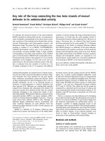
Fig. 1. Characterizations of AMP clay structure with FTIR spectrum explains the trioctahedral band at 670 cm
− 1
(A),
29
Si HPDEC NMR spectrum shows the T3 moiety for silane
structures (B), X-ray diffraction pattern characterize 060 reflection at 0.156 nm for the trioctahedral structure confirmation (C), and TEM images of exfoliated sheets of AMP clay in
distilled water with a layered edge view (D).
Table 1
The MIC values of AMP clay against microorganisms.
MIC (mg/ml)
Microorganism AMP clay
Buffer I
a
Buffer II
b
Gram (−) bacteria
E. coli 3.12 6.25
S. typhimurium 0.78 1.56
P. aeruginosa 3.12 6.25
Gram (+) bacteria
S. aureus 1.56 3.12
B. subtilis 3.12 6.25
L. monocytogenes 3.12 6.25
Fungal strain
C. albicans 1.56 3.12
Resistant strains
E. coli CCARM 1229
c
3.12 6.25
E. coli CCARM 1238
c
3.12 6.24
S. aureus CCARM 3108
d
0.78 1.56
S. aureus CCARM 3089
d
0.78 1.56
a
Buffer 1: 10 mM Sodium phosphate buffer, pH 7.2.
b
Buffer II: ph osphate buffered saline (1.5 mM KH
2
PO
4
, 2.7 mM KCl, 8.1 mM
Na
2
HPO
4
, 135 mM NaCl), pH 7.2.
c
Multidrug-resistant Escherichia coli strains.
d
Multidrug-resistant Staphylococcus aureus strains.
were withdrawn from the stock solution. The aliquots were further
subdivided into 300 μl each and stored at room temperature and 4 °C.
After a fixed number of days, the solutions were subjected to
antibacterial activity.
3. Results and discussion
3.1. Characterization of AMP clay
The synthesis of AMP clay was confirmed by FTIR,
29
Si NMR, XRD and
TEM. The FTIR spectrum of AM P clay, shown in Fig. 1A, exhibited
absorption bands corresponding to t e rminal NH
2
stretching (3600–
3200 cm
− 1
), NH
2
bending (1658 cm
− 1
), CH
2
stretching (2950–
2850 cm
− 1
), Si\O\Si stretching (1120 cm
− 1
), and Mg\O\Si bending
(670 cm
− 1
). The latter spectrum explains the surface property of the
clays. The
29
Si NMR s pectrum for AMP clay is s hown in Fig. 1B. Three
signals at − 66.5,−58.6, and−48.4 ppm are associated with R-SiO
3
-(T3),
R-SiO
2
-OH-(T2) and R-SiO-(OH)
2
-(T1) in the inorganic–organic b ackbone
structure. The T3 moiety predominates among the silane structures. The
XRD pattern of the AMP clay is characterized by a d
001
spacing at 1.44 nm
due t o t he presence of the a minopropyl organic chains between l ayers
(Fig. 1C). The position of the 060 peak indicates the presence o f a
trioctahedral Mg-phyllosilicate clay mineral although the d
060
value
(0.156 nm) is larger t han usually reported (Datta et al., 2007). TEM
investigations showed the presence of delaminated 30–150 nm ultrathin
sheets an d clay sheets edge indicat es the l ayered structure o f 20 nm in
thickness as shown in Fig. 1D(Ferreira et al., 2008 ; Holmström et al., 2007;
Lee e t al., 2010). Overall, AMP clay is h ydrophilic and possesses protonated
(R-NH
2
) groups, which create a number of bonding sites for ion exchange
within the inter laye r spaces and serve as s urf ace group on t he lamella,
which are driven by electrostatic forces for their antimicrobial effect.
3.2. Antimicrobial activity
Antibiotic resistance is increasing at a rate that exceeds by far the
development of new types of antibiotic agents. Therefore, there is an
increasing need to synthesize novel compounds with ant ibiotic
activity. To our knowledge, the antimicrobial properties of organically
modified derivatives of magnesium phyllosilicates have not been
reported so far. The AMP clay had MICs of 6.25 mg (both in low and
high ionic strength buffer) against many bacterial strains (Table 1).
However, this AMP clay showed twofold higher activity against S.
aureus and S. typhimurium than other bacteria. In addition, the MICs
for resistant E. coli and resistant S. aureus were 3.25 and 1.25 mg,
respectively, which were higher than their corresponding normal
strains. The AMP clay also exerted antifungal activity against C.
albicans cells. In contrast, this clay did not show antifungal activity
against plant fungi pathogens (Verticillium sp., Phytophthora sp., and
Fusarium wilt) (data not shown). Thus, the antimicrobial activity of
this AMP clay clearly suggests that it is significantly selective for most
microbial membrane cells except plant pathogens.
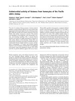
3.3. Radial diffusion assay (RDA)
To distinguish variable activities of AMP clay towards E. coli and S.
aureus, RDA experiments were performed. The results showed more
activity on S. aureus but slightly less active against E. coli (Fig. 2). The
zone of inhibition of the microorganisms is presented in Table 2.Itis
proposed that the AMP clay may serve as an antimicrobial agent for
the control of pathogenic organisms in the presence of salt conditions.
Generally, E. coli and S. aureus are human pathogens because they
cause urinary tract infections, respiratory system failure, dermatitis,
skin lesions, soft tissue infection and food poisoning. However, the
toxicity of AMP in humans must be elucidated in further studies.
Hemolytic and cytotoxicity assays have already shown that AMP clay
has little or no toxicity against human cells (data not shown).
Fig. 2. Radial diffusion assay: Panel A shows the results of testing at 6.25 and 12.5 mg/ml of AMP clay against E. coli; and Panel B shows the results of testing at 6.25 and 12.5 mg/ml of
AMP clay against S. aureus. The control well was the only buffer. The diameter (mm) of the inhibition zone was measured and averaged in triplicate.
Table 2
Measurements of the zone of the inhibitory effect of AMP clay on microorganisms using
radial diffusion assay.
Zone of inhibition (units)
Microorganism 6.25 mg/ml 12.5 mg/ml
E. coli 69
S. aureus 7–810–11
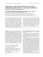
3.4. Scanning electron microscopy (SEM)
Visual observation by SEM micrographs of E. coli, S. aureus and C.
albicans cells before and after the treatment with AMP clay showed
extensive cell damage. The untreated E. coli cells had a smooth surface.
In contrast, when the AMP clay was applied at 1× MIC, membrane
damage was observed in the morphology of the treated bacteria, and,
at 2× MIC, the cells were damaged severely, which led to heavy
leakage and shape change with fragmentation. The fungal cells treated
with AMP clay at 1× MIC level shrank, and, at 2× MIC, degradation
Fig. 3. Scanning electron micrographs of untreated and treated of E. coli KCTC 1682 (A), S. aureus KCTC 1621 (B) and C. albicans cells KCTC 7270 (C) untreated cells with normal
smooth surfaces, shrunken and deformed cells at 1× MIC, and completely ruptured by AMP clay treated cells at 2× MIC.
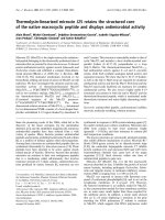
Fig. 4. Time course curves of membrane depolarization activities of the AMP clay against intact E. coli KCTC 1682 (A), S. aureus KCTC 1621 (B), and C. albicans KCTC 7270 (C).
Membrane depolarization was measured by an increase in the fluorescence of DiSC
3
-5 (excitation wavelength λex =620 nm; emission wavelength λem= 670 nm) after the
addition of AMP clay at different concentrations: 1/2× MIC (♦), 1× MIC (■), 2× MIC (▲). The fluorescence increase obtained using 0.1% Triton X-100 was taken as 100%. All the
experiments were preformed in triplicate, and the mean was taken for statistical analysis. Error bars represent the standard deviation.
occurred (Fig. 3). These phenomena suggest that the AMP clay leads to
swelling of the cells due to adsorption onto the bacterial cell surface
and membrane disruption, which leads to cell death. However, Brown
and Melling (1969) reported that Mg, Ca and Zn are present in the cell
wall of the bacteria. It is suggested that divalent cations are
responsible for the cross-linkages through the phosphate groups
present in the lipoprotein and lipopolysaccharides of the cell walls. It
therefore appears that this type absorption is closely linked with the
initial binding of AMP clay to bacterial membrane.
3.5. Membrane depolarization assay
To further determine the manner in which the interaction of this
AMP clay with the bacterial plasma membrane results in cell death, the
ability of the AMP clay to induce membrane depolarization in both
bacteria and fungi was evaluated. Membrane depolarization was
measured in intact microbial cells by monitoring the increase of
DiSC
3
-5 fluorescence after addition of the AMP clay. This indicates the
dose-dependent dissipation of the membrane potential, probablydue to
the leakage of protons and small ions (e.g., Na
+
and K
+
)(Fig. 4). There
was also a correlation between cytoplasmic membrane depolarization
and MIC values, indicating that the AMP clay induced maximum
dissipation of the membrane potential. This result indicated that the
selected AMP clay has a direct effect on the living (microbial) energized
cell membranes. The mechanism depends on the clay minerals involved
on the functional groups and the physicochemical properties of the
organic compounds. In normal conditions, the cell wall of the bacteria is
negatively charged due to the presence of lipoteichoic acid, lipoproteins
and lipopolysaccharides. The initial binding of bacteria and clays occurs
due to the electrostatic interactions between the cationic aminopropyl
groups on the clay lamellar surface and the anionic bacterial membrane
(Nagrete-Herrera et al.,2004). There are alsoreports on the antibacterial
effects of organoclays saturated with alkylammonium cations of
quaternary amines and quaternary ammonium groups (Malachova
et al., 2009; Özdemir et al., 2010, Rhim et al., 2009). Here, it is suggested
that cationic (hydrophilicity) property of aminopropyl groups controls
the antimicrobial properties of the AMP clay.
3.6. Analyses of leaking enzymes from cells
AST and ALT are members of the transaminase family of enzymes.
Except for structural injury of the cell wall or membrane permeability,
there were no intracellular enzymes released from the bacterial cell.
After treatment of the bacteria with AMP clay, the release rate of
intracellular material increased as was shown by determining the
cytoplasmic enzyme AST and ALT in the culture media (Fig. 5). This
assay can be used as a general indicator of bacterial membrane
disruption or injury (Korzeniewski and Callewaert, 1983; Ma et al.,
2010). Apparently, this AMP clay interacted with the cell surface and led
to penetration into the cytoplasmic barrier, causing larger membrane
permeability (leakage of AST and ALT components). Then, it caused
bacterial damage, which is consistent with the SEM observations. These
results clearly indicate a membranolytic mechanism. This result also
suggests that a sequence of steps is occurring at the membrane,
beginning with depolarization (small ion leakage), followed by larger
components leakage, which accompanies morphological changes in the
microbial surface and ultimately cell death (Fig. 3).
3.7. Time killing assay
The time-killing assay was performed as a final study to determine
whether the antimicrobial action of AMP clay was microbicidal or
growth inhibiting. Fig. 6 shows the time-killing studies of AMP clay
against E. coli (Fig. 6A) and S. aureus (Fig. 6B). At concentrations equal to
or above the MIC, the AMP clay kills the strain significantly faster at 2×
compared to 1× MIC concentrations. Also the AMP clay kills Gram
positive bacteria slightly more slowly than Gram negative bacteria. This
result is consistent with the membrane depolarization experiments
described above, which show the immediate effects of this AMP clay on
the integrityof the E. coli membrane compared toS. aureus.Variationsin
the cell wall structure and compositions between the Gram negative
and Gram positive bacteria may be attributed to the different damage
mechanism of the AMP clay. The AMP clay needs a longer time to reach
the plasma membrane of the Gram positive bacteria than the Gram
negative species due to their greater peptidoglycan layers. The fungi C.
Fig. 5. Percentage leakage of intracellular enzyme AST and ALT of the E. coli KCTC 1682
(black bars) and S. aureus KCTC 1621 (gray bars) microorganisms during the exposure
to AMP clay at 6.25 mg/ml. All the experiments were preformed in triplicate, and the
mean was taken for statistical analysis. Error bars represent the standard deviation.
Fig. 6. Kinetics of the bactericidal activity of the AMP clay against E. coli KCTC 1682 (A) and S. aureus KCTC 1621 (B). Bacteria treated with AMP clay were diluted at the appropriate
times and then plated on LB agar. The CFU was then counted after 16 h of incubation at 37 °C. White squares and blank squares are 1 and 2 times the MIC value, respectively; cells
(2× 10
5
CFU/ml) incubated in the absence of AMP clay served as controls. All the experiments were preformed in triplicate, and the mean was taken for statistical analysis. Error bars
represent the standard deviation.
albicans required more than 2 h for 80% killing (data not shown),
implying that this clay permeabilizes fungal membranes much more
slowly than bacterial membranes.
3.8. Stability and storage properties
The antibacterial activity of AMP clay did not change after storage
for 30 days at room temperature and at 4 °C. These results indicated
that the AMP clay was stable in PBS buffer in storage conditions for all
temperatures. Thus, the low cost and favorable storage stability AMP
clay make it an excellent carrier for antimicrobial agents in a variety of
environmental conditions.
4. Conclusions
Synthetic AMP clay showed antimicrobial and membrane perme-
ability activities. Based on the results obtained, a schematic
representation of the mechanism of action of AMP clay is proposed
(Fig. 7). AMP clay was organized in a perpendicular or parallel
orientation across the outer leaflet of the outer bacterial membrane.
The amino propyl group (positive charges) in the AMP clay interacted
electrostatically with the negatively charged lipid membrane when
the AMP clay was inserted into the inner leaflet of the bacterial inner
membrane. This interaction disrupted the higher tight of lipid bilayer,
encouraged membrane fusion events and promoted increased
membrane permeability, which culminated with the leakage of the
bacterial content. Characterization of the synthesized dispersed AMP
clay will necessarily precede the development of therapeutic agents.
In particular, the AMP clay may have some utility in dental caries
prevention, and these possibilities are currently being investigated.
Acknowledgments
This work was supported by the Mid-career Researcher Program
through an NRF grant funded by the MEST (No. 2010-0008596),
Republic of Korea.
References
Aguzzi, C., Cerezo, P., Viseras, C., Caramella, C., 2007. Use of clays as drug delivery systems:
possibilities and limitations. Appl. Clay Sci. 36, 22–36.
Boyd, S.A., Mortland, M .M., Chiou, C.T., 1988. Sorption characteristics of or ganic compounds
on hexadecyltrimethylammonium-smectite. Soil Sci. Soc. Am. J. 52, 652–657.
Brown, M.R., Melling, J., 1969. Role of divalent cations in the action of polymyxin B and
EDTA on Pseudomonas aeruginosa. J. Gen. Microbiol. 59, 263–274.
Burkett, S.L., Press, A., Mann, S., 1997. Synthesis, characterization, and reactivity of layered
inorganic–organic nanocomposites based on 2:1 trioctahedral phyllosilicates. Chem.
Mater. 9, 1071–1073.
Carrado, K.A., 2000. Synthetic organo-and polymer-clays: preparation, characterization,
and materials applications. Appl. Clay Sci. 17, 1–23.
Datta, K.K.R., Eswaramoorthy, M., Rao, C.N.R., 2007. Water-solubilized aminoclay-metal
nanoparticle composites and their novel properties. J. Mater. Chem. 17, 613–615.
Ferreira, R.B., da Silva, C.R., Pastore, H.O., 2008. Aminopropyl-modified magnesium
phyllosilicates: layered solids with tailored interlayer access and reactivity.
Langmuir 24, 14215–14221.
He, H., Yang, D., Yuan, P., Shen, W., Frost, R.L., 2006. A novel organoclay with antibacterial
activity prepared from montmorillonite and clorhexidini aetas. J. Colloid Interface Sci.
297, 235–243.
Holmström, S.C., Patil, A.J., Butler, M., Mann, S., 2007. Influence of polymer co-
intercalation on guest release from aminopropyl-functionalize d magnesium
phyllosilicate mesolamellar nanocomposites. J. Mater. Chem. 17, 3894–3900.
Jeong, N., Kim, J Y., Park, S C., Lee, J K., Gopal, R., Yoo, S., Son, B.K., Hahm, J.S., Park, Y.,
Hahm, K.S., 2010. Antibiotic and synergistic effect of Leu-Lys rich peptide against
antibiotic resistant microorganisms isolated from patients with cholelithiasis.
Biochem. Biophys. Res. Commun. 399, 581–586.
Johnsy, G., Datta, K.K.R., Sajeevkumar, V.A., Sabapathy, S.N., Bawa, A.S., Eswaramoorthy,
M., 2009. Aminoclay: a designer filler for the synthesis of highly ductile polymer-
nanocomposite film. ACS Appl. Mater. Interfaces 1, 2796–2803.
Korzeniewski, C., Callewaert, D.M., 1983. An enzyme-release assay for natural
cytotoxicity. J. Immunol. Methods 64, 313–320.
Lee, Y.C., Lee, T.H., Han, H.K., Go, W.J., Yang, J.W., Shin, H.J., 2010. Optical properties of
fluorescein-labeled organoclay. Photochem. Photobiol. 86, 520–527.
Ma, Y.L., Yang, B., Guo, T., Xie, L., 2010. Antibacterial mechanism of Cu
2+
-ZnO/
cetylpyridinium-montmorillonite in vitro. Appl. Clay Sci. 50, 348–353.
Malachova, K., Praus, P., Pavlickova, Z., Turicova, M., 2009. Activity of antibacterial
compounds immobilised on montmorillonite. Appl. Clay Sci. 43, 364–368.
Nagrete-Herrera, N., Letoffe, J M., Putaux, J L., David, L., Bourgeat-Lami, E., 2004.
Aqueous dispersions of silane-functionalized laponite clay platelets. A first step
toward the elaboration of water-based polymer/clay nanocomposites. Langmuir
20, 1564–1571.
Nordahl, E.A., Rydengard, V., Nyberg, P., Nitsche, D.P., Morgelin, M., Malmsten, M.,
Bjorck, L., Schmidtchen, A., 2004. Activation of the complement system generates
antibacterial peptides. Proc. Natl. Acad. Sci. 101, 16879–16884.
Oya, A., Banse, T., Ohashi, F., Otani, S., 1991. An antimicrobial and antifungal agent
derived from montmorillonite. Appl. Clay Sci. 6, 135–142.
Fig. 7. Schematic model of the action of AMP clay in bacterial cell membranes. AMP clay amino propyl group interacts electrostatically with the lipid membrane. Then, AMP clay is
able to moves to the space between the two bacterial membranes and induces fusion or hemifusion of the inner leaflet of the outer membrane and the inner membrane of bacteria.
These membrane fusions promote membrane permeability and leakage of the bacterial content and lead to cell death.
Özdemir, İ., Gürbüz, N., Doğan, Ö., Günal, S., Özdemir, İ., 2010. Synthesis and
antimicrobial activity of Ag(I)-N-heterocyclic carbene complexes derived from
benzimidazol-2-ylidene. Appl. Organomet. Chem. 24, 758–762.
Papo, N., Oren, Z ., Pag, U. , S ahl, H G., Shai, Y., 2002. The consequence o f seque nce altera tion
of an amphipathic alpha-helical antimicrobial p eptide and i ts diastereomers. J. B iol.
Chem. 277, 33913–33921.
Park, S.C., Kim, M.H., Hossain, M.A., Shin, S.Y., Kim, Y., Stella, L., Wade, J.D., Park, Y.,
Hahm, K.S., 2008. Amphipathic alpha-helical peptide, HP (2–20), and its analogues
derived from Helicobacter pylori : pore formation mechanism in various lipid
compositions. Biochim. Biophys. Acta 1778, 229–241.
Park, S.C., Lee, J.R., Shin, S.O., Park, Y., Lee, S.Y., Hahm, K.S., 2007. Characterization of a
heat-stable protein with antimicrobial activity from Arabidopsis thaliana. Biochem.
Biophys. Res. Commun. 362, 562–567.
Patil, A.J., Muthusamy, E., Mann, S., 2005. Fabrication of functional protein-organoclay
lamellar nanocomposites by biomolecule-induced assembly of exfoliated aminopropyl-
functionalized magn esium phyllosilicates. J. Mater. Chem. 15, 3838–3843.
Patil, A.J., Li, M., Dujardin, E., Mann, S., 2007. Novel bioinorganic nanostructures based
on mesolamellar intercalation or single-molecule wrapping of DNA using
organoclay building blocks. Nano Lett. 7, 2660–2665.
Pernyeszi, T., K asteel, R., Witthuhn, B ., Klahre, P., Vereecken, H., Klumpp, E., 2006. Organ oclays
for soil remediation: adsorption of 2,4-dichlorophenol on organoclay/aquifer material
mixtures studied under static and flow conditions. Appl. Clay Sci. 32, 179–18 9.
Reitman, S., Frankel, S.A., 1957. A colorimetric method for the determination of serum
glutamic oxalactic and glutamic pyruvic transminases. Am. J. Clin. Pathol. 28, 56–63.
Rhim, J W., Hong, S I., Ha, C S., 2009. Tensile, water vapor barrier and antimicrobial
properties of PLA/nanoclay composite films. Food Sci. Technol. 42, 612–617.
Whilto n, N.T., Burkett, S.L., Mann, S., 1998. Hybrid lamellar nanocomposites based on
organically functionalized magnesium phyllosilicate clays with interlayer
rea ctivity. J. Mater. Chem. 8, 1927–1932.
Xue, S., Pinnavaia, T.J., 2010. Methylene-functionalized saponite: a new type of
organoclay with CH
2
groups substituting for bridging oxygen centers in the
tetrahedral sheet. Appl. Clay Sci. 48, 60–66.