A study into the different mechanisms of anti cancer effect of docosahexanoic acid in colon cancer cells
Bạn đang xem bản rút gọn của tài liệu. Xem và tải ngay bản đầy đủ của tài liệu tại đây (897.05 KB, 134 trang )
A STUDY INTO THE DIFFERENT MECHANISMS OF ANTI-
CANCER EFFECT OF DOCOSAHEXANOIC ACID
IN COLON CANCER CELLS
MANAV
DEPARTMENT OF COMMUNITY, OCCUPATIOBNAL
AND FAMILY MEDICINE
NATIONAL UNIVERSITY OF SINGAPORE
2004
A STUDY INTO THE DIFFERENT MECHANISMS OF ANTI-
CANCER EFFECT OF DOCOSAHEXANOIC ACID
IN COLON CANCER CELLS
MANAV
(B. MED, GSMU, INDIA)
A THESIS SUBMITTED
FOR THE DEGREE OF MASTER OF SCIENCE
DEPARTMENT OF COMMUNITY, OCCUPATIOBNAL
AND FAMILY MEDICINE
NATIONAL UNIVERSITY OF SINGAPORE
2004
i
ACKNOWLEDGEMENTS
I would like to express my respect and gratitude to Professor Ong Choon Nam
and Professor Lee Hin Peng. As my supervisors, they ensured that I remained focused on
achieving my goal. Their observations and guidance helped me to establish the overall
direction of the research and to move forward with investigation in depth. What I learned
from them, especially their approach to a scientific question, is an invaluable lesson for
me not only in the academic perspective but also in my personal life.
I would also like to express my sincere thanks to:
Prof. David Koh, Head of the Department, for his support during the course of the study;
Dr. Shen Han Ming, for his advice and stimulating discussions;
Mr. Ong Her Yam, Madam Lee Bee Lan, Madam Jin Su, Madam New Ai Li and Mr Ong
Yeong Bing and Madam Zhao Min, for their guidance and help in the laboratory work.;
My seniors and colleagues Zhang Siyuan, Won Yen Kim, Shi Ranxin, Huang Qing and
Lu Guodong ; they were responsible for the fine-tuning of my techniques and ideas;
National University of Singapore, for providing me a research scholarship for this study.
Finally, I thank my family for their never-ending love and support. I dedicate this thesis
to them.
ii
TABLE OF CONTENTS
Acknowledgements i
Table of contents ii
Abbreviations vii
List of publications ix
Summary x
CHAPTER I: INTRODUCTION
1.1 Polyunsaturated Fatty Acids (PUFAs) 2
1.1.1 Structure of PUFAs 2
1.1.2 Synthesis of n-3 and n-6 PUFAs 2
1.1.3 Dietary sources 4
1.1.4 Intestinal absorption, metabolism 4
1.1.5 Functions of PUFAs 5
1.1.6 Competition between n-3 and n-6 PUFAs. 6
1.2 PUFAs and colorectal cancer 6
1.2.1 Epidemiological Studies 6
1.2.2 Animal studies 8
1.2.3 Intervention studies in humans 10
1.2.4 In vitro studies 10
1.3 Apoptosis 11
iii
1.3.1 Apoptosis - A brief introduction 11
1.3.2 PUFAs and apoptosis 13
1.4 Oxidative Stress 14
1.4.1 Reactive Oxygen Species (ROS) – Definition and source 14
1.4.2 Role of oxidative stress in carcinogenesis and apoptosis 15
1.4.3 PUFAs and oxidative stress 16
1.4.3.1 PUFAs and lipid peroxidation 16
1.4.3.2 PUFAs and ROS 17
1.5 Mitogen Activated Protein Kinases (MAPKs) 18
1.5.1 MAPK signaling pathways- Introduction 18
1.5.2 MAPKs and ROS 19
1.5.3 PUFAs and MAPKs 20
1.6 Peroxisome Proliferator-Activated Receptors (PPARs)
1.6.1 Functions of PPARs 21
1.6.2 PPARγ and colon cancer 22
1.6.3 PUFAs and PPARs 23
1.7 Objectives of the Study 24
CHAPTER II: MATERIALS AND METHODS 25
2.1 Cell lines and chemicals 26
2.2 Cell culture and fatty acid supplementation 27
2.3 Determination of cell viability - MTT assay 27
2.4 Evaluation of cell morphological alterations 28
iv
2.5 Determination of sub-G1 population 28
2.6 Measurement of cellular caspase-3 activity 29
2.7 Western Blotting 29
2.8 JNK in vitro kinase assay 30
2.9 Transient transfections 31
2.10 Luciferase assay 31
2.11 Measurement of ROS 32
CHAPTER III: EFFECT OF DHA ON VIABILITY OF COLON CANCER CELLS
3.1 Introduction 34
3.2 Results 35
3.2.1 Cytotoxic effects of PUFAs 35
3.2.1.1 Effect of different fatty acids on cell viability of HT-29 cells 35
3.2.1.2 Effect of DHA on cell viability of different colon cancer cell lines 35
3.2.1.3 Effect of metabolic pathway inhibitors on DHA-induced cell death 35
3.2.2 DHA induced cell death is apoptotic 36
3.2.3 DHA induced apoptosis is caspase mediated 37
3.2.3.1 Determination of caspase-3 activation 37
3.2.3.2 Effect of caspase-3 inhibitor on DHA-induced cell death 37
3.3 Discussion 37
CHAPTER IV: EFFECT OF DHA ON ROS GENERATION AND MAP KINASE
ACTIVATION
v
4.1 Introduction 50
4.2 Results 50
4.2.1 Effect of DHA on MAPK signaling pathways 50
4.2.1.1 DHA induces MAPK activation 50
4.2.1.2 Effect of MAPK inhibitors on DHA-induced cell death 51
4.2.1.3 Effect of JNK mutants on DHA-induced cell death 52
4.2.2 Role of ROS in DHA induced cell death 53
4.2.2.1 Generation of ROS by DHA 53
4.2.2.2 Effect of antioxidants and H
2
0
2
on DHA-induced MAPK activation 53
4.2.2.3 Effect of antioxidants on DHA-induced cell death 54
4.3 Discussion 54
CHAPTER V: DHA INHIBITS TNF-α INDUCED COX-2 EXPRESSION AND
NF-ΚB TRANSCRIPTION THROUGH THE PPARγ PATHWAY
5.1 Introduction 66
5.2 Results 67
5.2.1 DHA inhibits TNF-α induced COX-2 and NF-κB activation 67
5.2.2 DHA induces transcriptional activition of PPARγ 68
5.2.3 DHA regulates COX-2 expression and NF-κB activation through
PPARγ-dependent pathway 69
5.2.4 DHA induces apoptosis in colon cancer cells by a PPARγ-independent
mechanism 69
5.3 Discussion 70
vi
CHAPTER VI: DISCUSSION AND CONCLUSIONS
6.1 Chemotherapeutic actions of DHA 81
6.2 Chemopreventive role of DHA 85
6.3 Comparison of results from in vitro and in vivo studies 87
6.4 Conclusions 88
REFERENCES 90
vii
ABBREVIATIONS
AA Arachidonic acid
ALA Alpha-linolenic acid
APC Adenomatous polyposis coli
BADGE Bis-phenol A diglycidyl ether
BSA Bovine serum albumin
COX Cyclooxygenase
DAPI 4’, 6-diamidino-2-phenylindole
DHA Docosahexanoic Acid
DMEM Dulbecco’s Modified Eagle’s Medium
ERK Extracellular regulated kinase
FA Fatty acid
JNK c-Jun N-terminal kinase
LA Linoleic acid
LOX Lipoxygenase
MAPK Mitogen-activated protein kinase
MTT 3,(4,5-dimethylthiazol-2-yl)2,5-diphenyl-
tetrazolium bromide
NAC N-acetyl cysteine
NF-κB Nuclear factor- κB
PBS Phosphate buffered saline
PARP Polyadenosine diphosphate-ribose polymerase
PPAR Peroxisome Proliferator-Activated Receptor
viii
PPRE Peroxisome Proliferator-activated receptor response
element
PUFA Polyunsaturated fatty acid
ROS Reactive oxygen species
TNF-α Tumor necrosis factor-alpha
ix
LIST OF PUBLICATIONS
• Manav, Su, J., Hughes, K., Lee, H.P., and Ong, C.N. (2004). n-3 Fatty Acids and
Selenium as Coronary Heart Disease Risk Modifying Factors in Asian Indian and
Chinese Males. Nutrition. In press
• Manav, HM Shen and CN Ong. Involvement of c-Jun N-terminal kinase
activation in Docosahexanoic acid-induced apoptosis in colon cancer cell. In
preparation
• Manav, HM Shen and CN Ong . Docosahexanoic acid inhibits TNF-α induced
COX-2 expression in colon cancer cells: Role of PPARγ. In preparation
Conference Abstracts:
• Manav, Su, J., Hughes, K., Lee, H.P., and Ong, C.N. (2004). n-3 Fatty Acids and
Selenium as Coronary Heart Disease Risk Modifying Factors in Asian Indian and
Chinese Males. 5
th
MCBN - UNESCO/ COSTAM/ SFRR Workshop
x
SUMMARY
Colon cancer is one of the leading causes of cancer mortality in the developed
world. Evidence from epidemiological and animal studies indicates that the n-3
polyunsaturated fatty acids (PUFAs) may protect against colon cancer. However the
mechanisms of the anti-cancer effects of n-3 PUFAs have not been fully elucidated.
Docosahexanoic acid (DHA) (22:6, n-3), abundantly found in fish oil, is one of the
principal n-3 PUFAs. The aims of this study were (i) to investigate the effects of the n-3
PUFAs, and DHA in particular, on cell growth and apoptosis in colon cancer cell lines
(ii) to explore the signaling pathways responsible for the chemotherapeutic and
chemopreventive actions of DHA.
The growth inhibitory effects of DHA were demonstrated by the MTT assay.
DHA was more potent in inhibiting growth than the n-6 PUFAs, monounsaturated and
saturated fatty acids. The cytotoxic effect of DHA was due to apoptosis, as demonstrated
by nuclear condensation, sub-G1 assay and PARP cleavage. DHA also activated caspase-
3; and the caspase inhibitor z-VAD-fmk was able to block the DHA-induced apoptosis.
The effect of DHA on the mitogen-activated signaling kinases (MAPKs) was also
investigated. DHA activated JNK, ERK and p38 MAPKs. The activation of JNK played a
key role in the apoptotic action of DHA, as inhibition of JNK either by the specific JNK
inhibitor SP600125 or by overexpression of the dominant-negative mutants of JNK
significantly repressed both the JNK activity and apoptosis induced by DHA. It was
further shown that DHA induces oxidative stress but the JNK activity and apoptosis were
independent of the reactive oxygen species generation.
xi
DHA also induced the transcriptional activity of the Peroxisome Proliferator-
activated receptor γ (PPARγ). Activation of ERK interfered with ability of DHA to
activate PPARγ. DHA also inhibited TNF-α – induced COX-2 expression and NF-κB
activation. This inhibition was dependent on PPARγ as repressing the PPARγ expression
by transfection of antisense PPARγ oligonucleotides, decreased the inhibitory effect of
DHA on COX-2 expression. However, there were no significant changes in cell death
when PPARγ was inhibited, indicating the possibility that DHA induced apoptosis
through PPARγ-independent pathway.
In conclusion, this study provides an insight into the pathways involved in the role
of DHA in colon cancer. This study for the first time shows (i) activation of JNK plays a
key role in DHA-induced apoptosis (ii) DHA inhibits COX-2 by the activation of PPARγ.
Thus, different mechanisms seem to be involved in the anti-carcinogenic effect of DHA
in colon cancer.
1
CHAPTER I
INTRODUCTION
2
1.1 Polyunsaturated Fatty Acids
Polyunsaturated fatty acids (PUFAs) are ubiquitious biological molecules that
function as metabolic fuels, as covalent regulators of signaling molecules, and as
essential components of cellular membranes. However, alongside these essential
functions, fatty acids have also been identified as having an effect, both positive as well
as negative, on health and disease progression.
1.1.1 Structure
Fats form a large group, composed of different types of fatty acids and glycerol.
Fatty acids are hydrocarbon chains with a carboxyl group at one end. Fatty acids usually
contain an even number of carbon atoms and on the basis of their degree of saturation,
can be classified into saturated fatty acids (SFA), monounsaturated fatty acids (MUFA)
(with one double bond), and polyunsaturated fatty acids, PUFAs (with multiple double
bonds). According to conventional nomenclature of fatty acid molecules, PUFAs are
classified into different groups on the basis of the position of the first double bond from
the methyl terminus of the hydrocarbon chain of the molecule. Thus n-3 and n-6 PUFAs
are so named as they have their first double bond at the 3
rd
and 6
th
carbon respectively
(Nettleton, 1995).
1.1.2 Synthesis of n-3 and n-6 PUFAs
Most of the n-3 and n-6 PUFAs are metabolized from their precursors, linoleic
acid (LA; 18:2n-6) and alpha-linolenic acid (ALA; 18:3n-3) respectively. Both LA and
ALA are metabolized to longer chain PUFAs, largely in the liver, by a series of
3
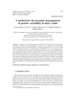
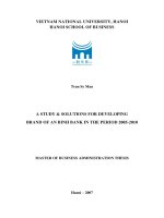
Fig 1.1 Structure and classification of fatty acids
elongation and desaturation reactions to yield longer, more unsaturated fatty acids. LA is
converted to AA (20:4n-6) while ALA is converted to eicosapentanic acid (EPA;20:5n-3)
and docosahexanoic acid (DHA; 22:6n-3). Mammalian organisms, unlike plants do not
possess the ∆12 and ∆15 desaturase enzymes required for LA and ALA synthesis.
Therefore these two PUFAs are considered as essential and must be supplemented in the
diet in order to maintain adequate body pools (Simopoulos, 1999).
4
1.1.3 Dietary sources of PUFAs
ALA is the major fatty acid in chloroplast lipids, and hence is the major n-3
PUFA from plant derived sources. Soybean, canola, perilla, linseed, and rapeseed oils are
rich sources of ALA, apart from green leafy plants. DHA and EPA are found exclusively
in aquatic animals as they are synthesized by phytoplankton and algae, which is then
consumed by fish, mollusks and crustaceans, and thereby concentrated in the aquatic food
chain (Nettleton, 1995). The relative amounts of DHA and EPA contained in fish oils
vary considerably between species, with deep sea fish containing more of these n-3
PUFAs compared to the freshwater ones (Childs et al., 1990). Among the n-6 PUFAs, LA
can be found in high proportions in many vegetable seeds and oils (safflower, soybean,
coconut, corn and sunflower). Dietary LA is considered to be the major source of tissue
AA, although lean meats and meat fat are the direct sources in the human diet (Li et al.,
1998).
1.1.4 Intestinal absorption and metabolism
Absorption of dietary n-3 PUFAs depends on the form in which they are ingested.
Fish and fish oil products are mostly ingested as triglycerides. In mammals, the relative
absorption of different forms of PUFAs varies as Free PUFA > triglyceride > ethyl ester
(Nelson and Ackman, 1988). Ingestion of n-3 PUFAs leads to their distribution to
virtually every cell in the body. The non-esterified fatty acids enter the cells via fatty acid
transporters and are rapidly converted to fatty acyl-CoA thioesters (FA-CoA) by acyl-
CoA synthetases. A major fraction of these lipids is bound to specific proteins, i.e. fatty
acid binding protein and FA-CoA-binding protein. FA-CoAs are substrates for neutral
5
lipid (triglycerides, cholesterol esters) and polar lipid (phospholipids, sphingolipids and
plasmalogens) synthesis (Jump, 2002). On exogenous supplementation, the phospholipid
pool comprising of phosphatidylcholine and phosphatidylethanolamine, is the major site
of PUFA incorporation in both cultured normal and transformed cells (de Bravo et al.,
1991).
1.1.5 Function of PUFAs
Dietary PUFAs have effects on diverse physiological processes impacting normal
health and chronic disease, such as the regulation of plasma lipid levels (Harris, 1997;
Mori et al., 2000), cardiovascular (Nilsen and Harris, 2004) and immune function
(Hwang, 2000). As structural phospholipids of cell membranes, they modulate membrane
fluidity, cellular signaling and cellular interaction. Moreover they play an important role
in the regulation of the immune system by acting as precursors for the synthesis of
eicosanoids. Arachidonic acid (AA) or EPA are mobilized from the cell membrane by the
action of the phospholipase enzymes especially phospholipase A
2
(PLA
2
) and C (PLC),
and subsequently metabolized by cyclooxygenase (COX) and lipoxygenase (LOX) into
prostaglandins (PGs), thromboxanes (TXs) and leukotrienes (LTs). As membrane
phospholipids normally contain much higher levels of arachidonic acid (AA) than of the
other 20-carbon PUFAs (Yaqoob et al., 2000), AA is the most common eicosanoid
precursor and gives rise to 2-series PGs and TXs and 4-series LTs. In contrast, EPA gives
rise to 3-series PGs and TXs and 5-series LTs, the difference being the presence of the
double bond in the structure (Johnson et al., 1983). n-3 PUFAs also play a role in the
regulation of gene expression (Jump and Clarke, 1999; Duplus et al., 2000). However
6
cell-specific lipid metabolism as well as the regulation of fatty acid regulated
transcription factors are likely to play an important role in determining how cells respond
to changes in PUFA composition.
1.1.6 Competition between n-3 and n-6 PUFAs
There is competition between n-3 and n-6 PUFAs for their metabolic conversion
via the desaturase and elongase enzymes, which are common to both pathways. However
these enzymes have a greater affinity for n-3 PUFAs such that when dietary n-3 intake is
high, they are preferentially metabolized (Jump, 2002). This leads to competitive
inhibition of n-6 PUFA metabolism, where linoleic acid desaturation and AA
concentration are significantly decreased after EPA or DHA supplementation, and
resulting in decreased generation of AA-derived eicosanoids (Yaqoob et al., 2000;
Caughey et al., 1996; Thies et al., 2001).
1.2 PUFAs in colon cancer
1.2.1 Epidemiological studies
Fat has been the focus of dietary studies on colorectal cancer more than any other
component. The initial insight into the relationship between dietary fat and cancer came
partly from epidemiological studies. With a few exceptions (Macquart-Moulin et al.,
1986; Tuyns et al., 1987), early case control studies showed a positive association
between the risk of colorectal cancer and the intake of fat and meat (Potter and
McMichael, 1986; Benito et al., 1990; Graham et al., 1988; Lee et al., 1989; La Vecchia
et al., 1988). Studies examining the effect of the degree of saturation of fats highlighted
7
saturated fat consumption, as an agent responsible for colorectal cancer (Burnstein, 1993;
Woutersen et al., 1999). On the other hand, a decrease in risk of colon cancer was
reported with an increase in the degree of unsaturation of fats (Lee et al., 1989;
Macquart-Moulin et al., 1987; Benito et al., 1991).
A reportedly lower incidence of thrombotic and immunologically mediated
diseases in Greenland Eskimos when compared with mainland Danish population,
aroused interest in the potential beneficial effects of marine lipiods (Bjerregaard and
Dyerberg, 1988). Lanier et al, (1976) in a 5-year survey from 1969-73 observed an
increase in cancer incidence of lung, colon and rectum among the Alaska Eskimos and
Aleuts. A further survey of cancer incidence for the years 1989-1993 found that there
were significant increases in the rates for cancers of prostate and colon in men (Lanier et
al, 1996). The diet of Eskimos of Alaska is high in fat but this comes largely from marine
animals and fish (Nobmann et al, 1992). Urbanization of the native population and the
decreasing trend of fish intake were suggested as contributing factors to the increase in
cancer rates, implying that fish and fish oils may have a protective risk modifying effect
in colorectal cancer. In an analysis involving 24 European countries, an inverse
correlation was found in males between colorectal cancer mortality and current fish
intake. There was evidence of a protective effect of a high fish intake relative to that of
dietary sources of n-6 PUFAs (Caygill and Hill, 1995). In a follow-up study, mortality
from colon cancer correlated with the consumption of animal fat and an inverse
correlation was observed with fish and fish oil consumption when expressed as a
proportion of total fat (Caygill et al., 1996). Fish and fish oil being rich sources of n-3
PUFAs, the findings of this analysis implied that lower levels of n-3 PUFAs and higher
8
levels of n-6 fatty acids in the body may be a predisposing factor in the causation of
colon cancer.
Prospective cohort studies also have shown positive association between fat, meat
and colorectal cancer. Willett et al, (1990) reported an almost 2-fold higher risk of
colorectal cancer among women in the highest quintile of red meat consumption in the
Nurses’ study. The Health Professionals Follow-Up Study, a cohort study of men, also
demonstrated a direct association between red meat consumption and risk of colon
cancer, but no association was observed with other sources of fat (Giovannucci et al.,
1994). The protective effects of fish consumption are seen only in areas where fish
consumption is high. Fishermen on the west coast of South Africa, having a significantly
higher (110 versus 30g/day) had six times lower colorectal cancer incidence than in white
urban dwellers (Schloss et al., 1997). In a study in Norway, intake of fish in general had
no protective effect against colorectal cancer, but the relative risk of people who ate five
or more fish meals per week was lower than that of people who ate fish less frequently
(Gaard et al., 1996). Taken together, these results indicate that various types of fat may
have opposite effects on the risk for colorectal cancer, with meat rich in n-6 PUFAs
promoting cancer and fish rich in n-3 PUFAs being protective.
1.2.2 Animal studies
The second line of evidence in understanding the influence of dietary fat on
colorectal cancer development came from rodent models. Laboratory animal studies
provided the evidence that not only the amount of fat but also the types of fat are
important factors in colon cancer development. Experiments with diets containing
9
saturated fatty acids, n-3 PUFA and n-6 PUFAs clearly show that n-3 PUFA rich diets
inhibit carcinogenesis (Nelson and Ackman, 1988; Reddy and Maruyama, 1986a; Reddy
and Sugie, 1988) whereas saturated fats and n-6 PUFA rich diets enhance tumor
production (Reddy and Maeura, 1984; Reddy and Maruyama, 1986b). In addition, the
stage of carcinogenesis at which the effect of dietary fat is exerted appears to depend on
the fatty acid composition, with the n-3 PUFAs being protective in both the initiation and
promotion phase of carcinogenesis (Reddy and Maruyama, 1986b; Reddy et al., 1991).
The effects of different n-3 and n-6 fatty acid ratios in experimental colon carcinogenesis
were studied by Deschner et al, (1990). The highest ratio of n-3:n-6 PUFAs inhibited
epithelial cell proliferation and induced S-phase arrest in the colonic cells, whereas the
lowest ratio of n-3:n-6 PUFAs produced the highest tumor incidence in azoxymethane
treated rats.
Germ line mutations of the murine Apc gene provide a model for human familial
adenomatous polyposis. The inhibitory effect of DHA and DHA-enriched fish oil were
also demonstrated in mouse models with a mutation at the Apc gene at codon 716 and
codon 850 respectively (Oshima et al., 1995; Paulsen et al., 1997). The administration of
DHA has also been shown to inhibit colon cancer cell metastasis with a reduction in the
matrix metalloproteinase activity (Suzuki et al., 1997; Iigo et al., 1997). These studies
tend to confirm the epidemiological evidence that n-3 PUFAs are protective, whereas n-6
PUFAs promote cancer formation.
10
1.2.3 Intervention studies in humans
Studies describing direct intervention with n-3 PUFAs in human subjects are not
many. The relative long periods of fatty acid supplementation makes the dietary
intervention studies difficult to conduct. Subjects, at high risk for colorectal cancer,
receiving fish oil supplementation showed changes in proliferation pattern of the rectal
mucosa similar to that observed in the low risk population (Anti et al., 1992; Bartoli et
al., 1993). In another study, patients with stage 1 or 2 colon carcinoma or adenomatous
polyps did not develop additional polyps after 12 months of n-3 PUFA supplementation
(Huang et al., 1996). In contrast, Akedo et al, (1998) reported that Familial Adenomatous
Polyposis (FAP) patients supplemented with DHA-enriched fish oil capsules, still
progressed to malignant lesions after 12 months. Though clinical intervention studies are
more relevant to the human in vivo situation, the existing data are not substantial. More
studies are needed to confirm the efficacy of clinical application of DHA in colon cancer.
1.2.4 In vitro studies
Compared to the numerous epidemiological and in vivo studies that have been
conducted on colon cancer to determine the role of fatty acids, few studies have tried to
look into the effect of PUFAs on colon cancer cell lines.(Tsai et al., 1998) showed that n-
3 PUFAs, DHA and EPA inhibited the proliferation of sigmoid colon cancer
transformants while having little effect on normal cells. In HCT-116 cells transfected
with inducible COX-2, both DHA and EPA inhibited cell proliferation, compared with
linoleic acid (Boudreau et al., 2001). In CaCo-2 cells, irrespective of the n-3/n-6 status,
the longer chain PUFAs - DHA, EPA and AA were more potent in inhibiting growth than
11
α-linolenic acid and linoleic acid (Dommels et al., 2003; Nano et al., 2003). In HCA-7
cell line, DHA acts synergistically with the selective COX-2 inhibitor, celecoxib, to
inhibit cyclooxygenase-2 and cell proliferation (Swamy et al., 2004). In HT-29 cells
DHA has been shown to inhibit growth and induce cell cycle arrest (Chen and Istfan,
2001), but the molecular mechanism of these actions still remain to be elucidated.
Apart from its effect on cellular proliferation, DHA also modulates other
functions in colon cells. In young adult mouse colonic cells DHA, compared with
linoleic acid inhibited Ras localization to the plasma membrane and GTP binding (Collett
et al., 2001).
Both n-3 and n-6 PUFAs increased the gap junctional intercellular
communication during spontaneous differentiation of Caco-2 (Dommels et al., 2002).
DHA increases the metabolism of all-trans-retinoic acid and CYP26 gene expression in
intestinal cells (Lampen et al., 2001) and selectively activates RXRalpha relative to n-6
PUFA in colonocytes (Fan et al., 2003). cDNA microarray analysis showed that in CaCo-
2 colon cancer cells, DHA down-regulated the inducible nitric oxide synthase, the
prostaglandin family of genes, as well as cyclooxygenase-2 expression and several cell
cycle-related genes, whereas it up-regulated genes associated with apoptosis (Narayanan
et al., 2003).
1.3 Apoptosis
1.3.1 Apoptosis – A Brief Introduction
Apoptosis or programmed cell death is a critical component of both normal
development and disease (Hengartner, 2000). First described by (Kerr et al., 1972), this
distinct type of cell death is characterized by cytoplasm swelling, blebbing of the plasma
membrane, chromatin condensation maintenance of organelle integrity, and condensation
12
and fragmentation of DNA, followed by orderly removal through phagocytosis. The
importance of apoptotic process can be assessed from the fact that the apoptotic
machinery has been highly conserved throughout evolution, with many similarities
between phylogenetically divergent groups including invertebrates and humans (Wyllie
et al., 1999). Defects in the apoptotic process can result in many pathological conditions
including cancer, Alzheimer’s disease, stroke and Acquired Immuno-deficiency
Syndrome (Webb et al., 1997).
One of the main executioners of the apoptotic pathway are the caspases. Caspases
are cysteine-specific proteases that are expressed as inactive precursors. Caspases are
activated early in the apoptotic cascade either by (i) processing by an upstream caspase
(ii) ligand binding to the death receptors (iii) or association with a regulatory subunit like
Apaf-1 (Hengartner, 2000). The initial activation of caspases is amplified by the caspase
cascade which also integrates the pro-apoptotic signals. Proteolytic cleavage of cellular
substrates by caspases largely determines the features of apoptosis. The caspase
substrates range from the single polypeptide chain enzymes, like polyADP-ribose
polymerase, to complex macromolecular structures like the lamin network (Creagh et al.,
2003).
Mitochondria not only serve as the major energy source in the living cells, but
they can also trigger or amplify the signals that lead to apoptosis (Green and Reed, 1998).
Alteration in mitochondrial membrane potential and permeabilisation of the
mitochondrial membrane often precede caspase activation and other manifestations of
apoptosis (Zamzami and Kroemer, 2001). Induction of permeability transition in the
inner mitochondrial membrane may be accompanied by the release of cytochrome c,