A study on pitting corrosion of stainless steels in halide solutions
Bạn đang xem bản rút gọn của tài liệu. Xem và tải ngay bản đầy đủ của tài liệu tại đây (2.32 MB, 114 trang )
A STUDY ON PITTING CORROSION OF STAINLESS
STEELS IN HALIDE SOLUTIONS
CHUA SHU ER SHERLYN
NATIONAL UNIVERSITY OF SINGAPORE
2011
A STUDY ON PITTING CORROSION OF STAINLESS
STEELS IN HALIDE SOLUTIONS
CHUA SHU ER SHERLYN
(B.Eng (Hons.), NUS)
A THESIS SUBMITTED
FOR THE DEGREE OF MASTER OF ENGINEERING
DEPARTMENT OF MATERIALS SCIENCE AND
ENGINEERING
NATIONAL UNIVERSITY OF SINGAPORE
2011
i
Acknowledgments
I would like to express my sincere thanks and appreciation to my supervisor A/P
Daniel Blackwood. He has shown utmost patience and optimism towards me during
the entire course of study. Most importantly, he is always ever ready to share his
knowledge and experiences not only in this project, but in other areas as well. He
displays no airs as a professor/supervisor and he was even willing to go down to the
laboratory to guide me in experiments. His encouragement, guidance and invaluable
insights have been the main motivation behind this thesis.
Special thanks also go to the laboratory staff in the Department of Materials
Science and Engineering. Amidst their busy schedule, they were always willing to fork
out time for equipment training. In particular, Mr. Henche Kuan had been very helpful
in the area of XPS and I deeply appreciate his thoughtful recommendations and advice.
Given that I was also holding on to teaching duties, I would also like to express my
sincere thanks to my fellow teaching assistants. They have been very tolerant of my
dual student/TA role and have been nothing but encouraging.
Laboratory work in E3A had been very enjoyable. When experimental results do
not go as planned, there were always laboratory mates to count on for advice,
encouragement, laughter and joy. These friendships we have forged will follow us all
the way – Chin Yong, Swee Jen, Chunhua, Wenlai, Dongqing, Gui Yang, Xuelian,
Yeru and many more from the E3A laboratories.
My last thanks go to my family and most importantly my best friend, Ho Pin. No
number of words can express my thanks. Simply to say, without her around, this thesis
would not materialize.
ii
Table of Contents
Acknowledgments i
Table of Contents ii
Summary iv
List of Tables vi
List of Figures viii
1. INTRODUCTION 1
1.1 General Overview of Pitting Corrosion 2
1.2 Stages of Pitting 5
1.2.1 Pit Initiation/Nucleation 5
1.2.2 Metastable Pitting 8
1.2.3 Stable Pit Growth 8
1.3 Determining Pitting Resistance in Stainless Steels 10
1.3.1 Pitting Resistance Equivalent Number (PREN) 10
1.3.2 Electrochemical Parameters of Pitting Corrosion in Stainless Steels 11
2. LITERATURE REVIEW 13
2.1 The Role of Molybdenum in Improving Pitting Resistance 13
2.2 Pitting Corrosion in Cl
-
and Br
-
solutions 14
2.3 Motivation and Organization of Thesis 17
3. EXPERIMENTAL DETAILS 19
3.1 Samples and Solutions 19
3.2 Electrochemical Experiments 21
3.2.1 Experimental Setup 21
3.2.2 Cyclic Potentiodynamic Polarization 22
3.2.3 Potentiostatic Metastable Pitting Tests 22
3.3 X-ray Photoelectron Spectroscopy 23
3.4 Scanning Electron Microscopy 24
4. RESULTS AND DISCUSSIONS 25
4.1 Effects of Temperature on Pitting Behaviour 25
4.1.1 Pitting and Repassivation Characteristics 25
4.1.2 Metastable Pitting 32
4.1.3 SEM Imaging 42
4.1.4 Studies on Passive Film – XPS 46
iii
4.2 Effects of Electrolyte Anion on Pitting Behaviour 55
4.2.1 Pitting and Repassivation Characteristics 55
4.2.2 Metastable Pitting 60
4.2.3 SEM Imaging 65
4.2.4 Studies on Passive Film – XPS 67
4.3 Effects of Electrolyte Cation on Pitting Behaviour 77
4.3.1 Pitting and Repassivation Characteristics 77
4.4 Correlation with PREN 83
5. CONCLUSION 85
6. FUTURE WORK 88
7. REFERENCES 90
iv
Summary
The role of Mo in the pitting behaviours of stainless steels in bromide solutions
is a matter of current debate. While Mo has been widely acknowledged to increase the
pitting resistance in chloride solutions, some authors have proposed that the beneficial
effects of Mo are compromised in bromide solutions. The work in this thesis was
initiated to shed further light on this controversial issue. The pitting behaviours of
austenitic 304L, 316L, SMO and duplex 329 stainless steels at different temperatures
in various solutions were investigated by traditional electrochemical techniques and
further characterized by scanning electron microscopy (SEM) and x-ray photoelectron
spectroscopy (XPS). With increasing temperature from 3 to 90°C, the pitting
resistances of the stainless steels decreased. The potentiodynamic and potentiostatic
tests showed that temperature had a greater effect on the nucleation and growth
compared to the repassivation and death of pits.
The temperature dependent pitting potentials of the stainless steels followed a
linear relationship in sodium bromide but an exponential relationship in sodium
chloride. Similarly, the temperature effect on the repassivation potentials was higher in
chloride compared to bromide solutions. The difference in pitting potential-
temperature relationships was proposed to be due to different rate-determining steps.
In chloride solutions, pitting corrosion due to MnS inclusions were more favoured
while pitting due to breakdown of passive film occurred more easily in bromide
solutions. A cross-over temperature T
c
was also established. Below T
c
, pitting
resistance was higher in chloride solution and above that pitting resistance was higher
in bromide solution. The estimated T
c
(22°C for 304L, 32°C for 316L, 52°C for 329
and >90°C for SMO) was observed to increase with the PREN of the stainless steels.
v
XPS results revealed the formation of molybdates MoO
4
2-
in the passive films
of SMO and 329 in chloride solutions, but the formation was compromised in bromide
solutions. The presence of the molybdates could be the main reason behind the high
pitting resistance of SMO in chloride solution. In addition, the XPS data indicated that
passive films formed on the stainless steels consisted of a surface hydroxide-oxide
layer, followed by a mixed iron-chromium oxide layer and a thick layer of Cr
2
O
3
.
The electrolyte cations are not typically involved in the pitting corrosion of
stainless steels. However, to further ascertain this point, the pitting potentials of 304L
were measured in LiBr, NaBr and KBr. The pitting potentials E
p
were found to be the
highest in LiBr, followed by NaBr and the lowest pitting resistance was in KBr. This
was proposed to be due to the different cation mobilities and diffusivities which will
then affect the rate at which the pit anolyte acidifies. Finally, it can be concluded from
this work that the pitting resistance number (PREN) is still a useful guide in predicting
the pitting resistances of the stainless steels in both chloride and bromide solutions at
different temperatures. It seems that the alloying of Mo is still beneficial in bromide
solutions.
vi
List of Tables
Table 1.1 Chemical reactions which occur during pitting corrosion. 4
Table 3.1 Composition of major alloying elements in stainless steels tested in
weight (%) 20
Table 4.1 Summary of pitting E
p
and repassivation E
r
potentials at 3, 22, 40, 60 and
80°C for 304L, 316L, SMO and 329 stainless steels in 1M NaCl and 1M
NaBr. 28
Table 4.2 Metastable pit radii and pit stability products calculated from the charge
passed in the metastable pitting events for 304L (50mV), 316L (200mV),
SMO (400mV) and 329 (300mV) in 1M NaBr at 22, 40, 60 and 80°C. A
hemispheric pit geometry was assumed. Potentials are quoted with
respect to Ag/AgCl (3.5M KCl, 25°C). 39
Table 4.3 Compositions of typical sulphide inclusions found on 304L and 316L. 43
Table 4.4 Range of pit sizes (at least 10 different pits) on 304L, 316L, SMO and
329 stainless steels after pitting (if any) had occurred at 22 and 80°C in
1M NaCl and 1M NaBr. 44
Table 4.5 Formation potentials of the potentiodynamic polarization tests prior to
XPS measurements (*SMO did not pit at 60°C, hence XPS spectrum was
taken for the sample at 80°C) 46
Table 4.6 Molar volumes of iron and chromium and their respective chlorides and
oxides. The molar volumes are in cm
3
per mole of metal atoms or ions
[96]. 48
Table 4.7 Thickness of passive films grown in 1M NaBr and 1M NaCl at 22 and
60°C as determined by XPS depth profiling (*SMO did not pit at 60°C in
1M NaCl, hence XPS spectrum was taken for the sample at 80°C). 53
Table 4.8 Calculated metastable pit sizes of 304L (0mV, 60°C), 316L (400mV,
22°C), SMO (350mV, 80°C) and 329 (200mV, 80°C) in 1M NaBr and
1M NaCl. The potentials and temperatures were specifically chosen such
that the stainless steels samples exhibited metastable pitting in both
solutions. 64
Table 4.9 Comparison of the thickness of passive films grown in 1M NaBr and 1M
NaCl (The passive films of 316L at 22°C were grown to different
vii
formation potentials in 1M NaBr and 1M NaCl, hence not listed here for
comparisons). 71
Table 4.10 Literature values for the 3d
5/2
peaks for Mo and its oxides [115,116] 73
Table 4.11 Ionic radii, mobilities and diffusion constants of cations [109,126]. 79
viii
List of Figures
Figure 1.1 Different pit morphologies adapted from [7]. 2
Figure 1.2 Schematic diagram illustrating the anodic and cathodic reactions inside
a pit. 3
Figure 1.3 Schematic of a potentiodynamic cyclic polarization curve indicating the
metastable pitting region, pitting potential E
p
, repassivation potential E
r
and corrosion potential E
corr
. 11
Figure 3.1 Electrochemical experimental setup, CE, WE and RE refer to counter,
working and reference electrode respectively. 21
Figure 4.1 Potentiodynamic cyclic polarization curves of 304L in (a) 1M NaBr and
(b) 1M NaCl at 3, 22, 40, 60 and 80°C. 26
Figure 4.2 Potentiodynamic cyclic polarization curves of 316L in (a) 1M NaBr and
(b) 1M NaCl at 3, 22, 40, 60 and 80°C. 26
Figure 4.3 Potentiodynamic cyclic polarization curves of SMO in (a) 1M NaBr and
(b) 1M NaCl at 3, 22, 40, 60 and 80°C 26
Figure 4.4 Potentiodynamic cyclic polarization curves of 329 in (a) 1M NaBr and
(b) 1M NaCl at 3, 22, 40, 60 and 80°C 27
Figure 4.5 Influence of temperature on the pitting potentials E
p
of 304L, 316L,
SMO and 329 in (a) 1M NaBr and (b) 1M NaCl. 27
Figure 4.6 Influence of temperature on the repassivation potentials E
r
of 304L,
316L, SMO and 329 in (a) 1M NaBr and (b) 1M NaCl. 28
Figure 4.7 Influence of temperature on the widths of the potentiodynamic cyclic
polarization hysteresis loop (E
p
– E
r
) of 304L, 316L, SMO and 329 in (a)
1M NaBr and (b) 1M NaCl. 29
Figure 4.8 Effect of temperature on the corrosion potentials E
corr
of 304L, 316L,
SMO and 329 in (a) 1M NaBr and (b) 1M NaCl. 30
Figure 4.9 Effect of temperature on the passivity regions (E
p
– E
corr
) of 304L, 316L,
SMO and 329 in (a) 1M NaBr and (b) 1M NaCl. 31
Figure 4.10 Effect of temperature on the stable passivity region (E
r
– E
corr
) of 304L,
316L, SMO and 329 in (a) 1M NaBr and (b) 1M NaCl. 31
ix
Figure 4.11 Potentiostatic current transient plots of 304L in 1M NaBr at 22°C at
150mV vs Ag/AgCl (3.5M KCl, 25°C). 32
Figure 4.12 Potentiostatic current transient plot of 304L in 1M NaBr at 40°C at
150mV vs Ag/AgCl (3.5M KCl, 25°C). 33
Figure 4.13 Potentiostatic current transient plot of 304L in 1M NaBr at 22°C at
300mV vs Ag/AgCl (3.5M KCl, 25°C). 33
Figure 4.14 Current transient of a single nucleation event. Adapted from Ilevbare
[30]. 34
Figure 4.15 Current transient of a metastable pitting event on 304L in 1M NaBr at
40°C at 150mV vs Ag/AgCl (3.5M KCl, 25°C). 35
Figure 4.16 Potentiostatic current transient plots of 316L in 1M NaBr at 200mV vs
Ag/AgCl (3.5M KCl, 25°C) at 22, 40, 60 and 80°C (from bottom to top).
Inset shows the current transients from 65 to 100 seconds. The plot of
80°C has been truncated as after 500s, there was a large steady increase
in current until more than 0.1mA, indicative that stable pitting had
occurred. The larger scale on the current axis means that the smaller
transients at 22°C are less easily distinguished 37
Figure 4.17 Potentiostatic current transient plots of 329 in 1M NaBr at 300mV vs
Ag/AgCl (3.5M KCl, 25°C) at 40, 60 and 80°C (from bottom to top).
Inset shows the current transients from 625 to 685 seconds. 37
Figure 4.18 Potentiostatic current transient plots of SMO in 1M NaBr at 400mV vs
Ag/AgCl (3.5M KCl, 25°C) at 40, 60 and 80°C (from bottom to top). 38
Figure 4.19 Analysis of the pit stability product against time for a single metastable
pitting event on 304L stainless steel in 1M NaCl at 22°C at 250mV vs
Ag/AgCl (3.5M KCl). 40
Figure 4.20 SEM images of typical inclusions found on (a) 304L and (b) 316L prior
to potentiodynamic polarization 42
Figure 4.21 SEM micrographs of (a) 304L, (b) 316L, (c) SMO and (d) 329 stainless
steels after pitting corrosion had taken place in 1M NaCl at 80°C. 43
Figure 4.22 Pit with incomplete lacy cover formed on 304L stainless steel at 80°C
in 1M NaCl. 45
x
Figure 4.23 XPS high resolution Cl 2p spectrum for 304L in 60°C 1M NaCl on the
surface and at a depth of 0.6, 1.2, 1.8 and 2.4nm. 47
Figure 4.24 XPS high resolution Cl 2p spectrum for 316L in 60°C 1M NaCl on the
surface and at a depth of 0.6, 1.2, 1.8 and 2.4nm. 47
Figure 4.25 XPS high resolution (a) Fe 2p and (b) Cr 2p spectra of the passive film
formed on 304L stainless steel in 1M NaBr at 22°C (surface, 0.6, 1.2,
1.8 and 2.4nm). Arrow indicates increasing depth into the stainless steel.
50
Figure 4.26 XPS high resolution (a) Fe 2p and (b) Cr 2p spectra of the passive film
formed on 304L stainless steel in 1M NaBr at 60°C (surface, 0.6, 1.2,
1.8 and 2.4nm). Arrow indicates increasing depth into the stainless steel.
50
Figure 4.27 XPS high resolution (a) Fe 2p and (b) Cr 2p spectra of the passive film
formed on SMO stainless steel in NaBr at 22°C (surface, 0.6, 1.2, 1.8
and 2.4nm). Arrow indicates increasing depth into the stainless steel. 51
Figure 4.28 XPS high resolution (a) Fe 2p and (b) Cr 2p spectra of the passive film
formed on SMO stainless steel in 1M NaBr at 60°C (surface, 0.6, 1.2,
1.8 and 2.4nm). Arrow indicates increasing depth into the stainless steel.
51
Figure 4.29 XPS high resolution O 1s spectra of the passive films formed on (a)
304L and (b) SMO in 1M NaBr at 60°C (surface, 0.6, 1.2, 1.8 and
2.4nm). Arrow indicates increasing depth into the stainless steel. 51
Figure 4.30 Concentration profiles of Fe and O of SMO in 1M NaBr at 60°C. 52
Figure 4.31 XPS compositional depth profiles of the major metallic alloying
elements (Fe, Ni, Cr, Mn) in 304L in 1M NaBr at (a) 22°C and (b) 60°C.
54
Figure 4.32 XPS Compositional depth profiles of the major metallic alloying
elements (Fe, Ni, Cr, Mo) in SMO in 1M NaBr at (a) 22°C and (b)
60°C. 54
Figure 4.33 Summary of pitting potentials E
p
against temperature for (a) 304L, (b)
316L, (c) SMO and (d) 329 stainless steels in 1M NaCl, 1M NaBr and
1M NaI. The data in Figure 4.33 is similar to Figure 4.5, however the
values have been re- plotted with a different legend. 55
xi
Figure 4.34 Summary of repassivation potentials E
r
against temperature for (a)
304L and (b) 316L in 1M NaCl and 1M NaBr. 57
Figure 4.35 Summary of repassivation potentials E
r
against temperature for (a)
SMO and (b) 329 stainless steels in 1M NaCl and 1M NaBr. 58
Figure 4.36 Solubility of FeBr
2
and FeCl
2
with temperature [109,110]. 58
Figure 4.37 Potentiostatic current transient plots of 316L in 1M NaBr and 1M NaCl
at (a) 22°C, 400mV (upper curve: NaCl) and (b) 80°C, 0mV (upper
curve: NaBr). Potentials are quoted against Ag/AgCl (3.5M KCl, 25°C).
60
Figure 4.38 Potentiostatic current transient plots of 304L in 1M NaBr (top) and 1M
NaCl (bottom) at 3°C and 300mV vs Ag/AgCl (3.5M KCl, 25°C). 61
Figure 4.39 Potentiostatic current transient plots of 329 in 1M NaBr and 1M NaCl
at 80°C, 200mV vs Ag/AgCl (3.5M KCl, 25°C) from (a) 0 to 1000s, (b)
63.5 to 77.5s and (c) 350 to 420s. 62
Figure 4.40 Potentiostatic current transient plot of SMO in 1M NaBr (top) and 1M
NaCl (bottom) at 80°C, 350mV vs Ag/AgCl (3.5M KCl, 25°C). 62
Figure 4.41 SEM images of pits formed on 304L at 80°C in 1M NaBr. 66
Figure 4.42 Potentiodynamic cyclic polarization curve of SMO at 80°C in 1M NaCl
and 1M NaBr, retrieved from Figure 4.3. 66
Figure 4.43 XPS high resolution O 1s spectra of the passive film formed on 316L
stainless steel in (a) 1M NaBr and (b) 1M NaCl at 60°C (surface, 0.6,
1.2, 1.8 and 2.4nm). Arrow indicates increasing depth into the stainless
steel. 68
Figure 4.44 XPS high resolution Fe 2p spectra of the passive film formed on 316L
stainless steel in (a) 1M NaBr and (b) 1M NaCl at 60°C (surface, 0.6,
1.2, 1.8 and 2.4nm). Arrow indicates increasing depth into the stainless
steel. 69
Figure 4.45 XPS high resolution Cr 2p spectra of the passive film formed on 316
stainless steel in (a) 1M NaBr and (b) 1M NaCl at 60°C (surface, 0.6,
1.2, 1.8 and 2.4nm). Arrow indicates increasing depth into the stainless
steel. 69
xii
Figure 4.46 XPS high resolution Fe 2p spectra of the passive film formed on 329
stainless steel in (a) 1M NaBr and (b) 1M NaCl at 60°C (surface, 0.6,
1.2, 1.8 and 2.4nm). Arrow indicates increasing depth into the stainless
steel. 69
Figure 4.47 XPS high resolution Cr 2p spectra of the passive film formed on 329
stainless steel in (a) 1M NaBr and (b) 1M NaCl at 60°C (surface, 0.6,
1.2, 1.8 and 2.4nm). Arrow indicates increasing depth into the stainless
steel. 70
Figure 4.48 XPS high resolution (a) Fe 2p and (b) Cr 2p spectra of the passive film
formed on SMO stainless steel in 1M NaCl at 80°C (surface, 0.6, 1.2,
1.8 and 2.4nm). Arrow indicates increasing depth into the stainless steel.
70
Figure 4.49 XPS high resolution Mo 3d peaks of 329 in (a) 1M NaBr and (b) 1M
NaCl at 60°C (surface, 0.6, 1.2, 1.8 and 2.4nm). Arrow indicates
increasing depth into stainless steel. 71
Figure 4.50 XPS high resolution Mo 3d peaks of the passive film formed on 329 in r
and (b) 1M NaCl at 60°C (surface, 0.6, 1.2, 1.8 and 2.4nm). Arrow
indicates increasing depth into stainless steel. 72
Figure 4.51 XPS high resolution Mo 3d peaks of the passive film formed on SMO
in (a) 1M NaBr at 60°C and (b) 1M NaCl at 80°C (surface, 0.6, 1.2, 1.8
and 2.4nm). Arrow indicates increasing depth into stainless steel. 72
Figure 4.52 XPS composition depth profiles of Cr, Fe and O in the passive films
formed on SMO in (a) 1M NaBr at 60°C and (b) 1M NaCl at 80°C. 75
Figure 4.53 Potentiodynamic cyclic polarization curves of 304L, 316L, 329 and
SMO (increasing Mo content) stainless steels in (a) 1M NaCl at 80°C
and (b) 1M NaBr at 60°C. Inset of (a) and (b) show the curves from -
200 to 600mV. The current density in the active region decreases with
increasing Mo content of the stainless steel (enlarged in inset). 75
Figure 4.54 Potentiostatic current transient plots of 304L, 316L, 329 and SMO
stainless steels at 80°C in (a) 1M NaCl at 0mV and (b) 1M NaBr at
50mV vs Ag/AgCl (3.5M KCl). 76
Figure 4.55 (a) Potentiodynamic polarization curves for 304L at 22°C in LiBr, NaBr
and KBr and (b) summary of pitting potentials E
p
against temperature
for 304L in LiBr, NaBr and KBr. 78
xiii
Figure 4.56 Summary of repassivation potentials E
r
against temperature for 304L
stainless steel in 1M LiBr, 1M NaBr and 1M KBr. 80
Figure 4.57 Summary of pitting potentials E
p
against PREN in 1M NaCl and 1M
NaBr at 80°C 83
1
1. INTRODUCTION
The total annual cost due to corrosion in the United States was estimated to be
3-5% of the gross national product [1,2]. Due to their high corrosion resistivity,
engineering alloys like stainless steels have very high practical interest in many
applications and fields. Their uses range from simple household appliances, cooking
wares and surgical instruments to large scale construction scaffolds, marine equipment
and automotive structures. The high corrosion resistivity is largely attributed to the
thin layer (nanometer scale) of passive oxide film formed on the surface of the
stainless steel. This passive film isolates the metal/alloy from the environment,
provides a powerful barrier towards ionic migration and greatly reduces the dissolution
or corrosion rate of the alloy [2]. For instance, a 1mm thick plate of stainless steel can
resist the action of the corrosive environment for several thousands of years. However,
in certain aggressive environments these passive films are susceptible to localized
breakdown, leading to accelerated dissolution/corrosion of the underlying metal [3,4].
There are generally two kinds of localized corrosion – if the attack occurs at an
occluded site, it is known as crevice corrosion; if it occurs on an open surface, such
localized metal degradation is termed pitting corrosion [4]. Cavities, or better known
as pits, formed on the surface of stainless steels are often quite small and are easily
hidden by apparently inoffensive corrosion products. Hence pits appear less severe
than they actually are and often remain undetected until leaks or cracks result from the
perforation of structural components [5]. Pitting corrosion is insidious, unpredictable
and it was reported that a third of the chemical plant failures in the United States are
attributed to localized corrosion [6].
2
1.1 General Overview of Pitting Corrosion
Since pitting corrosion occurs due to a localized breakdown in the protective
passive film, the corrosive media must be sufficiently oxidizing to favour passivity. In
addition, pitting corrosion almost only occurs in the presence of aggressive anionic
species, the most common being the chloride ion, others include bromide, iodide and
sulphide [2,4].
Pitting corrosion typically takes place at regions of local imperfections, such as
surface scratches, grain boundaries and, most commonly in stainless steels,
compositional inhomegeneities such as non-metallic inclusions [3]. Depending on the
chemistry of the environment and the metallurgy of the alloy, the resulting pits can
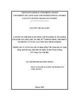
become wide and shallow or narrow and deep. Figure 1.1 illustrates some classic pit
morphologies [7].
Once a pit is formed and begins to grow, pitting becomes self-sustaining or
autocatalytic in nature where conditions develop such that further pit growth is
promoted. Within the pit, the oxygen supply (cathodic reagent) becomes depleted
hence shifting the cathodic reaction to the exposed surface. The anodic corrosion
reaction inside the pit is then supported by the external cathodic reaction. In the local
Figure 1.1 Different pit morphologies.
Narrow and Deep
Elliptical Wide and Shallow
Subsurface
Undercutting
Horizontal Vertical
3
pit environment, metal cations are first produced by the dissolution of metal. The
hydrolysis of the metal cations produces H
+
ions. To maintain charge neutrality within
the pit, negative anions such as Cl
-
diffuse into the pit. This further increases the
aggressive anion concentration and causes the pH to fall further. With the decrease in
pH, the dissolution rate of the metal increases and the whole process becomes
autocatalytic or self-sustaining. The anodic metal dissolution liberates electrons which
are then consumed by the cathodic reaction taking place on the exterior surface
(cathode) adjacent to the pit. A schematic illustration is given in Figure 1.2 and a
summary of the chemical reactions taking place is listed in Table 1.1.
The pit electrolyte is very acidic, with pH as low as 1-2 and the chloride
concentration can also be up to ten times that of the bulk solution. The anodic reaction
products form a layer of salt film at the base of the pit which prevents repassivation.
The remnants of the passive film form a pit cover, acting as a diffusion barrier,
retaining a sufficiently high concentration of Cl
-
and H
+
ions inside the pit. This creates
a highly acidic and concentrated environment suitable for stable pit growth.
Figure 1.2 Schematic diagram illustrating the anodic and cathodic reactions inside a pit.
4
Table 1.1 Chemical reactions which occur during pitting corrosion.
Inside the pit (anode)
Passive surface adjacent to the pit
(cathode)
Metal dissolution:
Fe → Fe
2+
+ 2e
-
Hydrolysis of cations, producing an acidic
environment within the pit:
Fe
2
+ + H
2
O → Fe(OH)
+
+ H
+
Fe
3+
+ 2H
2
O → Fe(OH)
2
+
+ 2H
+
Reaction between cation and Cl
-
, causing
pH to decrease further:
Fe
3+
+ 3Cl
-
+ 3H
2
O → Fe(OH)
3
+ 3HCl
Formation of salt film:
Fe(OH)
2
+
+ OH
-
→ H
2
O + FeOOH
Fe
2+
+ 2Cl
-
+ 2H
2
O → FeCl
2
+ 2H
2
O
O
2
+ 2H
2
O + 4e
-
→ 4OH
-
5
1.2 Stages of Pitting
The evolution of corrosion pits on stainless steels in halide solutions can be
divided into three stages – nucleation, metastable growth and stable growth. In this
section, the different proposed pit initiation/nucleation mechanisms are first introduced,
followed by the growth of metastable pits and lastly, the formation and propagation of
stable pits.
1.2.1 Pit Initiation/Nucleation
There have been many models introduced to explain pit nucleation. It is widely
believed that sulphide inclusions are very detrimental towards pitting resistance of
stainless steels [8-12], but there has yet to be a universally accepted mechanism. The
most common argument is that the dissolution of the sulphide inclusions exposes the
bare metal and creates an aggressive local environment due to the dissolution products
[6,13]. Williams et al. [14] suggested that the dissolution of MnS inclusions leads to a
local decrease in pH and the deposition of a sulphur-rich crust in a ring around the
inclusion. Moreover, electromigration through the sulphur crust is essential to support
the high dissolution current. This leads to local accumulation of chloride under the
crust which in turn catalyzes the dissolution of the inclusion. In 2010, Williams et al.
once again put forth an argument that a thin porous metal-deficient polysulphide skin
forms between the bulk of the inclusion and the steel, where a pit can be triggered [11].
Ryan et al. proposed that instead of the dissolution of the inclusion causing pit
nucleation, it was the depletion of Cr around the sulphide inclusions which resulted in
pits forming around the inclusions [15]. This argument was disputed by Meng et al.
[16] who found no such evidence.
6
Several groups proposed that pit nucleation is caused by a mechanical
breakdown of the film thus exposing parts of the bare metal surface to the electrolyte
[17-21]. Hoar claimed that when the passive film is in contact with an aggressive
electrolyte, the film becomes mechanically stressed and damaged by pores and flaws
as a result of changes in the interfacial forces [20]. In contrast, Fromhold suggested
that the electrochemical potential gradients inside the film give rise to large stress
values which are high enough to produce mechanical breakdown of the passive film
[22]. According to Sato, mechanical stresses resulting from electrostriction and surface
tension effects cause localized breakdown in the passive film [18,19]. He further
presented another model introducing the formation of a through-going pore which
leads to the electrocapillary breakdown of the passive film [17]. Alternatively, Xu et al.
proposed that the localized breakdown of the passive film takes place preferentially on
the concave regions of a metal surface by the concentration of electrostatic stresses
[21].
Other than the mechanical breakdown theory, the idea of the thinning of the
oxide film as a possible cause to the localized breakdown was also introduced [23-26].
The basis of the theory is that the adsorbed aggressive anions form soluble transitional
complexes with the cations on the oxide surface. Under a constant anode potential, the
electrical field increases at the thinned point of the passive film, resulting in film
dissolution until the bare metal surface.
Another well-known pit initiation mechanism is the ion penetration mechanism.
The thickness of the passive layer is typically a few nanometers and thus there exists a
very high electric field across the passive layer, on the order of 10
6
Vcm
-1
[27]. With
the assistance of the electric field, Cl
-
ions are able to penetrate the passive film, as
advocated by Evans [28], Hoar et al. [29] and Ilevbare et al. [30]. Ilevbare and
7
Burstein suggested that chloride ions (along with oxygen ions) can be drawn through
the passive film under this high electric field and at the metal-oxide interface, metal
chloride is formed. Since metal chloride has a larger molar volume than the metal
oxide, an internal pressure builds up, resulting in the rupture of the passive film [30].
Interestingly, there have been works by various authors who found the incorporation of
chloride ion into the passive film of stainless steels [31-36]; however, at the same time,
there are others who report its absence [37-38]. The contradictory results may likely
be due to differences in the experimental methodology, sample preparation and
sensitivity of the surface characterization technique.
In contrast to the penetration model, Macdonald et al. introduced the point
defect model, which is based on the migration of point defects (oxygen and metal
vacancies) [41]. In this model, the chloride ion is absorbed into the oxygen vacancies
at the outer layer of the passive film, hence increasing the local cation vacancy
concentration. This increases the electromigration-dominated flux of cation vacancies
from the outer layer of the passive film to the barrier/metal interface. The cation
vacancies at the metal/barrier interface are then annihilated by an oxidative injection of
cation from the metal into the passive film. If the rate of annihilation is slower than the
enhanced flux of cation vacancies, the accumulation of cation vacancies at the
metal/passive oxide leads to a collapse of the film. These collapse sites act as pit
nucleation sites [41]. This point defect model has continuously been adopted and
optimized to provide an analytical description of the breakdown of the passive film
and subsequent pitting activities [42,43].
Following the initiation stage would be the growth of pits which occurs in 2
stages – metastable and stable growth.
8
1.2.2 Metastable Pitting
Metastable pitting was first observed by Hisamatsu et al. in 1971 where small
current transients (< 20μA cm
-2
) were measured in potentiostatic tests at low potentials.
They attributed these current transients to the dissolution and repassivation at sites of
film breakthrough [44]. Following that, Sato suggested that the breakdown of the
passive film provided initiation sites for pitting and described these current transients
as unstable pit embryos [17,45]. In 1987, Frenkel et al. introduced the term
“metastable pitting” which described pits which nucleated but did not achieve stable
growth [46].
Once a pit has nucleated, its growth is sustained by the development of a highly
aggressive anolyte inside the pit [47]. As mentioned in Section 1.1, the pit anolyte is
highly acidic, with a high metal salt concentration. At any point of time, should the pit
contents be diluted, repassivation would be favoured and the pit stops growing [45,48].
These pits which stopped growing are termed metastable pits. Metastable pits do not
cause serious damage to the metals in real-life scenarios as the size of these pits are
typically on the order of a few micrometres. The study of metastable pits however, can
reveal information about the formation of stable pits as there is little difference
between metastable growth and the initial growth of a pit which steadily propagates
without repassivation [49].
1.2.3 Stable Pit Growth
Pistorius and Burstein introduced the concept of Pit Stability Product (i·a), the
product of the pit radius (a) and its dissolution current density (i) [47,50]. For stable pit
growth, the product i·a of the pit has to exceed a critical value (for example, for 304L
steel in chloride solution at ambient temperature, this value was determined to be 3mA
9
cm
-1
). If the pit anolyte is maintained at a high acidity level and saturated metal salt
concentration required to sustain metal dissolution, pit growth can be sustained and
repassivation is prevented [17,47,51]. It was suggested that metastable pits relied on a
porous cover to maintain the highly aggressive pit environment. If this pit cover is lost
prematurely before the critical pit stability product can be reached, repassivation will
occur and the pit does not reach stable growth. Nevertheless, when the cover is no
longer required for sustained propagation and the pit depth itself acts as a sufficient
diffusion barrier, the pit has achieved stability. From then onwards, pit growth is
effectively indefinite.
10
1.3 Determining Pitting Resistance in Stainless Steels
1.3.1 Pitting Resistance Equivalent Number (PREN)
There are over 100 different types of stainless steels and they can be classified
based on their microstructures and compositions into five main categories: austenitic,
ferritic, duplex (mixture of austentic and ferritic), martensitic and precipitation
hardening martensitic [52]. The more common alloying elements include chromium,
vanadium, molybdenum, tungsten, nickel, manganese and sulphur. Extensive work on
the effects of different alloying metals on the pitting resistance of stainless steels have
been reviewed by many authors and it has been widely accepted that while S is
detrimental, Cr, V, Mo, W and N are generally beneficial towards pitting resistance
[53-54]. A common way to rank the pitting corrosion resistance of stainless steels is to
compute and compare the Pitting Resistance Equivalent Number (PREN) [55,56]. A
higher PREN indicates a greater corrosion resistance. The PREN can be calculated as:
PREN = %Cr + 3.3%Mo + 16%N
(1.1)
PREN is a widely recognized theoretical relationship to compare the pitting
resistances of stainless steels based on the chemical compositions of the alloy. Various
multipliers for N (12.8 – 30) have been used in equation 1.1, with the larger values
used for the austenitic stainless steels grades. For super-duplex stainless steels
containing tungsten, the effects of W can also be included in the PREN to
acknowledge its positive effects on pitting resistance.
PREN = %Cr + 3.3%(Mo + 0.5%W) + 16%N
(1.2)
Ni and Mn are not believed to directly influence the pitting resistance. In
general, stainless steels with PREN larger than 26 are suitable for biomedical
applications while those larger than 40 are typically used in stagnant seawaters.