Effect of snake venoms on blood coagulation
Bạn đang xem bản rút gọn của tài liệu. Xem và tải ngay bản đầy đủ của tài liệu tại đây (28.59 MB, 201 trang )
EFFECT OF SNAKE VENOMS ON
BLOOD COAGULATION
YAU YIN HOE
(B.Sc., NUS)
A THESIS SUBMITTED
FOR THE DEGREE OF DOCTOR OF PHILOSOPHY
DEPARTMENT OF BIOCHEMISTRY
NATIONAL UNIVERSITY OF SINGAPORE
2007
AGKNOWLEDGEMENTS
I would like to express my heartfelt gratitude to Associate Professor Khoo
Hoon Eng, who has been my inspiring supervisor, a wise consultant, my role model
and a trusted friend throughout these few turbulent and challenging years of mine. I am
truly honoured and lucky to come under the tutelage of the most outstanding and
admirable person I have met. Without her trust and acceptance, I would have never had
the chance to further my degree. My gratitude to everything she has done for me can
never ever be expressed with mere words.
I am also greatly indebted to my co-supervisor, the world renowned
toxinologist Professor Ponnapalam Gopalakrishnakone, for his vast knowledge and
experience, one who has introduced me to the fascinating field of venoms and toxins. I
thank him wholeheartedly for imparting me his unsurpassed knowledge and
experimental skills in this field.
I would like to thank Associate Professor Kini Majunatha for his critical
advices, valuable recommendations and kind permission to use his Fibrometer in my
initial coagulation assays. I also want to thank his then post-graduate student, Dr Pung
Yuh Fen, for her help and friendship during my initial two gruelling research years.
I am thankful to have walked through these few years with many remarkable
people who have made lasting footprints alongside mine. My gratitude goes to:
Ms Beatrice Goh Hwey Nei, honours and post-graduate student, who shared
most of our candidature years together both in work and play. There were many
occasions where we debated over our research findings as well as over silly matters
ranging from global politics to computer games during our many meals together. I
i
greatly appreciate her companionship during both weekdays and weekends,
particularly in frustrating moments when experiments did not turn out smoothly;
Mr Sun Wentian, whom I may need to address as ‘Dr Sun’ now, a most senior
post-graduate student in the laboratory we called our ‘clan leader’ and one who helped
me tremendously with much necessary translations of Chinese articles for my research.
I am also thankful to have him sharing his expertise in badminton and photography
with us during much of our meal-time discussions;
Mr Wu Feiyi, who has graduated with an M. Sc. and taught me good tennis
strokes besides other skills;
Dr Hon Wei Min, Dr Ng Hian Cheong, Dr Wei Changli, Dr Julia Sung, Dr
Yap Lai Lai, Ms Liew Huei Chun, Mr Gregory Tan Ming Yong and Mr Yim Onn
Siong for giving me many pleasant memories and being such great friends;
Ms Ong Lai Chun, a diligent, amiable and jovial honours student for her
assistance in helping me conduct most in vivo and histological work presented herein;
Ms Tan Liting and Ms Teoh Kit Yee (Undergraduate Research Opportunity
Programme (UROP) students) for performing various routine assays.
I also thank Dr Stephen Koh (Department of Obstetrics and Gynaecology,
NUH) and his capable team members, Raymond, Seok Eng and Bee Lian, for their
expert help and assistance in thrombelastography and Euglobulin clot time.
I am grateful for the support and encouragement I received from my parents,
Mr Yau Kee and Mdm Wong Fei Ching, for their providing of my education that
served as an important foundation for my subsequent academic pursuit. I would not
become a man I am today without them teaching me the virtues – love, patience,
ii
perseverance, determination, and last but not least, dedication. Essentially, I would
simply cease to exist without them.
Most importantly, I dedicate this degree to my loving wife, Mdm Leong Kee
Mei, for her steadfast support and trust in my pursuant of dream, for her understanding
and empathy in easing my household workload, and for all her sacrifices in being my
livelong partner and providing me a blissful marriage, a lovely home, a happy family,
a conducive working condition as well as bearing our first beautiful child, Mas Yau
Zhi Yeong, in year 2005. They gave me the reason for me to strife and work hard
everyday and I regard myself as the luckiest person in this world.
iii
TABLE OF CONTENTS
Page
Acknowledgements
Table of contents
i
iv
List of figures
viii
List of tables
xi
List of abbreviations
xii
Summary
xv
CHAPTER 1: Introduction
1.1
Pit vipers from the genus Agkistrodon
1
1.2
Pit viper envenomation
4
1.3
Mammalian blood coagulation system
8
1.4
Prothrombinase and fibrinolysis pathway
10
1.5
Thrombosis and embolism
13
CHAPTER 2: Purification and Characterisation of agkistase
2.1
Introduction
16
2.1.1 Liquid chromatography
2.1.2 Proteins and peptides isolated from snake venom
2.2
Methods and materials
19
2.2.1 Snake venom and materials
2.2.2 Purification of anti-coagulant protein, agkistase (AFg)
2.2.3 Ion-exchange chromatography
2.2.4 Size exclusion chromatography
2.2.5 Reverse phase HPLC
2.2.6 SDS-PAGE (reducing/non-reducing) and Tris-Tricine gel
2.2.7 Isoelectric focusing (IEF)
2.2.8 Capillary electrophoresis
2.2.9 Mass spectrophotometry
2.2.10 Mass protein finger-printing
2.2.11 Edman degradation protein sequencing
iv
2.3
Data and results
27
2.3.1 Preliminary screening and general activities of Agkistrodon halys
halys venom
2.3.2 Purification of agkistase from Agkistrodon halys halys venom
2.3.3 Homogeneity determination of agkistase
2.3.4 Molecular sequencing and homology alignment of agkistase with
other snake venom proteins
2.3.5 Physical properties of agkistase
2.4
Discussion
42
CHAPTER 3: In vitro Studies of agkistase in Reference to Blood Coagulation
3.1
Introduction
48
3.1.1 Snake venom toxins and the coagulation system
3.1.2 Fibrinogen-targetting toxins
3.2
Methods and materials
52
3.2.1 Blood collection and storage
3.2.2 Proteolytic assays on chromogenic substrates
3.2.3 Haemolytic assay
3.2.4 Thrombin time assay
3.2.5 Prothrombin time assay
3.2.6 Recalcification time assay
3.2.7 Platelet aggregation assay
3.2.8 Fibrino(geno)lytic assay
3.2.9 Fibrinolytic
3.2.10 Fibrin plate lysis
3.2.11 Euglobulin lysis time
3.2.12 Thrombelastography
3.3 Data and results
59
3.3.1 Enzymatic properties of agkistase
3.3.2 Effects of agkistase on blood coagulation system
3.3.2.1 Haemolysis assay
3.3.2.2 Platelet aggregation assay
3.3.2.3 Recalcification, prothrombin and thrombin assay
3.3.3 Effects of agkistase on prothrombinase complex and fibrin
formation
v
3.3.4 Effects of agkistase on fibrinolytic pathway
3.3.5 Effects of agkistase on human haemostasis system
3.4
Discussion
79
3.4.1 Characterisation of the enzymatic properties of agkistase using
synthetic peptides
3.4.2 Determination of
coagulation system
agkistase’s
target
molecule
in
plasma
3.4.3 Fibrinogenolytic and fibrinolytic assays for agkistase
3.4.4 Agkistase is a new serine protease
3.4.5 Clinical implications of agkistase α, β-fibrinogenase activity
CHAPTER 4: In vivo Studies of agkistase in Reference to Thrombosis and
Coagulopathy
4.1
Introduction
90
4.1.1 Thrombosis and coagulopathies
4.1.2 Thrombolytic agents from toxins
4.2
Methods and materials
95
4.2.1 In vivo assays and animal species
4.2.2 Blood collection and treatment
4.2.3 Microplate-based coagulation assay
4.2.4 D-dimer assay
4.2.5 Platelet count
4.2.6 Collection and histological sectioning of organs
4.2.6.1 Haematoxylin eosin (H & E) staining
4.2.6.2 Masson’s trichrome staining (MTS)
4.2.6.3 Microscopy and histological pictures
4.2.6.4 Data processing and statistical analyses
4.2.7 In vivo haemorrhagic effects and systemic toxicity
4.2.8 In vivo effects of agkistase concentration
4.2.9 In vivo effects of agkistase exposure time
4.2.10 Thromboembolic model challenge
4.3
Data and results
101
4.3.1 In vivo haemorrhagic effects and systemic toxicity
4.3.2 In vivo studies of agkistase effects on administrative concentration
and exposure time
vi
4.3.3 Thromboembolic challenge and clinical therapeutic evaluation of
agkistase
4.4
Discussion
127
CHAPTER 5: Implications and Future Studies
5.1
Contributions of snake toxins
134
5.2
Defibrinating proteins from snake venoms
136
5.3
Agkistase as a new source of fibrinogenase
147
5.4
Future work
149
List of publications
151
References
152
Appendix I: Taxonomic status of the Agkistrodon complex
177
Appendix II: Permission to reproduce copyright material
179
vii
LIST OF FIGURES
Chapter 1
1.1
A picture of Agkistrodon halys halys
1.2
Schematic diagram of cell-based haemostatic system
1.3
Schematic model of a fibrinogen molecule (NDS knot)
1.4
Scheme of lysis of the fibrinogen molecule by plasmin
Chapter 2
2.1
Purification profile of Agkistrodon halys halys venom
2.2
Homogeneity determination of agkistase
2.3
SDS-PAGE of agkistase
2.4
HPLC and MALDI-TOF analyses of agkistase
2.5
The chemistry of Edman degradation
2.6
Output display for tandem mass spectroscopy
2.7
Short sequences obtained from Edman and de novo MS/MS sequencing
2.8
BLAST results of agkistase sequences
2.9
Sequence alignment of similar BLAST proteins
2.10
Isoelectric focusing of agkistase
2.11
CE analysis of agkistase
2.12
SDS-PAGE analysis on ion-exchange column peaks
2.13
Partial sequence of agkistase aligned from Edman and MS/MS results
2.14
Phylogenetic tree of similar proteases in BLAST
Chapter 3
3.1
A simplied diagram of thrombelastography
3.2
Chromogenic peptides and their chemical structures
3.3
Kinetic profile and colour absorbance range for chromogenic substrates
3.4
Haemolysis assay performed on human erythrocytes
3.5
Platelet aggregation assay
3.6
Concentration-dependent anticoagulation response of agkistase
3.7
Prothrombin activation assay
3.8
Fibrinogenolytic assay (incubation time)
3.9
Decrease in band intensity in fibrinogenolytic activity
3.10
Stability of agkistase fibrinogenolytic activity under different pH and
viii
salt concentration
3.11
Plasminogen activation assay
3.12
Clot dissolution resolved on SDS-PAGE
3.13
Fibrin plate lysis
3.14
Determination of EC50 on clinical coagulation assays
3.15
A typical thrombelastogram showing the important parameters
3.16
Thrombelastograms of Ca2+ effects on venom
3.17
Thrombelastograms of Ca2+ effects on agkistase
3.18
Thrombelastograms on effects of concentration on venom
3.19
Thrombelastograms on effects of concentration of agkistase
3.20
Thrombelastograms of exposure duration of venom
3.21
Thrombelastograms of exposure duration of agkistase
3.22
Schematic diagram of cell-based haemostatic system
3.23
Domain structure of fibrinogen molecule
Chapter 4
4.1
Haemorrhagic effect and systemic toxicity of Agkistrodon venom and
agkistase
4.2
Histological sections of mouse organs injected with venom under
haematoxylin and eosin staining (H & E)
4.3
Histological sections of mouse organs injected with venom and agkistase
under Masson’s trichrome staining (MTS)
4.4
Concentration-dependent effects of agkistase
4.5
Depletion of circulating fibrinogen with increasing concentration of
agkistase
4.6
Time-dependent effects of agkistase
4.7
Heart sections (concentration-dependent effects of agkistase)
4.8
Lung sections (concentration-dependent effects of agkistase)
4.9
Liver sections (concentration-dependent effects of agkistase)
4.10
Kidney sections (concentration-dependent effects of agkistase)
4.11
Spleen sections (concentration-dependent effects of agkistase)
4.12
Heart sections (time-dependent effects of agkistase)
4.13
Lung sections (time-dependent effects of agkistase)
4.14
Liver sections (time-dependent effects of agkistase)
4.15
Kidney sections (time-dependent effects of agkistase)
4.16
Spleen sections (time-dependent effects of agkistase)
ix
4.17
Mouse thromboembolic model challenge
4.18
Heart sections (mouse thrombosis model challenge)
4.19
Lung sections (mouse thrombosis model challenge)
4.20
Liver sections (mouse thrombosis model challenge)
4.21
Kidney sections (mouse thrombosis model challenge)
4.22
Spleen sections (mouse thrombosis model challenge)
4.23
Histological sections of necrotic kidney from venom-injected mice
4.24
Histological sections of lung from agkistase-injected mice showing
rethrombosis at 72 hr
x
LIST OF TABLES
Chapter 1
1.1
Agkistrodon halys halys directives as described by Gloyd and Conant
(1990)
Chapter 2
2.1
Screening of venoms for proteolytic and defibrinating activities
2.2
Purification table of agkistase
Chapter 3
3.1
Enzymatic efficiency of agkistase against other venom proteinases
3.2
Enzymatic activity of agkistase against all substrates
3.3
Fibrometer clotting times with agkistase
3.4
Inhibitory studies on agkistase
3.5
Euglobulin lysis time
Chapter 5
5.1
Well-defined and purified SVTLEs with clinical and therapeutic use
5.2
Fibrinogenases from snake venoms
xi
LIST OF ABBREVIATIONS
< Glu-Phe-Lys-pNA.HCl
L-pyroglutamyl-L-phenyl-L-lysine-p-nitroaniline hydrochloride
< Glu-Pro-Arg-pNA.HCl
L-Pyroglutamyl-L-prolyl-L-arginine-p-nitroaniline hydrochloride
α2-PI
alpha 2-plasmin inhibitor
ACN
acetonitrile
AFg
agkistase
Ahh
agkistrodon halys halys
APS
ammonium persulphate
aPTT
activated partial thromboplastin time
ARF
acute renal failure
Bz-CO-Ile-Glu-(-OR)-Gly-
N-Benzoyl-L-isoleucyl-L-glutamyl-glycyl-L-arginine-p-nitroaniline
Arg-pNA.HCl
hydrochloride and its methyl ester
BLAST
basic local alignment search tool
CIEF
capillary isoelectric focusing
cm
centimetre
CV
column volume
CZE
capillary zone electrophoresis
Da
Dalton
DD
D-dimer
DIC
disseminated intravascular coagulation
DTT
dithiothreitol
DVT
deep vein thrombosis
EC50
median effective concentration (required to induce a 50% effect)
EDTA
ethylenediaminetetraacetic acid
ESI-MS
electrospray ionization – mass spectrophotometry
EST
expressed sequence tag
FDP
fibrin degradation product
FPA
fibrinopeptide A
xii
FPB
fibrinopeptide B
H-D-Ile-Pro-Arg-pNA.2HCl
H-D-Isoleucyl-L-prolyl-L-arginine-p-nitroaniline dihydrochloride
H-D-Phe-Pip-Arg-pNA.2HCl
H-D-Phenylalanyl-L-pipecolyl-L-arginine-p-nitroaniline
dihydrochloride
H-D-Pro-Phe-Arg-pNA.2HCl
H-D-Prolyl-L-phenylalanyl-L-arginine-p-nitroaniline dihydrochloride
H-D-Val-Leu-Lys-pNA.2HCl
H-D-Valyl-L-leucyl-L-lysine-p-nitroaniline dihydrochloride
HPMC
hydroxypropylenemethylene cellulose
hr
hour
IEF
isoelectric focusing
k
k time (thrombelastography)
kg
kilogramme
MA
maximum amplitude (thrombelastography)
MALDI-TOF
matrix-assisted laser desorption ionisation – time of flight
mg
milligramme
MI
myocardiac infarction
min
minute
ml
millilitre
Mr
relative molecular weight
MTS
Masson’s trichrome stain
ng
nanogramme
PBS
phosphate-buffered saline
PE
pulmonary embolism
PMSF
phenylmethylsulphonyl fluoride
pNA
p-nitroaniline
PPP
platelet-poor plasma
PRP
platelet-rich plasma
PT
prothrombin time
PVDF
polyvinylidine difluoride
r
reaction time (thrombelastography)
RP-HPLC
reverse phase-high performance liquid chromatography
xiii
RT
recalcification time
s
sec
SBI
soybean trypsin inhibitor
SDS
sodium dodecyl sulphate
SDS-PAGE
sodium dodecyl sulphate-polyacrylamide gel electrophoresis
SVTLE
snake venom thrombin-like enzyme
TEMED
N,N,N’,N’-Tetramethylethylenediamine
TFA
trifluoroacetic acid
tPA
tissue-type plasminogen activator
Tris
tris[hydroxymethyl]aminomethane
TT
thrombin time
µg
microgramme
uPA
urokinase-type plasminogen activator
VTE
venous thromboembolism
xiv
SUMMARY
Snake venoms are rich collections of enzymes, proteins, peptides and other
components that can cause a wide range of physiological, neurological and
haemostatic effects on their prey in an attempt to immobilise them and aid in digestion.
Among these effects, the venom components that affect mammalian haemostasis have
been most well studied for more than 150 years. They have contributed to elucidation
of the detailed mechanisms of the coagulation cascade (e.g. platelet aggregation and
inhibition, mechanism of defibrination, DIC, various coagulopathies, etc), elucidation
of various clinical disorders (e.g. congenital haemorrhagic disorder, various blood
factor deficiencies, etc), development of many diagnostics (e.g. Styphen or Reptilase®
time) and therapeutics (e.g. ancrod and batroxobin). Therefore venoms have been
regarded as ‘gold mines’ for researchers, pharmaceutical companies, clinical analysts
as well as medical practitioners and surgeons.
An anticoagulant proteinase, named agkistase, was isolated from the venom of
pit viper, Agkistrodon halys halys, through successive ion-exchange and size exclusion
liquid chromatography. Its purity was checked by high resolution HPLC, capillary
electrophoresis and mass spectrophotometry. It is a serine protease with α,βfibrinogenase activity, which cleaves plasmatic fibrinogen chain α and β rendering it
unclottable by thrombin. Agkistase was found to be fibrinogenolytic with slight
fibrinolytic activity and did not affect other coagulation factors nor cause platelet
aggregation. This fibrinogenase activity is unaffected by pH 5 ~ 9 and salt
concentrations up to 0.8 M NaCl but was inhibited by serine protease inhibitors (e.g.
PMSF and aprotinin). It has an α-fibrinogenase and β-fibrinogenase activities of 15.1
xv
and 0.25 mg min-1 mg enzyme-1, respectively. Thrombelastographic analyses revealed
a prolongation in r and k values but unchanged MA, with extension in incubation with
whole blood samples. This observation is usually seen for haemophilic blood samples,
evident of abnormalities in one or more coagulation factors – in this case, fibrinogen.
Human blood coagulation assays on agkistase showed that it has EC50 values of
5.1 ± 1.5, 0.26 ± 0.1 and 0.5 ± 0.2 µg/ml on prothrombin time, recalcification time and
thrombin time, respectively, in vitro. In vivo studies on C57BL mice showed that it is
not toxic, haemorrhagic or thrombogenic up to 1.67 mg/kg when injected
intravenously. These mice also showed no signs of thrombocytopenia. Evaluation of
its anti-thrombotic potential on a thrombotic mouse model exhibited positive results in
both reductions in thrombi occurrence and size, at agkistase concentration of only
50 ng/kg mouse when injected intravenously. We concluded that it will be a promising
new anti-thrombotic drug different from current available snake venom thrombin-like
enzymes (SVTLEs) due to: (i) its low effective concentration with no observable
negative effects, (ii) fast and specific fibrinogenolytic activities, (iii) presence of mild
secondary fibrinolytic activities, and (iv) successful demonstration of its antithrombotic capability on a mouse model.
xvi
CHAPTER 1: INTRODUCTION
1.1 PIT VIPERS FROM THE GENUS AGKISTRODON
Vipers and pit vipers are mainly classified into the taxonomic subfamily
Viperinae and Crotalinae (Order: Squamata; Suborder: Serpentes; Infraorder:
Alethinophidae; Family: Viperidae). This group of snakes were first studied in the late
1950s by a renowned American herpetologist, the late Prof Howard Kay Gloyd (19021978). After Prof Gloyd’s death in 1978, the colossal task of completing their
identification and taxonomic organisation was passed onto the late Isabelle Hunt
Conant.
The description of the genus Agkistrodon as well as its subspecies A. halys
halys in this dissertation was based mainly on the work of Gloyd and Conant (1990),
who obtained and studied over 6000 specimens found on several continents spanning
over 11,000 kilometres. This is by far the most number of specimens studied
unsurpassed by any other field taxonomists or herpetologists to-date. However,
taxonomic classification is not the major focus in this dissertation. Hence only
simplistic reference is presented to aid and/or resolve identification.
A. halys complex has confused many taxonomists for almost a century. Three
full species, A. blomhoffii, A. halys and A. intermedius, and all their many subspecies
were conveniently categorised as “halys” for decades. According to the classification
of Gloyd, the genus Agkistrodon halys complex was split into four separate and
distinct genera – Deinagkistrodon, Agkistrodon, Calloselasma and Hypnale. Of these
genera, Agkistrodon is presumed to comprise the largest species and has the greatest
diversity – 7 species were reported in Asia and 3 from North America. The North
American species were thought to have evolved from an ancestral lineage from the
1
Asian species that crossed the Bering Land Bridge during the late Oligoscene or early
Miocene. This remarkable diversification of Agkistrodon snakes, which created many
controversies of identification before the 1950s, was attributed to intergradation
between species.
Snakes of the Agkistrodon complex are reported to possess the following
characteristics: (i) anal plate is single (not divided), (ii) a single pair of enlarged chin
shields (although remnants of a posterior pair are occasionally evident),
(iii) nine symmetrically arranged scutes or plates, (iv) pre-oculars are almost always
two in number, (v) presence of a maxillary (facial) pit, which is a chief characteristic
of crotaline snakes, and (vi) triads (a group of large dark spots occupying a
ventrolateral position in conjuction with each dark dorsal crossband) only
characteristic of the North American forms.
A summarised directive for identification of A. h. halys is presented in Table 1.1
below.
Table 1.1: Agkistrodon halys halys directives as described by Gloyd and Conant (1990).
Males
Females
N
Range
Mean N
Range
Mean
Ventrals
5 164-173 169.4 5 171-178 174.0
Subcaudals
5
45-49
47.2 5
42-45
42.8
Undivided subcaudals
5
0
5
0
Scale rows at midbody*
14
22.9
Supralabials*
20
7.8
Infralabials*
20
10.7
Postoculars plus suboculars*
26
2
Crossbands
5
33-41
37.5 5
31-47
39.3
Ratio of tail length to total length (%) 5
14-15
14.6 5
12-15
12.8
Snout-vent lengths (mm)
450
590
Total lengths (mm)†
530
590
Dentary tooth counts*
4
11
Pterygoid tooth counts*
4
10.5
Palatine tooth counts*
4
3
*
Sexes were not determined in the reported specimens, † longest measured was 750 mm
2
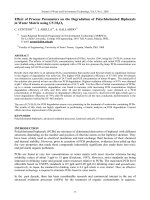
A. h. halys was first reported by Prof Peter Simon
Pallas (1741-1811), a German botanist and zoologist. The
pit viper, which was named A. h. Pallas, was discovered in
Southern Siberia and Mongolia. Due to variation of
morphology and great diversity of this subspecies, it was
given many other names – Coluber halys Pallas (Pallas
1776), Vipera halys (Latreille, 1802), Echidna aspis, var.
Prof Peter Simon Pallas
(1741-1811)
Courtesy of Wikipedia
()
pallasii (Merrem, 1820), Trigonocephalus halys (Lichtenstein, 1823), Halys pallasii
(Günther, 1864), Trigonocephalus intermedius (Strauch, 1876), Ancistrodon halys
(Boulenger, 1896), Agkistrodon halys (Stejneger, 1907), Ancistrodon halys halys
(Nikol’skii, 1916), Agkistrodon halys intermedius (Schmidt, 1927), and finally
Agkistrodon halys halys (Mertens and Müller, 1928). Hence Mertens and Müller were
the first to name this species the present-day name of Agkistrodon halys halys. The
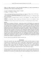
Mongols referred to this snake as
mogoi, their name for serpents in
general; the Tungus as abù; the
Mongol
name
given
by
Obst
(Obst, 1963) was uulyn mogoj; and
the Chinese named it
first
properly
蝰科蝮蛇. The
documented
indications showed that halys was
Yenisey river, Russia
Courtesy of EarthTrends ()
first discovered on the Upper Yenisey by Strauch in 1873. The Yenisey river in Russia
has its origin in Mongolia flowing due north into the Kara sea.
3
The geographical distribution of A. h. halys was mainly confined to Asia, in
southern Siberia, Inner Mongolia, Mongolia and several provinces in central and upper
China (Gloyd and Conant, 1990; Pope, 1935; Zhao and Adler, 1993; Zhao, 1990). This
species thrived in rocky, sunny and arid deserts and mountains commonly
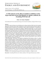
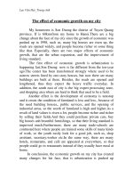
uninhabitable by other snakes. A picture of A. h. halys is presented in Figure 1.1.
Figure 1.1: A picture of Agkistrodon halys halys.
Courtesy of Venomous snakes in China (Zhao, 1990).
1.2 PIT VIPER ENVENOMATION
Snake bites are a serious medical problem, especially in the Southeast Asia
region (Warrell, 1989), causing many lethalities as well as a host of clinical symptoms
including local tissue injury, flaccid paralysis, systemic myolysis, cardiotoxicity, renal
damage and failure as well as haemorrhage and coagulopathy (White et. al., 1992;
White, 2004; White and Fassett, 1983; White and Williams, 1989; Williams and White,
1997; Yatziv et. al., 1974). It is estimated that global venomous snakebites affects
greater than 2.5 million humans annually, of whom more than 100,000 die (Chippaux,
1998). Such a high rate of morbidity and mortality is greater in the rural tropics (Laing
4
et. al., 1995; Lalloo et. al., 1995) than other localities. Each of the venom components
may cause a number of clinical symptoms and secondary effects with potential
morbidity and mortality. Any single species of snake may show activity in one or more
of these categories – haemorrhagic, neurogenic, myotoxic, etc. In the past it was
believed that vipers cause local and/or haemorrhagic effects whereas elapids cause
purely systemic, non-haemorrhagic effects.
Viperid bites are now considered to cause medically significant effects, i.e.
coagulopathy, haemorrhage and thrombosis with deep vein thrombosis (DVT) and
pulmonary embolism (PE), on the haemostatic system with their abundant disintegrins
and haemorrhagins (White, 2005). Certainly, the diverse clinical symptoms reported
reflects the numerous venom components found in each species. These symptoms can
essentially be categorised into: (i) reduced coagulability of blood, resulting in an
increased tendency to bleed, (ii) bleeding due to damage of the blood vessels,
(iii) secondary effects of increased bleeding, ranging from hypovolemic shock to
secondary organ damage, such as intracerebral haemorrhage, anterior pituitary
haemorrhage or renal damage, (iv) direct pathologic thrombosis and its consequences,
particularly pulmonary embolism. Viperid venoms are found to harbour many
components that mainly cause such clinical symptoms, whether directly or indirectly.
They are the procoagulants (e.g. thrombin-like enzymes) (Markland, 1998a),
anticoagulants (e.g. fibrinogenases) (Markland, 1998a), platelet effectors (Clemetson
et. al., 2005; Kamiguti, 2005) and haemorrhagins (e.g. HTa and HTb from Bitis
gabonica) (Marsh et. al., 1995).
5
Srilekha Karthik from St John’s Medical College Hospital, Bangalore, India
reported a 12-year old boy, who was admitted with oliguric acute renal failure (ARF),
showed all clinical symptoms of coagulopathy including oedema, micro-angiopathic
haemolytic anaemia, thrombocytopenia, prolonged coagulation parameters and
disseminated intravascular coagulation (DIC), 4 days after a reported snakebite (snake
could not be identified) (Karthik and Phadke, 2004). He was discharged after 17 days
with a normal coagulation profile and with improving renal function. Another report
from Hung Dong-Zong, Institute of Toxicology and Pharmacology, National Taiwan
University involves two cases of Russell’s viper bites where, unfortunately, one patient
underwent amputation and the other died of complications. The first patient was a 67year-old male farmer who developed haemolysis, rhabdomyolysis, acute renal failure,
thrombocytopenia,
coagulopathy
and
bleeding
from
the
genitourinary
and
gastrointestinal tracts, which later extended into drowsy consciousness, left upper limb
flaccid paralysis and multiple ecchymosis patches over his trunk. He was discharged
after 61 days in the hospital with amputated toes due to gangrenous tissues. The other
patient was a 52-year-old female field worker who hovered between consciousness,
developed high blood pressure, haematuria and bloody vomits. Despite efforts to
reverse her deterioration of renal function, pulmonary oedema, myocardial ischaemia,
arterial thrombosis, DIC and haemorrhage, the patient died of multiple septic condition
after 49 days of hospitalisation (Koo et. al., 2002).
Snakebites from Agkistrodon halys were reported to have similar symptoms and
severity to those reported above. However, due to the difficulty in capture of the
snakes and identification, precise reports of envenomation by this species are scanty.
One such case was reported from Guangxi Medical University, China involving a
6
27-year-old male who was showing local oedema and bleeding symptoms; subsequent
laboratory results showed a reduction in platelet aggregation rate (37-52%), reduced
anti-thrombin III activity (56-84%), reduced α2-PI activity (30-49%), low fibrinogen
(0-131 mg/dL) and presence of fibrin degradation products (FDP) (< 2.5 µg/ml)
suggesting DIC (Li et. al., 2000). An analysis of cDNA library construction, EST
sequencing and clustering on Agkistrodon sp performed by Qinghua et. al. (Qinghua et.
al., 2006) estimated the composition of putative cellular proteins in venom to comprise
mainly of metalloproteinases (32.08%), C-type lectins (5.22%), bradykininpotentiating peptides (0.90%), serine proteases (0.51%), nucleotidase and nuclease
(0.41%), phospholipase A2 (0.30%), disintegrins (0.05%), cytokine-like molecules
(0.06%) and other proteins (0.63%) (Liu et. al., 2006). This finding helps to explain the
predominant clinical symptoms exerted by viper venoms which comprise a variety of
coagulation-related complications as more than 40% identified ESTs are known to
affect coagulation to some degree.
7
1.3 MAMMALIAN BLOOD COAGULATION SYSTEM
The mammalian blood coagulation system is an intricate but tightly-regulated
process involving innumerable serine proteases, coenzymes, phospholipids, blood cells,
platelets, vessel walls, etc – all interrelated and affecting each other directly or
indirectly in a dynamic manner (Jenny and Mann, 2002). Haemostasis can generally be
divided into three main components, namely vessel wall, thrombocytes and plasmatic
coagulation system. Consequently, haemostatic disorders are clinically categorised into
vasculopathies, thrombocytopathies and coagulopathies, respectively, according to
their primary defect. The vessel wall plays a double role in haemostasis: (i) a
neurogenic contraction after an injury, lasting 20 to 30 sec, which permits the
formation of a primitive platelet plug and the activation of the plasmatic clotting
system, and (ii) injured or irritated endothelial cells release chemical signals that
reversibly activate thrombocytes and the plasmatic clotting system. Thrombocytes, or
platelets, are small anuclear corpuscles derived from megakaryocytes. Under
physiological conditions blood contains 200 – 400 x 109 platelets per litre of blood. In
their inactive form thrombocytes have an oval, disk-shaped form with an equatorial
diameter of 2 – 4 µm and a thickness of 1 – 2 µm. In this form they are unable to
adhere to an intact vascular wall, to other cells, or to each other. But when
thrombocytes are exposed to agonists, e.g. during an injury, they undergo rapid and
dramatic changes in cell shape, converting from discs into spiny forms within seconds.
At the same time platelets also undergoes an exocytosis of storage granules, releasing
mediators that enhance platelet plug formation by attracting additional platelets to the
surface of the wound (aggregation) and initiating cellular repair reactions (signalling).
Lastly, the plasmatic coagulation system consists of 13 major coagulation factors,
mainly proteases that activate its downstream targets in an amplification manner
8