Regulation of intestinal inflammation by mitogen activated protein kinase phosphatase 3
Bạn đang xem bản rút gọn của tài liệu. Xem và tải ngay bản đầy đủ của tài liệu tại đây (9.8 MB, 78 trang )
REGULATION OF INTESTINAL INFLAMMATION BY
MITOGEN-ACTIVATED PROTEIN KINASE
PHOSPHATASE-3
SUZAN SAIDIN
(Bachelor of Science (Biotechnology) (Hons.), Monash University)
A THESIS SUBMITTED
FOR THE DEGREE OF MASTER OF SCIENCE
YONG LOO LIN SCHOOL OF MEDICINE
NATIONAL UNIVERSITY OF SINGAPORE
2013
ACKNOWLEDGEMENT
First and foremost I would like to thank my supervisor Dr. Zhang Yongliang for
giving me an opportunity to work on this project and for his guidance throughout the
duration of my study. I would also like to express my deep gratitude towards Dr. Png
Chin Wen for his mentorship, advice and suggestions, which contributed significantly
to this project. I would like to thank Fiona and Chein Sze who have kindly given
permission for their work to be included in the supplementary figures.
I would like to thank Weiliang and Emi for their excellent administrative
support. Thank you to Huipeng, Jenny, Madhu, Wu Di, Danke, Jennifer, Heng Boon,
Mei Xing, Hong Ying, Siyuan, Edward, Han Jian and Yi Xiong for their great
company in the lab.
I am very lucky to have known Hoey Lit, Yen-ling and Narisa who have been
very supportive friends. Their sheer company provided pleasant distractions from the
unexplainable results and failed experiments. I would like to thank Hong Ting for
making sure to feed me with a little bit of alcohol whenever I am distressed and also
Ban Xiong for reading this thesis.
I am sincerely grateful to Eng Kwan, Dana and Grace for helping me in going
through my difficult times.
Last but not least I would like to thank my parents and my sisters for their
support.
i
TABLE OF CONTENTS
ACKNOWLEDGEMENT ...........................................................................................i!
TABLE OF CONTENTS ............................................................................................ii!
LIST OF ABBREVIATIONS ....................................................................................iv!
ABSTRACT.............................................................................................................. viii!
1.! Introduction...........................................................................................................1!
1.1.! Inflammatory Bowel Diseases (IBD)........................................................................1!
1.1.1.! Causes and Pathogenesis of IBD..........................................................................2!
1.1.2.! Dextran Sulphate Sodium (DSS)-induced Murine Model of Colitis ...................5!
1.2.! The Role of Immune Response in Intestinal Inflammation ...................................5!
1.3.! The Role of Intestinal Epithelium in Intestinal Inflammation ..............................7!
1.4.! Mitogen-Activated Protein Kinases (MAPKs)......................................................10!
1.4.1.! The Role of MAPKs in the Immune Response ..................................................12!
1.4.2.! The Role of MAPKs in Cell Proliferation and Survival ....................................14!
1.5.! MAPK Phosphatases (MKPs).................................................................................16!
1.5.1.! MAPK Phosphatase-3 (MKP-3).........................................................................18!
1.6.! Study Rationale and Objectives .............................................................................19!
2.! Materials and Methods.......................................................................................21!
2.1.! Cell Culture ..............................................................................................................21!
2.2.! RNA Isolation and Analysis....................................................................................22!
2.3.! Western Blotting ......................................................................................................24!
2.3.1.! Protein Extraction...............................................................................................24!
2.3.2.! SDS-PAGE and Protein Transfer.......................................................................25!
2.3.3.! Protein Detection and Analysis ..........................................................................25!
2.4.! Wound Healing Assay .............................................................................................26!
ii
2.5.! Proliferation Assay ..................................................................................................27!
2.6.! Animal Studies .........................................................................................................27!
2.6.1.! Histological Analysis .........................................................................................28!
2.6.2.! Ki67 Immunohistochemistry..............................................................................29!
2.7.! Statistical Analysis ...................................................................................................29!
3.! Results..................................................................................................................30!
3.1.! MKP-3 Negatively Regulates Inflammatory Response in CMT93 cells .............30!
3.2.! MKP-3 Regulates Epithelial Cell Proliferation and Migration In Vitro ............32!
3.3.! Loss of MKP-3 Results in Less Severe Colitis.......................................................36!
3.4.! Increase in IEC-Associated Proliferation Genes in MKP-3-/- Mice after DSS
Treatment ...........................................................................................................................40!
4.! Discussion ............................................................................................................43!
5.! Future Directions ................................................................................................50!
6.! Conclusion ...........................................................................................................52!
REFERENCES ..........................................................................................................53!
APPENDICES............................................................................................................65!
iii
LIST OF ABBREVIATIONS
AOM
Azoxymethane
APC
Antigen presenting cells
ASK1
Apoptosis signal-regulating kinase 1
BAD
B-cell lymphoma-2-associated death protein
BAX
B-cell lymphoma-2-associated X protein
Bcl-2
B-cell lymphoma-2
BMDM
Bone marrow-derived macrophages
BSA
Bovine serum albumin
CD
Crohn’s disease
CDK
Cyclin-dependent kinase
Cox2
Cyclooxygenase-2
DEPC
Diethylpyrocarbonate
DLK
Dual leucine zipper-bearing kinase
DMEM
Dulbecco’s Modified Eagle’s Medium
DNA
Deoxyribonucleic acid
DSS
Dextran sodium sulphate
DUSP
Dual-specificity phosphatase
DUSP2-/-
DUSP-2 knockout
EDTA
Ethylenediaminetetraacetic acid
EGF
Epidermal growth factor
ER
Endoplasmic reticulum
ERK
Extracellular signal-regulated kinases
FasL
Fas ligand
FBS
Fetal bovine serum
iv
FGF
Fibroblast growth factor
Foxp3
Forkhead box P3
GDP
Guanosine diphosphate
Grb-2
Growth factor receptor-bound protein-2
GM-CSF
Granulocyte macrophage colony-stimulating factor
GTP
Guanosine triphosphate
GWAS
Genome-wide association studies
H and E
Hematoxylin and Eosin
HRP
Horse radish peroxidase
IBD
Inflammatory bowel diseases
IEC
Intestinal epithelial cells
IFN-!
Interferon-!
IL
Interleukin
iNOS
Inducible nitric oxide synthase
IRF3
Interferon regulatory transcription factor 3
JNK
c-Jun NH2-terminal kinases
KLF
Krüppel-like transcription factor
KLF4-/-
KLF4 knockout
KLF5+/-
Heterozygous KLF5 knockout
LPS
Lipopolysaccharide
M1
Macrophage 1
MAPK
Mitogen-activated protein kinase
MCP-1
Monocyte chemotactic protein-1
MKK
Mitogen-activated protein kinase kinase
MKKK
Mitogen-activated protein kinase kinase kinase
v
MKP
Mitogen-activated protein kinase phosphatase
MKP-1-/-
MKP-1 knockout
MKP-3-/-
MKP-3 knockout
MKP-5-/-
MKP-5 knockout
MLK3
Mixed lineage kinase 3
mRNA
Messenger ribonucleic acid
MSK
Mitogen- and stress-activated protein kinase
MyD88
Myeloid differentiation primary response 88
NF-"B
Nuclear factor kappa-light-chain-enhancer of activated B cells
PAMP
Pathogen-associated molecular pattern
PCR
Polymerase chain reaction
PRR
Pattern recognition receptor
PVDF
Polyvinylidene fluoride
RO
Reverse osmosis
ROS
Reactive oxygen species
SDS
Sodium dodecyl sulphate
SOS
Son of sevenless
SPF
Specific pathogen-free
TAK1
Transforming growth factor-# activated kinase 1
TBS
Tris buffered saline
TBST
Tris buffered saline tween
TCR
T cell receptor
TIRAP
Toll-interleukin 1 receptor domain containing adaptor protein
TPL2
Tumour Progression Locus 2
vi
TRAM
Toll/interleukin-1 receptor-domain-containing adapter-inducing
interferon-!-related adaptor molecule
TRIF
Toll/interleukin-1 receptor-domain-containing adapter-inducing
interferon-!
TGF-#
Transforming growth factor-#
Th
T helper cells
TLR
Toll-like receptor
TNF-$
Tumour necrosis factor-$
Treg
Regulatory T cells
UC
Ulcerative colitis
UPR
Unfolded protein response
vii
ABSTRACT
During intestinal inflammation, the disruption of the intestinal mucosa barrier
enabled the luminal microbiota to come into direct contact with the underlying
immune cells, which results in inflammatory response. The recognition of the luminal
microbiota by the Toll-like receptors (TLRs) activates downstream signalling
pathways such as the mitogen-activated protein kinase (MAPK). The MAPK pathway
is essential in regulating immune response and its negative regulation is controlled by
MAPK phosphatases (MKPs). MKP-3, also known as DUSP6, is a MAPK
phosphatase with high specificity towards ERK, which is known to regulate cell
proliferation. The role of MKPs in immune cells has been widely studied but the role
of MKPs in the intestinal epithelial cells (IEC) during intestinal inflammation has yet
to be explored. In this study, we utilised CMT93 cells overexpressing MKP-3 and
dextran sodium sulphate (DSS)-induced colitis model in mice deficient in MKP-3 to
investigate the role of MKP-3 in intestinal inflammation. The results showed that
overexpression of MKP-3 downregulated the phosphorylation of all three major
groups of MAPKs (ERK, JNK and p38) and pro-inflammatory gene expression upon
DSS and LPS stimulation. In addition, the overexpression of MKP-3 retarded cell
proliferation and wound healing ability of CMT93 cells. MKP-3-/- mice subjected to 7
days of DSS treatment developed less severe colitis compared to the wildtype mice.
mRNA and protein analysis showed that the expression of pro-inflammatory genes
was reduced and the phosphorylation of ERK was increased in the colon of MKP-3-/mice. The expression of proliferation genes and Krüppel-like transcription factor 5
(KLF5) protein was also elevated in the colon of MKP-3-/- mice. In addition, Ki67
staining showed increased IEC proliferation in the colon of MKP-3-/- mice. These
results suggested the role of MKP-3 in IEC during intestinal inflammation by
affecting inflammatory gene expression, IEC proliferation and restitution.
viii
1. Introduction
1.1. Inflammatory Bowel Diseases (IBD)
Inflammatory bowel diseases (IBD) is a term used to describe the chronic
relapsing inflammatory condition in the gastrointestinal tract. The two major
conditions of IBD are ulcerative colitis (UC) and Crohn’s disease (CD). UC is
characterised by continuous inflammation and superficial ulceration in the mucosa
and submucosa of the colon accompanied by crypt abscesses. The primary infiltrates
in UC are neutrophils and lymphocytes, which are densely present in the lamina
propria. In contrast of UC which is only limited to the colon, the inflammation in CD
can be anywhere along the gastrointestinal tract. The involvement of various parts of
the gastrointestinal tract in CD causes a broader range of clinical manifestation such
as nutritional deficiency. In addition, the ulceration in CD is deeper into the intestinal
wall and the inflammation is usually segmented, whereby alteration between normal
and inflamed region is commonly found. The primary infiltrates in CD are
macrophages and lymphocytes which often form non-caseating granulomas 1–4.
IBD is mostly prevalent in North America, Northern Europe and the UK
although the rate of incidence is rising in Southern Europe, Asia and most developing
countries 5. Despite the understanding of the characteristics of IBD, the exact causes
of the disease remain elusive. There are several multiple causes of IBD which are
known to contribute towards the pathogenesis of the disease. These key contributing
factors are genetics predisposition, environmental factors and factors affected by
lifestyle such as host/mucosa immune response, mucosa epithelial barrier and gut
microbiota 6.
1
1.1.1. Causes and Pathogenesis of IBD
Higher prevalence of IBD in certain ethnic groups such as Jewish becomes a
supporting evidence that genetic makeup contributes to the development of the
disease. In addition, family history of IBD is a strong contributing risk factor towards
the development of the disease in an individual 5. Genome wide association studies
(GWAS) have identified several susceptibility genes across several regions in the
chromosome. These studies revealed several pathways that are affected by the
identified genes, which may play important roles in the pathogenesis of IBD. These
pathways involve intestinal epithelia integrity, innate and adaptive immune responses,
autophagy, endoplasmic reticulum (ER) stress and numerous other regulatory
pathways that are yet to be elucidated 7,8. These pathways may also affect each other,
which further shows IBD as a polygenic disease 9.
It is also interesting to note that despite being born with the predisposed genetic
background, most individuals do not develop IBD symptoms until later in life. This
shows that besides genetic predisposition, environmental and lifestyle factors play a
role in the development of IBD in individuals. Smoking is one of the lifestyle factors
that exacerbates CD but on the contrary protects against UC
10
. Several hypotheses
have also suggested exposure to environmental antigens and access to hygiene and
sanitation as factors that contribute to the development of IBD. Growing up in highly
sanitised environment may impair the development of the immune system by
restricting antigen exposure, which in turn causes the immune system to have
exaggerated response to the antigen later in life 5.
As with the uncertain cause of the disease, the pathogenesis of IBD is still
unclear. The recent understanding of the disease speculates that IBD is a result of the
2
shift in intestinal microbial content, breakdown of the protective barrier of the
intestinal epithelia and/or a dysregulation in the immune system 4,11. The composition
of the intestinal microflora has been associated with human IBD. For example,
adherent-invasive E. coli was found in colonic lesions from CD patients and has been
associated with ileal mucosa of CD patients 12,13. The presence of the bacteria in early
recurrent lesions indicates that it plays a role in the initiation of inflammation. In
addition, its ability to survive and replicate within macrophages has been thought to
contribute to its ability to spread within the intestinal mucosa and trigger chronic
inflammatory response 13.
Besides the intestinal microflora, the loss of intestinal epithelial integrity and
function is an early event in IBD pathogenesis
11
. Increased and sustained intestinal
epithelial ER stress caused by unfolded protein response (UPR) can trigger apoptosis.
UPR is a cytoprotective mechanism to prevent the accumulation of misfolded protein
in the cell. Accumulation of misfolded protein causes sustained ER stress, which in
turn result in apoptosis of the intestinal epithelial cells (IEC). Studies in mice with
MUC2 missense mutation showed increased ER stress in the IEC, which causes
susceptibility to colitis
14
. Besides ER stress, other factors such as dysregulation in
transcription factors involved in IEC regeneration and breakdown in tight junction
proteins such as ZO-1 and ZO-2 have also been linked to the pathogenesis of IBD 4.
Similar to many other autoimmune diseases, the pathogenesis of IBD involves
the dysregulation of T cell responses. Earlier findings have suggested CD and UC as a
result of T helper 1 (Th1) and T helper 2 (Th2) response, respectively 15. However this
paradigm remains controversial as other observations showed mixed cytokine profiles
in ex vivo cultures from IBD patients
16,17
. In addition, increased level of IL-17A in
the intestinal mucosa of IBD patients has also indicated the involvement of T helper
3
17 (Th17) responses in the disease
18
. Previous studies have shown plasticity in the
late stage development of Th17 and regulatory T (Treg) cells, and that each T cell
requires TGF-# for differentiation
19
. As TGF-# is abundant in the intestines, this
finding may indicate the involvement of the balance between these two T cell
responses in IBD. The presence of inflammatory cytokines, such as IL-6 or IL-21,
promotes the development of Th17 in the intestines, which otherwise would
predominate towards Treg differentiation to maintain the state of tolerance 4,20.
In summary, the pathogenesis of IBD involves a complex interplay of these
three factors, and it is most likely that the occurrence of one would trigger the others.
An illustrated summary of the pathogenesis of IBD is shown in Figure 1.1.
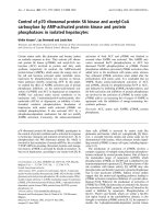
Figure 1.1 Schematic diagram illustrating the pathogenesis of UC 21. (A) The
pathogenesis of UC could initiate from the disruption in intestinal epithelial barrier.
(B) This causes the luminal microbiota to come in contact with underlying immune
cells in the lamina propria. Upon recognising the antigenic luminal microbiota,
antigen presenting cells (APCs) such as macrophages and dendritic cells become
activated and secrete pro-inflammatory cytokines (innate immunity). (C) The antigens
are presented by the APCs to the naïve T cells, which results in T cell responses
(adaptive immunity).
4
1.1.2. Dextran Sulphate Sodium (DSS)-induced Murine Model of Colitis
There are numerous animal models of colitis that have been used to mimic IBD
in humans. In this study, the DSS-induced murine model of colitis was employed.
DSS is a sulphated polymer of glucose with varying molecular weight depending on
the length of the polymer chain. In this study, the mice were administered DSS with
molecular weight of approximately 36-50 kDa, which is known to affect the distal
colon 22. Therefore, the phenotypic change of the mice administered with DSS closely
mimics the clinical features of UC in humans. The mechanism by which DSS causes
intestinal inflammation has not been precisely understood but the chemical is believed
to affect the intestinal epithelial barrier function. A study showed that DSS penetrated
and caused disruption to the intestinal mucus layer. Damage in the mucus layer
allowed the luminal bacteria to come into contact with the intestinal epithelia, which
then triggered an immune response
23
. Another study suggested that the sulphate
group in DSS form electrostatic interaction with medium- or long-chained fatty acid
in the intestines, which then enables the complex to enter the epithelial cells and
trigger pro-inflammatory response in those cells 24.
1.2. The Role of Immune Response in Intestinal Inflammation
The gastrointestinal tract contains more than 500 species of microbiota which
are constantly in contact with the luminal wall 5. In normal individuals, the intestinal
epithelia layer provides a physical barrier that separates the microbiota from the
immune cells in the lamina propria. In addition, the immune system is anergic
towards this population of commensal microorganisms. However during intestinal
inflammation, the barrier of epithelia layer is disrupted and the immune system form
an exaggerated response towards these microorganisms, which causes chronic
inflammation 25.
5
Underneath the epithelia layer of the intestines lies the lamina propria where
resident innate immune cells such as dendritic cells and macrophages reside. These
innate immune cells constantly sample the antigens from the microbiota in the
intestinal lumen and maintain immune tolerance towards them. Resident macrophages
in the lamina propria are known to produce IL-10, which prevents inflammation by
inducing and sustaining expression of Foxp3 transcription factor in regulatory T cells
(Treg)
26
. In addition, it has also been suggested that IL-10 suppresses pro-
inflammatory response to pathogen-associated molecular pattern (PAMP) in
macrophages through suppression of IL-12p40 expression
27
. Aside from producing
IL-10, these macrophages are also hyporesponsive due to reduced expression of Tolllike receptor (TLR) and other surface receptors that are required for macrophage
activation 28. Meanwhile, CD103+ dendritic cells in the intestines are also capable of
inducing Foxp3 expression in Treg cells by producing retinoic acid and TGF-# in order
to sustain the state of tolerance 29.
However when intestinal inflammation occurs, the epithelia mucosa barrier is
disrupted and the innate immune cells mount inflammatory response towards the gut
microbiota. Once the epithelia and immune cells are exposed to the gut microbiota,
they are recognised by the TLRs. This results in the recruitment of intracellular
adaptor molecules such as Myeloid differentiation primary response 88 (MyD88),
Toll-interleukin
1
receptor
domain
containing
adaptor
protein
(TIRAP),
Toll/interleukin-1 receptor-domain-containing adapter-inducing interferon-! (TRIF)
and Toll/interleukin-1 receptor-domain-containing adapter-inducing interferon-!related adaptor molecule (TRAM), which activate downstream signalling pathways.
The TRIF-dependent pathway of the TLR signalling leads to the activation of Nuclear
factor kappa-light-chain-enhancer of activated B cells (NF-"B) and Interferon
6
regulatory transcription factor 3 (IRF3) and results in the induction of type I
interferon and inflammatory cytokines. Meanwhile, the MyD88-dependent pathway
leads to the activation of NF-"B and MAPK and results in the induction of
inflammatory cytokines 30.
In the intestines, one of the early responses in intestinal inflammatory
conditions such as IBD is mounted by neutrophils. The infiltrating neutrophil
produces antimicrobial peptides and reactive oxygen intermediates which causes
tissue damage. Besides causing tissue damage, the neutrophils also recruit other white
blood cells such as macrophages by producing TNF-$, IL-1#, IL-6 and IL-8 31. The
accumulation of macrophages at the inflamed site results in further tissue damage via
secretion of TNF-$, IL-1# and proteases
32
. In addition, activated macrophages
produce more cytokines, which drives T cell polarization and differentiation into T
helper cells. In IBD patients, this inflammation does not resolve and results in
vascular leakage and further exposure of the luminal microbiota to the immune cells
lying underneath the epithelium, thus causing the chronic inflammation in the
intestine.
1.3. The Role of Intestinal Epithelium in Intestinal Inflammation
The intestinal epithelia layer provides a physical and physiological barrier
between the luminal microbiota and the immune cells in the lamina propria. The
intestinal epithelia layer is covered by a thick mucus layer consisting of mucin
glycoprotein, phospholipids and antimicrobial peptides, which are mostly secreted by
cells in the epithelia. Besides preventing direct contact between the luminal
microbiota with the epithelial cells, the mucus layer also hydrates the epithelia layer
and provides lubrication for the smooth flowing of the luminal content, as well as
7
homes secreted IgA which binds to luminal bacteria that are trapped in the mucus
layer
33
. When the mucus layer is disrupted, the epithelial cells are exposed to the
luminal microbiota. Upon recognizing the antigens on the luminal bacteria, the
epithelia cells produce pro-inflammatory cytokines and chemokines in defensive
response to the bacteria. Recognition of the bacterial antigens by epithelia cells,
likewise in innate immune cells like macrophages and dendritic cells, is also mediated
by the pattern recognition receptors (PRR) such as TLRs
34
. Secretion of pro-
inflammatory cytokines and chemokines by the intestinal epithelia results in the
recruitment of immune cells to the inflammation site and epithelial damage.
Upon injury to the epithelia layer, there are several mechanisms of which the
epithelia layer restores its barrier integrity. After an injury, the adjacent epithelia cells
migrate to the site of injury to re-establish the barrier continuity in a process called
restitution. This mechanism does not require cell proliferation and occurs within
minutes to hours after epithelial injury. During this process, the nearby epithelia cells
dedifferentiate into pseudopodia-like structure and migrate to the wound site. Upon
reaching the wound site, the cells would then reorganize their cytoskeleton and
redifferentiate to complete the wound closure
35–37
. There are numerous regulatory
factors that affect epithelia restitution in the intestine: growth factors, cytokines,
neuropeptides, polyamines, luminal peptides and probiotics
37
. These factors are
produced and secreted by not only the epithelia cells but also the underlying immune
cells, as well as contribution from the luminal content 36.
Another mechanism that accompanies epithelial restitution is cell proliferation
and differentiation. In order to replenish the decreased epithelial cell pool due to
injury, the epithelia layer needs to regenerate more cells. Nevertheless, apart from
intestinal epithelia injury, cell proliferation and differentiation by itself is a part of
8
normal epithelia physiology. The cells on the epithelia layer are constantly being shed
off into the lumen and replaced with new cells as a part of normal epithelia turnover.
In the colon, the cellular proliferation occurs at the base of the crypt. As the daughter
cells migrate to the surface of the epithelia, they differentiate into epithelia cells 38. In
the event of epithelia injury where more cells are shed off than usual, cell
proliferation and differentiation becomes a very important process to re-establish the
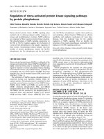
barrier integrity. A schematic diagram illustrating intestinal epithelial wound healing
process is shown in Figure 1.2.
Figure 1.2 A schematic diagram showing wound healing process in the intestines.
The lack of intestinal epithelia integrity has been implicated in intestinal
inflammation. Numerous studies using animal models of gene knockouts have shown
the importance of epithelial integrity in colitis whereby absence of genes that affect
the regulation of wound healing, proliferation, differentiation and apoptosis in
intestinal epithelia have been shown to play crucial role in the phenotypic outcome in
the animals
39–41
. In IBD patients, lack of intestinal epithelia integrity and increase in
intestinal barrier permeability has been thought to be the primary cause, if not the
exacerbating factor, of the disease 33. Since the epithelial barrier separates the luminal
microbiota from the immune cells, the breach of this barrier would expose the luminal
9
bacteria to the immune cells and cause or even exacerbate the already existing
inflammation.
The epithelial barrier is important in maintaining the intestinal homeostasis by
constantly undergoing repair through cell proliferation and restitution to replace aged
or damaged epithelia cells. Upon injury, the epithelia layer senses the invasion of
intestinal bacteria through the TLRs, which culminates in the activation of the NF-"B
and MAPK pathways. While the role NF-"B and its regulation have been
characterised in the gut
39,42
, the role of MAPK phosphatases, the major negative
regulator or MAPKs, and its regulation towards MAPKs in the gut have not been fully
understood.
1.4. Mitogen-Activated Protein Kinases (MAPKs)
The MAPK signalling pathway is an evolutionarily conserved pathway that
conveys extracellular stimuli from receptors on the cell surface to the target genes in
the cell. This pathway regulates a wide range of cellular processes including cell
proliferation,
differentiation,
motility,
survival,
apoptosis,
metabolism
and
inflammation 43–45. The activation of MAPKs occurs through phosphorylation on their
threonine and tyrosine residues in a three-tiered cascade of kinases. The MAPKs are
activated by MAPK kinase (MKKs), which in turn are activated by MAPK kinase
kinases (MKKKs)
46,47
. Once activated, MAPKs will then act on their substrate (eg.
protein kinases, cytoskeletal proteins, phospholipases or transcription factors) to exert
their effects on cellular processes 47.
There are three major groups of MAPKs in mammals: extracellular signalregulated protein kinases (ERK), c-Jun NH2-terminal kinases (JNK) and p38 proteins.
They each differ in their tripeptide motif (Thr-Glu-Tyr for ERK, Thr-Pro-Tyr for JNK
10
and Thr-Gly-Tyr for p38), which allows specific activation by their upstream MKKs
46,47
. The ERK pathway is activated from Raf serine/threonine kinases at the MKKK
level. Raf activates MKK1 and MKK2 which in turn activates ERK1 and ERK2
48
.
Meanwhile, the JNK pathway was found to be activated from several upstream
kinases such as transforming growth factor-# activated kinase 1 (TAK1), MEKK3,
Tumour Progression Locus 2 (TPL2) and Mixed Lineage Kinase 3 (MLK3).
Activated forms of these kinases in turn activates MKK4 and MKK7 which then
activates JNK
49
. In the p38 pathway, upstream kinases at the MKKK level such as
Apoptosis signal-regulating kinase 1 (ASK1), TAK1, Dual Leucine Zipper-bearing
Kinase (DLK) and MEKK4 have been identified to regulate p38 activation through
MKK3, MKK4 and MKK6
50
. Both JNK and p38 pathway share several upstream
kinases (eg. MKK4), and are known to be activated simultaneously under certain
stimuli 45. The JNK and p38 pathways are normally more responsive to environmental
stress, while the ERK pathway is preferentially activated by growth factors. The
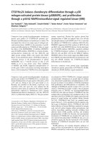
different levels of protein kinases that activate the MAPKs are illustrated in Figure
1.3.
11
Figure 1.3 A simplified schematic diagram of MAPK signalling pathway 45,48–50.
1.4.1. The Role of MAPKs in the Immune Response
TLRs are important PRRs found on both cell surface and in the cytoplasm.
These receptors recognize PAMPs present in pathogens and help to mount immune
response against invading pathogens. The binding of TLR ligands results in activation
of several signalling cascades to regulate the expression of numerous genes which
would help to fight against pathogens and to further recruit more immune cells 51. One
of the signalling cascades activated downstream of the TLRs is the MAPK pathway.
The MAPK pathway is an important signalling cascade in the mediation of
innate immune response. In macrophages, ERK is essential for the production of
cytokines including TNF-$, IL-1# and IL-10 upon TLR stimulation. JNK activation
is important for the macrophage M1 polarization which have enhanced production of
12
inflammatory cytokines such as TNF-$, IL6 and IL-12 51. Meanwhile, p38 activation
by several TLRs stimulation results in the regulation of several pro-inflammatory
factors such as TNF-$, IL-6, Cox2 and iNOS
52
. Downstream p38 targets such as
MSK1 and MSK2 also play a role in pro-inflammatory negative feedback loop in
macrophages by inducing the expression of anti-inflammatory cytokine IL-10 and
MAPK phosphatase-1 which deactivates both p38 and JNK 53.
MAPKs have also been shown to play an important role in adaptive immunity.
The Ras/ERK pathway is activated downstream of T cell receptor (TCR) engagement
by antigen presenting cells (APC) and is essential in thymocyte development, T cell
proliferation and IL-2 production 48. ERK has also been shown to be important in Th2
cells differentiation. Inhibition of ERK in naïve T cells negatively affects the early IL4 expression upon TCR stimulation and the subsequent Th2 differentiation
54
. JNK
activation can be observed soon after T cell activation, indicating its role in this
process. CD4+ T cells from JNK2-/- mice had impaired Th1 differentiation and IFN-!
production, while CD4+ T cells from JNK1-/- mice had enhanced Th2 differentiation.
This suggests that JNK2 may be important for Th1 differentiation while JNK1 is a
negative regulator for Th2 differentiation. In T cell effector function, JNK activation
was observed after restimulation of Th1 cells, but the activation was minimal in
restimulated Th2 cells 52. The activation of p38 has also been shown to be important
for T cell differentiation. p38 activation plays a role in Th1, but not Th2,
differentiation and cytokine production. This was shown where the inhibition of p38
resulted in the inhibition of IFN-! production by Th1 cells, but had no effect on IL-4
production by Th2 cells
46
. It was also shown in vitro that suppression of p38 in
induced Treg cells results in enhanced proliferation of the cells and possibly loss of
anti-inflammatory action of the cells 52.
13
1.4.2. The Role of MAPKs in Cell Proliferation and Survival
As discussed in section 1.3., cell proliferation and restitution are important in
maintaining intestinal epithelial barrier. One of the mechanisms that regulate these
processes is the activation of ERK. The ERK pathway is one of the important
signalling pathways in cell proliferation
55
. Dysregulation in the ERK pathway has
been implicated in human colorectal cancer, which shows that this pathway plays an
important role in regulating intestinal cell proliferation and homeostasis 56. There are
several extracellular growth factors that exert their effects through the ERK pathway.
One of such growth factor stimuli is the epidermal growth factor (EGF). Binding of
EGF to its receptor on the cell surface causes auto-phosphorylation of the cytoplasmic
tail of the receptor. This phosphorylation allows recruitment of an adaptor protein,
growth factor receptor-bound protein 2 (Grb-2), which further recruits son of
sevenless (SOS). This complex then activates Ras by removing guanosine
diphosphate (GDP) and allowing loading of guanosine triphosphote (GTP) to Ras.
Activated Ras ultimately lead to the activation of ERK through the Raf/MKK/ERK
kinase cascade 57. Upon phosphorylation, ERK translocates from the cytoplasm to the
nucleus to target gene expression. The activation of ERK has been shown to induce
the expression of cyclin D1, which promotes cell division
58
. In addition, the
Ras/Raf/MKK/ERK pathway also regulates numerous molecules that regulate cell
cycle progression such as cyclin-dependent kinase (CDK) inhibitors, p16Ink4a, p15Ink4b
and p21Cip1
56,59
. In cell survival, ERK was shown to be anti-apoptotic by
phosphorylating pro-apoptotic Bim and subjecting it to ubiquitination 59.
The role of JNK in cell proliferation differs between JNK1 and JNK2 in
regulating JUN, a positive regulator of cell cycle progression. While JNK1
phosphorylates and stabilizes JUN, JNK2 targets JUN for degradation 45. Meanwhile,
14
in cell survival, JNK has been shown to be pro-apoptotic. JNK is a mediator of TNF$ dependent apoptosis through the caspase-8 activation pathway. JNK is also
involved in the expression of several pro-apoptotic molecules such as TNF-$, Fas
ligand (FasL), Bcl-2-associated X protein (BAX), Bcl-2-associated death promoter
(BAD) and 14-3-3 protein 45,60.
p38 negatively regulates cell proliferation through modulation of several factors
in cell cycle progression. During G1/S and G2/M transition, p38 has been shown to
upregulate CDK inhibitors and downregulate cyclins 45. In primary fibroblasts, p38
can prevent tumorigenesis by inducing cell senescence through phosphorylating p53
and upregulation of p16, a CDK inhibitor
45,61
. As a responder to several external
stress stimuli, p38 pathway is also involved in apoptosis. In immortalized cell lines,
p38 has been shown to induce apoptosis in response to reactive oxygen species (ROS)
production driven by oncogenes. p38 can also promote cell survival by induction of
cell differentiation and production of anti-apoptotic inflammatory cytokine IL-6. In
addition, p38 can trigger cell cycle arrest and DNA repair during the G2/M
checkpoint which contributes to resistance towards apoptotic drugs in cancer cells 45.
Although the activation of MAPK is important in regulating cell proliferation
and growth, prolonged activation of MAPK can lead to disease phenotypes such as
cancer. Activating Ras mutations are most commonly found in human carcinomas,
melanomas and myeloid malignancies
62,63
. Being a downstream effector of Ras,
constantly activated ERK can result in uncontrolled cell proliferation and growth,
which leads to tumour development. Therefore in order to regulate ERK signalling, a
negative feedback mechanism is needed. Such negative regulation is provided by
MAPK phosphatases.
15