Characterization and comparison of oncogene transgenic zebrafish in three different transgenic systems
Bạn đang xem bản rút gọn của tài liệu. Xem và tải ngay bản đầy đủ của tài liệu tại đây (2.1 MB, 120 trang )
CHARACTERIZAION AND COMPARISON OF
ONCOGENE TRANSGENIC ZEBRAFISH IN THREE
DIFFERENT TRANSGENIC SYSTEMS
Liang Bing
(B.Sc.) Wuhan University
A THESIS SUBMITTED FOR THE DEGREE OF
MASTER OF SCIENCE
DEPARTMENT OF BIOLOGICAL SCIENCES
NATIONAL UNIVERSITY OF SINGAPORE
2009
Acknowledgements
Acknowledgements
I would like to express my heartfelt thanks to my supervisor, Prof. Gong Zhiyuan,
who has been extremely kind and patient to teach me throughout my project. The
opportunities that he has provided for me to study under his guidance tremendously
enriched my knowledge in term of how to do research independently and how to
present research results professionally. All his kind help and patient instruction make
it possible for me to complete this degree.
I would like to give my special thanks to Huiqing and Li Zhen, who have helped me a
lot in the bench work as well as the experiment design as a senior student in the lab.
Also I would like to thank the people who have made the laboratory an extremely
warm and friendly place filled with lots of pleasant memories and life-long bond of
friendship. They are Zhengyuan, Jianguo, Weiling, Choong Yong, Vivien, Yin Ao,
Grace, Tina, Handrien, Myintzu, Anh Tuan and Lili. Plus also to the secretaries,
administrators and technicians who have made it possible to discover so much outside
the degree.
In addition, I would like to thank my family and friends for supporting my interest in
biology research. Special thanks to my girl friend, who has been always supporting
and understanding during my project.
i
Table of Contents
Table of Contents
Acknowledgements
ⅰ
Table of Contents
ⅱ
Summary
ⅴ
List of Tables
ⅶ
List of Figures
ⅷ
Chapter 1
Introduction
1
1.1 Zebrafish as an excellent model for vertebrate developmental studies
1.2 Zebrafish as an emerging cancer model
1.2.1 The neoplasm of zebrafish
1.2.2 Cancer genes in zebrafish
1.2.3 Techniques in studies of zebrafish cancer genetics
1.2.3.1 Forward genetics
1.2.3.2 Reverse genetics
1.2.3.3 Conditional transgenic systems in zebrafish
1.2.3.3.1 Tetracycline responsive system
1.2.3.3.2 Cre-lox system
1.2.3.3.3 GAL4-UAS system
1.2.3.3.4 Heat-shock inducible system
1.2.4 Zebrafish as a model for small-molecule screening
1.2.5 Limitations of using zebrafish as a cancer model
1.3 Oncogene utilized in the transgenic lines
1.3.1 Xmrk oncogene
1.3.1.1 The Xiphophorus melanoma model
1.3.1.2 The Xmrk oncogene in the Tu locus
1.3.1.3 Oncogenic signal transduction of Xmrk
1.3.1.4 Xmrk oncogene in transgenic animal models
1.3.2 MYC oncogene
1.3.2.1 The discovery of MYC
1.3.2.2 The structure and function of MYC
1.3.2.3 Myc oncogene in transgenic animal models
1.3.2.3.1 Myc in transgenic mouse models
1.3.2.3.2 Myc in transgenic zebrafish models
1.4 Main objectives and significance of the study
2
2
2
4
7
7
10
11
12
15
17
17
18
19
20
20
20
22
24
25
28
28
29
30
30
32
34
ii
Table of Contents
Chapter 2
Materials and Methods
37
2.1 Maintenance of zebrafish and embryos
2.2 Preparation of plasmid DNAs
2.2.1 Retransformation
2.2.2 Minipreparation of plasmid DNA
2.3 RNA preparation
2.3.1 RNA extraction
2.3.2 Measurement of RNA concentration
2.3.3 Formaldehyde RNA gel electrophoresis
2.4 Reverse transcription of RNA to cDNA
2.5 Polymerase chain reaction
2.6 One-Step reverse transcription PCR
2.7 Whole mount in situ hybridization on zebrafish larva
2.7.1 Probe synthesis
2.7.2 Preparation of staged zebrafish embryos
2.7.3 Proteinase K treatment
2.7.4 Prehybridization
2.7.5 Hybridization
2.7.6 Post-hybridization washing
2.7.7 Incubation with antibody
2.7.7.1 Preparation of preabsorbed DIG-AP antibody
2.7.7.2 Incubation with preabsorbed anti-DIG-AP antibody
2.7.8 Staining
2.7.9 Mounting and photography
2.8 Quantitive real-time PCR
2.9 Histological analysis
2.9.1 Fixation
2.9.2 Dehydration and infiltration
2.9.3 Embedding
2.9.4 Sectioning
2.9.5 Staining
2.10 E2 treatment of Tg (mvtg1:mMyc-GFP) zebrafish lines
2.11 Oncogene transgenic lines used in the present project
38
38
38
40
41
41
42
42
42
43
44
46
46
46
47
48
48
48
49
49
49
50
50
51
52
52
53
53
54
54
55
55
Results and Discussion
Chapter 3
Characterization of Tg (lfabp:rtTA; Tre:mMyc-GFP)
57
transgenic lines
3.1 Test of the functionality of the Tet-on system
3.2 Tumorigenesis after Dox treatment
3.2.1 Several putative Myc downstream genes showed obvious up-regulation
iii
59
62
64
Table of Contents
3.2.2 The abnormality was diagnosed by histopathology as neoplasm
3.3 Discussion
Chapter 4
66
68
Characterization of Tg(mvtg1:mMyc-GFP) transgenic
71
lines
4.1 mvtg1 gene promoter is E2-inducible in Tg(mvtg1:mMyc-GFP)
zebrafish
4.2 Leaky expression of mMyc at early stage
4.3 The expression level of mMyc is much lower than zvtg1 in zebrafish liver
4.4 Putative mMyc downstream genes are activated with mMyc expression
4.5 Discussion
4.5.1 The mvtg1 gene promoter is inducible by E2 in transgenic zebrafish
liver
4.5.2 The expression level of mMyc under mvtg1 promoter is too low for
tumorigenesis
4.5.3 Comparison of study on Tg(lfabp:rtTA;Tre:mMyc-GFP) transgenic
lines and Tg(mvtg1::mMyc-GFP) transgenic lines
Chapter 5
72
75
77
79
81
81
82
83
Characterization of Tg (lfabp:Xmrk) transgenic lines
85
5.1 Expression of Xmrk in Tg(lfabp:Xmrk) transgenic lines
5.2 Xmrk does not affect the early stage development of Tg(lfabp:Xmrk)
line 40
5.3 Crossing of Tg(lfabp:Xmrk) line 40 with tp53M214K mutant transgenic
line did not increase abnormal incidence at early stages
5.4 Discussion
86
90
Chapter 6
93
96
Major conclusions and future directions
98
6.1 Major conclusions
6.1 Future directions
99
102
References
105
iv
Summary
Summary
In the present study, three types of oncogene transgenic zebrafish lines were
characterized: two inducible expression lines with oncogene mouse c-myc (mMyc)—
Tg(lfabp:Tre/rtTA-mMyc-GFP) and Tg(mvtg1:mMyc-GFP), and one direct expression
line with oncogene Xmrk—Tg (lfabp:Xmrk).
Tg(lfabp:Tre/rtTA-mMyc-GFP) lines utilized Tet-on inducible system, so the
expression of the transgene can be activated with Dox treatment. To investigate the
potential to develop tumors, the fish were treated with Dox (30 ug/ml &60 ug/ml)
from 21 dpf. Around 20 days post-treatment, all the treated fish developed an enlarged
belly. Fish from 60 ug/ml group had a severer phenotype than 30 ug/ml group, and
were later diagnosed as hepatocellular hyperplasia and hepatocellular adenoma by
histopathology analysis.
Tg(mvtg1:mMyc-GFP) line utilized the Medaka vitellogenin 1 (mvtg1) gene promoter,
and we found that this mvtg1 gene promoter was also E2-inducible in transgenic
zebrafish, as in Medaka. By measuring the absolute concentrations of zvtg1 and mMyc
RNAs, we found that the efficiency of the mvtg1 gene promoter is quite low, which
probably explained why Tg(mvtg1:mMyc-GFP) line failed to develop abnormal
phenotypes as the Tg(lfabp:Tre/rtTA-mMyc-GFP) lines.
v
Summary
Tg (lfabp:Xmrk) lines are direct expression lines, which means that oncogene Xmrk is
constitutively expressed in the fish liver. However, no obvious abnormality was
observed from F1 to F4 generations up to 1.5 years of age, while the survival rate at
the early stages is also normal in compared with wild type fish. The study to cross Tg
(lfabp:Xmrk) lines with p53214K mutant line is still in process, and from the
preliminary results of this study we found that the survival rate of the Xmrk (+/-)
p53(+/-) double transgenic progeny is still normal.
vi
List of Tables
List of Tables
Table No.
1
2
Title of Table
Page
Summary information on transgenic lines characterized in the 56
present study
Summary of characterization of oncogene transgenic zebrafish 101
lines in the present study.
vii
List of Figures
List of Figures
Fig. No.
1
2
3
4
5
6
7
8
9
10
11
12
13
14
15
16
Title of Figure
Adenocarcinoma of the pancreas in zebrafish and humans
Schematic representation of large-scale two-generation
genetic screens
Schematic outline of the Tet regulatory systems
Known signaling pathways of Xmrk that induce different
characteristics of the neoplastic phenotype
mMyc expresses in Tg(lfabp:rtTA;Tre:mMyc-GFP) after Dox
treatment
Abnormal phenotype observed in
Tg(lfabp:rtTA;Tre:mMyc-GFP) progeny after Dox treatment
using 60 μg/ml concentration
Examination of several putative c-myc downstream genes by
semi-quantitive RT-PCR
Histopathological analysis of abnormal transgenic progeny of
Tg(lfabp:rtTA;Tre:mMyc-GFP) lines
Tissue distribution of zvtg1 and mMyc mRNAs in male,
female and E2 treated male fish of Tg(mvtg1:mMyc-GFP)
Expression of mMyc in Tg(mvtg1:mMyc-GFP) transgenic
lines at early stage
Quantification of zvtg1 and mMyc mRNAs using real-time
RT-PCR
Expression log fold change of 4 putative downstream genes of
mMyc
Expression of Xmrk mRNA in Tg (lfabp:Xmrk) transgenic
lines
Expression level of Xmrk in Tg(lfabp:Xmrk) line 40
Survival Rate of Tg (lfabp:Xmrk) line 40
Survival rate after crossing of Tg (lfabp:Xmrk) line 40 with
tp53M214K mutant transgenic line at early stage
Page
6
9
14
26
61
63
65
67
74
76
78
80
87
89
92
95
viii
Introduction
Chapter 1
Introduction
1
Introduction
1.1 Zebrafish as an excellent model for vertebrate developmental studies
The zebrafish (Danio rerio) is a small tropical freshwater species originated from
northern India. As early as the 1970s, George Streisinger and colleagues described the
use of zebrafish as a model organism for studying embryogenesis (Detrich et al. 1999;
Streisinger et al. 1981), and it has become a popular and useful model organism for
studying vertebrate development and gene function. They may supplement higher
vertebrate models, such as rats and mice. However, as an experimental animal model
in this area, the zebrafish has many innate advantages. Firstly, when zebrafish mate,
they produce large numbers (100–200) of external, transparent embryos. Secondly, in
these embryos, cleavage divisions, gastrulation, morphogenesis and organogenesis
occur within 24 hours. Although the overall generation time of zebrafish is
comparable to that of mice, zebrafish embryos develop rapidly, progressing from eggs
to larvae in less than three days. Thirdly, the embryos are large, robust, and
transparent and develop externally to the mother, which all facilitate experimental
manipulation and observation (Dahm et al. 2006).
1.2
Zebrafish as an emerging cancer model
1.2.1
The neoplasm of zebrafish
Fish has been used to study cancer for almost a century. Since Gaylord started the
study of thyroid cancer in trout as early as the 1910s (Rettig et al. 2000). Other early
2
Introduction
studies on Xiphophorus have showed that melanomas develop when Xiphophorus
helleri (sword tails) are mated with a hybrid fish that is created via artificial
insemination from two different species, Xiphophorus helleri and Xiphophorus
maculates (Gordon et al., 1931; Walter et al., 2001). Over the years, many fish other
than trout and Xiphophorus have been used in experimental carcinogenesis studies,
including medaka, top minnow, sheepshead minnow, western mosquitofish, guppy
and zebrafish (Law et al., 2001).
The zebrafish was the first fish species used as a chemical carcinogenesis model. In
the 1960s, Stanton et al. (1965) exposed zebrafish to diethylnitrosamine, and found
that they developed hepatic neoplasms. During the following years, researchers
started to use similar approaches in other fish species and the medaka became one of
the best-characterized small fish for carcinogenesis studies (Bunton et al., 1990;
Bunton et al., 1996). In the 1980s and 1990s, the rise of zebrafish genetics gave
zebrafish the momentum as a model of chemical carcinogenesis (Hendricks et al.,
1996; Tsai et al., 1996).
A common observation, regardless of the species, is that fish have a very low
incidence of spontaneous cancers, but a high rate of tumorigenesis after carcinogen
treatment. In most cases, the neoplasms of fish are quite relevant to human cancer
biology. At the level of histopathology, fish neoplasms are strikingly similar to human
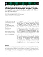
cancers (Howard et al., 2003). Figure 1 illustrates a zebrafish pancreatic cancer that
3
Introduction
developed spontaneously in the offspring of an ENU (ethylnitrosourea)-mutagenized
line. Histologically, the zebrafish tumour shows the same nuclear-atypia, haphazard
gland arrangement, desmoplastic stromal response and locally invasive behavior as
human pancreatic adenocarcinomas. Wide range of carcinomas, sarcomas and other
tumours are also observed in zebrafish.
1.2.2
Cancer genes in zebrafish
To date, little is known about fish neoplasia at molecular level. However, some
evidence indicates that certain key players in human cancers are involved in fish
tumorigenesis. For instance, p53 is a transcription factor which in humans is encoded
by the TP53 gene and it regulates the cell cycle and thus functions as a tumor
suppressor that is involved in preventing cancer. It coordinates the cell’s response to
genotoxic stress in mammalian systems, and is regulated by the inhibitor Mdm2,
which ubiquitylates p53, leading to its degradation. The regulation of apoptosis by
p53 has been examined in zebrafish embryos by using antisense morpholino
oligonucleotides to ―knockdown‖ or reduces the p53 expression in zebrafish embryos
(Langheinrich et al., 2002). When these embryos with low levels of p53 expression
were exposed to DNA-damaging stimuli, such as ultraviolet irradiation, ionizing
irradiation or the chemotherapeutic drug camptothecin, they had a reduced apoptotic
response compared with control embryos, indicating that in zebrafish p53 activates
the apoptotic response to DNA damage, as what happens in human. Furthermore,
4
Introduction
when anti-mdm2 morpholinos were injected into embryos, they underwent high levels
of apoptosis. However, this phenotype could be rescued by co-injection with anti-tp53
morpholinos, indicating that the apoptotic phenotype in the absence of Mdm2 is
mediated by p53. This result is, again, in agreement with the known regulation of p53
by Mdm2 in mammalian systems.
Cancer genes in zebrafish can also be studied by looking for orthologues of common
human oncogenes and tumour-suppressor genes in zebrafish genome. Many
orthologues have been found for most cancer genes, although few have been cloned
and verified to be functional, and much work still remains to be done in the future
study.
5
Introduction
A
B
Figure 1. Adenocarcinoma of the pancreas in zebrafish and humans. (A) human
pancreatic carcinoma (B) Zebrafish pancreatic carcinoma arose spontaneously in the
offspring of an ethylnitrosourea (ENU)-mutagenized line. In both human and
zebrafish pancreatic tissue, a mass of haphazardly arranged, irregularly shaped glands
can be detected, along with nuclear pleomorphism and increased mitotic activity. The
glands invade adjacent pancreatic tissue and induce a desmoplastic stromal reaction.
All of these features are histological hallmarks of malignancy. Normal pancreatic
tissue is visible at the top of the field (P). Insets show high-power views of neoplastic
glands. (Adapted from Howard et al., 2003)
6
Introduction
1.2.3
Techniques in studies of zebrafish cancer genetics.
To study cancer genetics in zebrafish, a powerful approach is the creation of fish with
alterations in specific cancer genes. To achieve this goal, several methods have been
developed for genetic manipulation of zebrafish, including both forward and reverse
genetic strategies.
1.2.3.1 Forward genetics
Forward genetics is also known as genetic screen, which is a procedure or test to
identify and select individuals that possess a phenotype of interest. Since unusual
alleles and phenotypes are rare, geneticists use a mutagen, such as a chemical or
radiation, to generate mutations in chromosomes. In the early 1990s, it has been
reported that ethylnitrosourea (ENU) can induce point mutations in the zebrafish
genome (Grunwald et al., 1992) and large-scale forward genetic screens have been
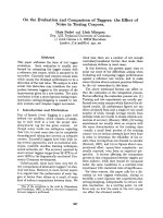
performed for developmental mutants. Approximately 2,000 genetic mutants have
been generated by these screens with specific defects that affect virtually every aspect
of embryogenesis (Fig. 2) (Driever et al., 1996; Eisen et al., 1996; E. Elizabeth Patton,
2001). Forward genetics screens for zebrafish developmental defects have already
revealed very interesting angiogenesis mutants related to cancers and subsequent
screening for mutants relative to genomic instability and cell-cycle regulation have
also been conducted. Viable mutants obtained in these screens can be further analyzed
in well-established zebrafish carcinogenesis assays to directly test if the mutation
7
Introduction
alters cancer incidence or tumour spectrum.
Insertional mutagenesis is another forward genetic method which has been quite
successful in zebrafish. In this approach, a mouse retrovirus is used as the mutagen
and at least 500 mutants with an embryonic phenotype have been identified
(Amsterdam et al., 1999; Golling et al., 2002). In these mutant zebrafish lines,
Amsterdam et al. (2004) have identified 12 lines with elevated cancer incidence,
which primarily develop malignant peripheral nerve sheath tumors (MPNSTs). It is
found that 11 of the 12 lines were each heterozygous for a mutation in a different
ribosomal protein (RP) gene, suggesting that although association of cancers with
ribosomal genes in mammals is rare, many RP genes may act as haploinsufficient
tumor suppressors in fish. A more recent study has showed that p53 is not synthesized
apparently due to insufficient ribosomal proteins in the ribosomal protein gene
mutants, thus making the fish be prone to tumors (MacInnes et al., 2008).
8
Introduction
Figure 2. Schematic representation of large-scale two-generation genetic screens.
In F2 screens, a mutagen, such as ethylnitrosourea (ENU), is used to generate
hundreds of point mutations in the male pre-meiotic germ cells (spermatogonia).
ENU-treated males are crossed to wild-type females to produce the F1 heterozygous
progeny. F1 fish are then crossed to siblings to create F2 families, half of which are
genetically heterozygous for a specific mutation (m), whereas the other half are wild
type. F2 siblings are crossed, and the resulting F3 progeny are 25% wild type (+/+),
50% heterozygous (+/m) and 25% homozygous (m/m) for a recessive mutation.
Together, the Boston and Tübingen screens, starting from about 300 ENU founder
males, involved raising more than 5,000 F2 families, analysing more than 6,000
mutagenized genomes and selecting more than 2,000 new developmental mutants for
characterization. (Adapted from Elizabeth et al., 2001)
9
Introduction
1.2.3.2 Reverse genetics
Reverse genetics is an approach to discover the function of a gene by examining the
possible phenotypes that may derive from a specific genetic sequence. Transgenic
technology has been widely used in this area. Transgenic zebrafish are created by
injecting a DNA construct into one-cell stage embryos. There are two transgenic
approaches: transient transgenic expression and stable transgenic lines. The transient
transgenic approach is to analyze gene expression and function immediately after the
introduction of the foreign gene into embryos. Although this system is rapid and
convenient, differential and mosaic gene expression from the same transgenic
construct are frequently observed among injected embryos due to mosaic segregation
of injected DNA during cleavage stage. In contrast to the transient transgenic
expression system, stable transgenic lines refer to germline transmitted transgenic
organisms. Offspring from the same transgenic founder usually present an identical
pattern of transgene expression as the transgene is already stably integrated into the
host genome. Therefore the approach of stable transgenic lines offers a large number
of transgenic individuals with the same expression pattern in repeated analyses. In a
microinjection experiment, typically, 50–75% of injected embryos express the
transgene, while only 1–10% of these undergo stable germline transmission (Long et
al., 1997; Picker et al., 2002). Given the optical clarity of zebrafish embryos, such
transgenes are frequently coupled to a fluorescent protein tag such as green
fluorescent protein (GFP), to visualize transgene expression in vivo. For example,
10
Introduction
Langenau et al. (2003) have successfully established a transgenic zebrafish model
which developed Myc-Induced T Cell Leukemia by expressing mouse c-myc under
control of the zebrafish Rag2 promoter.
In reverse genetics, there is another important technique which can help researchers to
isolate zebrafish with specific gene disruptions called targeting induced local lesions
in genomes (TILLING) (McCallum et al., 2000). TILLING is a method in molecular
biology that allows directed identification of mutations in a specific gene. Specifically,
ENU-mutagenized libraries of live fish or frozen sperm are screened for specific gene
alterations. For example, researchers have used TILLING to identify zebrafish that
carried a disruption in the rag1 gene (Wienholds et al., 2002) , which is a mediator of
V(D)J recombination in lymphocyte and the TILLING strategy was also used to
identify zebrafish with p53 mutations, so this technique could be applied to any
cancer gene (Howard et al., 2003).
1.2.3.3 Conditional transgenic systems in zebrafish.
In transgenic zebrafish, if the oncogene is constitutively expressed, the transgenic
lines could be prone to tumors or other diseases and the fish may not survive to sexual
maturity to produce the next generation, making it difficult to maintain stable
transgenic lines for further applications such as detailed characterization of tumor
formation and small molecule suppressor screens. To overcome these problems,
11
Introduction
conditional gene activation systems are desired. Nowadays transgenic technology has
been revolutionized by the development of techniques that allow temporal-spatial
control of gene deletion or expression in transgenic animals.
1.2.3.3.1
Tetracycline responsive system
The tetracycline transactivator system has been established as a reliable tool for
regulated transgene expression by pioneering work of Gossen (1992). Tetracycline
repressor (tetR) is a protein that binds specifically to tetracycline operator (tetO)
sequences within the promoter, rendering the gene transcriptionally silent. However,
tetracycline can avidly binds tetR to relieve the repression. In this way, tetracycline
resistance is controlled in a simple on/off manner by tetracycline itself (Gossen et al.,
1992). Afterwards, two modifications have been made to suit transgenic purposes.
First, tetR has been converted into a transcriptional activator by fusing it with the
activation domain of the herpes simplex virus VP16 protein, which is a virus-encoded
factor that recruits cellular transcription factors and potently activates transcription in
eukaryotic cells (Herr et al., 1998). This hybrid molecule is termed the tetracycline
transcriptional activator (tTA). The second modification is the use of a
cytomegalovirus (CMV)-derived minimal promoter, fused with tetO sequences to
control transgene expression. All these form the original so-called ―tet-off‖ system,
which means that the chimaeric promoter is inhibited by the presence of tetracycline.
Reverse tTA (rtTA) is a mutagenised version which binds tetO in the presence of
12
Introduction
doxycycline (Dox, a derivative of tetracycline) and activates transcription (Gossen et
al., 1995), which is called ―tet-on‖ system, meaning that the promoter is activated in
the presence of tetracycline or doxycycline. The major advantage of the latter system
is that gene induction occurs rapidly because the low levels of doxycycline required
for transcriptional activation can be readily achieved. In contrast, the kinetics of gene
induction by tTA is somewhat slower, since clearance of doxycycline can take days in
animals. However, the original tet-on system has a low level of basal expression
because rtTA retains some affinity for tetO sequences even in the absence of
doxycycline, which may not be acceptable in some kind of experiment (e.g.
expression of toxins). To overcome this problem, several attempts have been taken
and finally the variant RtTA2s-M2 has been generated. This has virtually no
background activity, enhanced doxycycline sensitivity and improved transcript
stability (Urlinger et al., 2000a; Urlinger et al., 2000b). Finally, substitution of the
VP16 moiety of rtTA with the transactivation domain of the mammalian transcription
factor E2F4 appears to be tolerated better by mammalian cells (Akagi et al., 2001).
Doxycycline and anhydrotetracycline, which are the analogues of the tetracycline and
have higher tTA binding affinities and lower toxicities, tend to be used in preference
to tetracycline itself (Gossen et al., 1993; Efrat S et al., 1995; A-Mohammadi et al.,
1997). For instance, Tet-on system using the cardiac myosin light chain 2 promoter in
zebrafish has been reported by Chiu-Ju Huang et al., 2005). They have compared
various transactivators in the zebrafish fibroblast cell line, including tTA, rtTA,
rtTA-M2, rtTA-S2 of prokaryotic origin, the humanized codons
13
Introduction
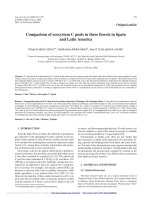
A
B
Figure 3. Schematic outline of the Tet regulatory systems. (A) The mode of action
of the Tc-controlled trans-activator (tTA). tTA, a fusion protein between the Tet
repressor of the Tn10 Tc resistance operon from E. coli and the C-terminal portion of
VP16 from herpes simplex virus, binds in the absence of the effector molecule
doxycycline (Dox) to multiple tet operator sequences (tetO) placed upstream of a
minimal promoter and activates transcription of gene x. Addition of Dox prevents tTA
from binding and thus the initiation of transcription. (B) The mechanism of action of
the reverse Tc-controlled trans-activator (rtTA). rtTA is identical to tTA with the
exception of 4 amino acid substitutions in the TetR moiety, which convey a reverse
phenotype. rtTA requires Dox for binding to tetO sequences in order to activate
transcription of gene y. (Adapted from Udo Baron et al., 2000)
14
Introduction
rtTA2S-S2 and rtTA2S-M2. All transactivators display a regulated capacity of
activating luciferase expression and rtTA-M2 and the humanized rtTA2S-S2 mutant
has the best performance in terms of increase of luciferase activity after induction.
1.2.3.3.2
Cre-lox system
The Cre-lox system is a useful genetic tool to control site specific recombination
events in genomic DNA. The system consists of Cre recombinase and loxP sites. The
Cre is originally a recombinase of the P1 bacteriophage directs recombination
between loxP (locus of X-over P1) sites. Its function is to maintain phage-encoding
plasmids as monomers. In other words, Cre is a site-specific DNA recombinase,
which can catalyze the recombination of DNA between loxP sites in a DNA molecule.
When cells that have loxP sites in their genome express Cre, a reciprocal
recombination event will occur between the loxP sites, resulting in deletion,
duplication, integration, inversion or translocation of sequences, according to the
orientation of the recombination sites and the number of molecules involved.
Specifically, for two loxP sites on the same chromosome arm, inverted loxP sites will
cause an inversion, while a direct repeat of loxP sites will lead to a deletion event. If
loxP sites are on different chromosomes, it is possible for translocation events to be
catalysed by Cre induced recombination. This system has allowed researchers to
manipulate a variety of transgenic organisms to control gene expression, delete
undesired DNA sequences and modify chromosome architecture. For instance, to test
15
Introduction
the Cre/loxP recombination system in zebrafish, our lab has generated a stable
transgenic zebrafish line by using a floxed (loxP flanked) GFP (green fluorescent
protein) gene construct under the muscle-specific mylz2 promoter, and the new
transgenic line expresses GFP reporter faithfully in fast skeletal muscles to the same
intensity like our previous non-floxed GFP transgenic line under the same promoter.
After injection of in vitro synthesized Cre RNA into embryos of floxed GFP
transgenic zebrafish, a dramatic reduction of GFP expression has been observed,
indicating the excision of floxed GFP transgene, as confirmed by the following PCR
and sequencing information. Thus we have demonstrated that the Cre/loxP system can
function efficiently and accurately in the zebrafish system.
Another example of the application of Cre/LoxP system in zebrafish is from Dr.
Thomas Look’s group. Previously, they have created a stable transgenic
rag2:GFP-mMyc zebrafish line that develops GFP-labeled T cell acute lymphoblastic
leukemia (T-ALL). However, this line can only be maintained by in vitro fertilization
because the consistent myc expression makes T-ALL develop very rapidly. So they
created a conditional transgene in which the GFP-mMyc oncogene is preceded by a
floxed dsRED2 gene and have generated stable rag2:loxP-dsRED2-loxP-GFP-mMyc
transgenic zebrafish lines, which have red fluorescent thymocytes and do not develop
leukemia. By injecting Cre RNA into one-cell-stage embryos of the transgenic
progeny of these lines, T-ALL can be induced to develop (Langenau et al., 2005). This
work also demonstrated the invaluable utility of the Cre/lox system in the zebrafish.
16