Development and validation of a generic assay to detect compounds acting via an aggregation based mechanism
Bạn đang xem bản rút gọn của tài liệu. Xem và tải ngay bản đầy đủ của tài liệu tại đây (1016.65 KB, 86 trang )
DEVELOPMENT AND VALIDATION OF A GENERIC ASSAY TO DETECT
COMPOUNDS ACTING VIA AN AGGREGATION-BASED MECHANISM
SUKRITI MALPANI
(B. Sc. (Hons.) Biological Sciences)
National University of Singapore
A THESIS SUBMITTED FOR THE DEGREE OF
MASTER OF SCIENCE IN INFECTIOUS DISEASES,
VACCINOLOGY AND DRUG DISCOVERY
DEPARTMENT OF MICROBIOLOGY
YONG LOO LIN SCHOOL OF MEDICINE,
NATIONAL UNIVERSITY OF SINGAPORE
AND
BIOZENTRUM, UNIVERSITÄT BASEL
2011
Acknowledgements
I would like to thank my supervisor, David Beer, for the opportunity to carry
out my project at the Screening Unit of the Novartis Institute for Tropical Diseases
His guidance, support, and encouragement made my time at NITD an invaluable
learning experience.
I am indebted to Pornwaratt Niyomrattanakit for her mentorship, forbearance,
and untiring enthusiasm in guiding me. Special thanks to her for critically reviewing
this manuscript. I am very grateful to Christophe Bodenreider and Wan Kah Fei for
their help and support during the course of this project. I would also like to thank the
other members of the Screening Unit, Jessie Lim, Balbir Chaal, Amelia Yap and
Nurdiana Abas, for creating a wonderful working environment.
I would like to thank all my classmates from the Joint Masters programme for
making this entire experience so memorable. I am very thankful to all my friends for
being a constant source of support. I am eternally grateful to my parents and brothers
for being my voice of reason. Their guidance and encouragement at every step has
been a great source of inspiration and motivation.
i
Table of Contents
Acknowledgements .....................................................................................................................i
Summary ...................................................................................................................................iv
List of Tables.............................................................................................................................vi
List of Figures ......................................................................................................................... vii
List of Abbreviations.................................................................................................................ix
1. Introduction ............................................................................................................................1
1.1 Introduction to High Throughput Screening ....................................................................1
1.2 Steps involved in setting up a high throughput screen .....................................................3
1.2.1 Assessment of assay quality ......................................................................................3
1.2.2 Primary screen ...........................................................................................................3
1.3 Hit to lead phase ...............................................................................................................4
1.3.1 Selectivity ..................................................................................................................5
1.3.2 Evaluation of potential lead candidates .....................................................................5
1.4 Sources of false positives in high throughput screening ..................................................6
1.4.1 Interference in assay readout .....................................................................................6
1.4.2 Inhibition of detection system ...................................................................................8
1.4.3 Aggregation-based enzymatic inhibition in biochemical assays ...............................9
1.5 Aim of the project ..........................................................................................................18
2. Materials and Methods .........................................................................................................20
2.1 β-Lactamase primary screen and secondary assays .......................................................20
2.1.1 Primary screen .........................................................................................................20
2.1.2 Secondary assays using chromogenic substrate ......................................................20
2.1.3 Secondary assays with fluorometric readout ...........................................................21
2.1.4 Data analysis ...........................................................................................................21
2.1.5 Dynamic light scattering analysis............................................................................22
2.2 DENV RdRp assay principle, hit selection and follow-up assays..................................22
2.2.1 Assay principle, compound screening and hit selection ..........................................22
2.2.2 Testing inhibition potency of hits in different detergents .......................................24
2.2.3 Testing inhibition potency of hits at varying enzyme concentrations .....................25
2.2.4 Effect of Triton X-100 on kinetic constants of DENV RdRp .................................25
2.3 Selection of compounds from PanK hit list....................................................................26
2.4 Measurement of change in meniscus..............................................................................27
ii
3. Results ..................................................................................................................................29
3.1 β-Lactamase primary screen and follow-up assays ........................................................29
3.1.1 Hit Selection and re-confirmation ...........................................................................29
3.1.2 Detergent sensitivity of inhibition potency of β-Lactamase hits .............................30
3.1.3 Enzyme-concentration sensitivity of inhibition potency of β-Lactamase hits ........34
3.1.4 Dynamic light scattering analysis of β-Lactamase hits ...........................................35
3.2 Follow-up of DENV RdRp pilot screen .........................................................................39
3.2.1 Detergent sensitivity of inhibition potency of DENV RdRp hits ............................39
3.2.2 Enzyme-concentration sensitivity of inhibition potency of DENV RdRp
hits ....................................................................................................................................42
3.2.3 Effect of Triton X-100 concentration on enzyme kinetics ......................................43
3.3 Investigation of inhibition of unrelated enzymes or a model enzyme as means
of identification of aggregation-based inhibition .................................................................44
3.4 Development and validation of change in meniscus shape as a generic assay for
detection of aggregate formation..........................................................................................48
4. Discussion ............................................................................................................................52
4.1 Choice of β-Lactamase as model enzyme ......................................................................52
4.2 Design and implementation of compound library screening for inhibitors of βLactamase .............................................................................................................................54
4.2.1 Prediction of aggregation-based inhibition by β-Lactamase hits based on
sensitivity to detergent .....................................................................................................55
4.2.2 Prediction of aggregation-based inhibition by β-Lactamase hits based on
sensitivity to enzyme concentration .................................................................................56
4.2.3 Prediction of aggregation-based inhibition on the basis of particle size
measurements of β-Lactamase hits using Dynamic Light Scattering ..............................57
4.3 Determination of specificity of DENV RdRp hits .........................................................60
4.3.1 Assessment of classification of specificity of DENV RdRp hits based on
detergent sensitivity of inhibition potency .......................................................................60
4.3.2 Assessment of classification of specificity of DENV RdRp hits based on
sensitivity of inhibition potency to enzyme concentration ...............................................62
4.4 Steepness of dose-response curves as an indicator of aggregate-based inhibition .........64
4.5 Target specificity of aggregate-forming inhibitors.........................................................65
4.6 Viability of change in meniscus assay as a generic assay for detection of
aggregation ...........................................................................................................................66
4.7 Concluding remarks .......................................................................................................69
5. References ............................................................................................................................71
iii
Summary
High throughput screening (HTS) has emerged as a reliable
component of the drug discovery process. It is now recognized that a large number of
compounds inhibit their target enzyme via an aggregation-based binding mechanism
leading to false positive results in HTS assays. Aggregate-forming compounds act
non-competitively; show little relation between structure and activity; have steep
dose-response curves and are reported to inhibit multiple unrelated enzymes
(McGovern et al. 2002; McGovern et al. 2003; Feng et al. 2007). Removal of these
compounds from screening hit lists is desirable as they are not good starting points to
initiate medicinal chemistry programs. There are many techniques currently in use to
identify aggregation-based inhibition such as dynamic light scattering (DLS), testing
sensitivity of inhibition potency to detergent or enzyme concentration, and
measurement of meniscus curvature changes in high density multi-well plates
associated with colloidal changes in solution.
To evaluate the feasibility of large-scale identification of aggregate-based
inhibition, hits from three enzyme screens (β-Lactamase, DENV RdRp and
Pantothenate kinase) were analysed for signs of aggregate-based inhibitions using
various techniques. For a majority of non-specific hits, characteristic features of
aggregate-based inhibition such as steep dose-response curves, presence of aggregate
particles in solution and inhibition of unrelated enzymatic targets were not found to
be associated with detergent or enzyme-concentration sensitive inhibition. Particle
size measurements by DLS were inconsistent for many compounds. Steepness of
dose response curves depended on buffer composition and assay format employed.
iv
Aggregate-based inhibitors displayed target specificity towards their respective target
enzymes rather than ‘promiscuous’ inhibition of multiple targets.
Different detergents often yielded conflicting results and required derivation
of new cut-offs for different enzyme systems or different assay conditions. For
example, while the sensitivity of inhibition potency to detergent was not dependent
on the nature of the detergent for hits of β-Lactamase, this was not the case for hits of
the DENV RdRp enzyme. The inhibition potencies of the hits of DENV RdRp were
found to have different degrees of sensitivity to different detergents. Furthermore, the
results of the enzyme-concentration sensitivity tests for the DENV RdRp hits did not
seem to correlate with the detergent-sensitivity results. It was observed that the
interaction between the enzyme and its substrate possibly confounded the effect of
varying the enzyme concentration.
The measurement of changes in meniscus curvature, as a means of
identification of aggregate-forming small molecule compounds, has been used for the
first time in an actual HTS campaign, as reported in this study. The meniscus
measurements of hits from all screens correlated well with detection of aggregationbased inhibition based on measurement of changes in inhibition potency. A
classification scheme is presented that can be used to rapidly characterize the hits
from high throughput screens and eliminate compounds with a non-specific
mechanism of inhibition. In summary, the meniscus-based aggregation assay is
simple, cost-effective, and a reliable method to identify and eliminate compounds that
inhibit a specific target enzyme via an aggregation-based mechanism.
v
List of Tables
Table 1: Differences in allowed parameters between laboratory “bench top” and
HTS assays ................................................................................................................................2
Table 2: IC50 values of hits from β-Lactamase screen in the absence and presence
of detergent. ............................................................................................................................31
Table 3: IC50 values of hits from β-Lactamase screen in the fluorometric assay
format. ....................................................................................................................................34
Table 4: IC50 values of DENV RdRp hits in the presence of different detergents
in the assay buffer. .................................................................................................................40
Table 5: Changes in IC50 values of DENV RdRp hits at higher concentrations of
detergent..................................................................................................................................41
Table 6: Enzyme-concentration dependent changes in IC50 values of DENV
RdRp hits. ...............................................................................................................................42
Table 7: The apparent Km and Vmax of the 3’UTR-U30 RNA substrate at different
Triton X-100 concentrations. ................................................................................................44
vi
List of Figures
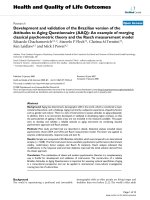
Figure 1: Historical comparison of number of leads found by HTS study
participants. ..............................................................................................................................1
Figure 2: Illustration of steps involved in the initial drug discovery process .....................4
Figure 3: Aggregating compounds visualized by transmission electron
microscopy ..............................................................................................................................10
Figure 4: (A) Model of aggregate and enzyme binding. (B) Mechanism of action
of small-molecule aggregators. ..............................................................................................13
Figure 5: Z-factor trend across assay plates used in the primary β-Lactamase
screen .......................................................................................................................................29
Figure 6: Histogram of normalized inhibition data of compound library tested
against β-Lactamase. ..............................................................................................................30
Figure 7: Dose-response curves of A) BZBTH2B, a reference inhibitor of E.
cloacae β-Lactamase and B) Tetraiodophenolphthalein.....................................................32
Figure 8: Dose-response curves showing inhibition of β-Lactamase by A) BLAC11 and B) BLAC-13. ...............................................................................................................33
Figure 9: DLS correlogram of BLAC-1 at A) 20µM and B) 66µM as measured
with a Malvern Zetasizer Nano ZS dynamic light scattering instrument in assay
buffer. ......................................................................................................................................36
Figure 10: DLS correlogram of BLAC-2 at A) 20µM and B) 66µM as measured
with a Malvern Zetasizer Nano ZS dynamic light scattering instrument in assay
buffer. ......................................................................................................................................38
Figure 11: Effect of Triton X-100 on apparent Km and Vmax values of DENV
RdRp........................................................................................................................................43
vii
Figure 12: Comparison of primary screens of various enzymes. .......................................45
Figure 13: Distribution of DENV RdRp hits. ......................................................................46
Figure 14: Distribution of Pantothenate Kinase hits. .........................................................47
Figure 15: Relative fluorescence of β-Lactamase hits measured as the ratio of
top-read fluorescence intensity in assay buffer to control buffer. .....................................49
Figure 16: Relative fluorescence of DENV RdRp hits measured as the ratio of
top-read fluorescence intensity in assay buffer to control buffer. .....................................50
Figure 17: Relative fluorescence of MTB PanK hits measured as the ratio of topread fluorescence intensity in assay buffer to control buffer. ............................................51
viii
List of Abbreviations
Acetyl CoA
Acetyl coenzyme A
BBT
2′-[2-benzothiazoyl]-6′-hydroxybenzothiazole
BCS
Biopharmaceutical Classification System
BSA
Bovine Serum Albumin
BZBTH2B
Benzo(b)thiophene-2-boronic acid
CIP
Calf Intestinal Alkaline Phosphatase
CMC
Critical Micelle Concentration
DENV
Dengue virus
DLS
Dynamic Light Scattering
EC50
Half maximal Effective Concentration
HTS
High Throughput Screening
IC50
50% Inhibitory Concentration
LC-MS
Liquid chromatography-mass spectrometry
NMR
Nuclear Magnetic Resonance
NS5
Non-structural protein 5
PanK
Pantothenate Kinase
RdRp
RNA-dependent RNA polymerase
RNA
Ribonucleic acid
SAR
Structure Activity Relationship
TEM
Transmission Electron Microscopy
UTR
Untranslated region
ix
1. Introduction
1.1 Introduction to High Throughput Screening
High throughput screening (HTS) is a widely used approach to discover
novel chemical entities for drug design. In concert with the generation of large
libraries of chemically diverse small molecules, the advancements in automation
technologies have lead to growth of HTS programs in academia and industry (Inglese
et al. 2007; Shelat and Guy 2007). A recent worldwide study involving 58 HTS
laboratories has reported increasing numbers of leads identified by HTS over the
years (Fig. 1) and documented 104 clinical candidates and four marketed products
that have emerged from these leads (Fox et al. 2006).
Figure 1: Historical comparison of number of leads found by HTS study
participants. Reprinted with permission from “High-throughput screening:
update on practices and success” by Fox et al in J Biomol Screen, 2006
11(7):864-869. Copyright 2006 by Sage Publications.
HTS methodology enables expeditious screening of sizeable chemical
libraries to identify leads that act on a biological target of interest, e.g., as inhibitors
of target enzymes, as competitors for binding of a natural ligand to its receptor, as
agonists or antagonists of receptor-mediated intracellular processes, and so forth.
HTS assays involve a variety of strategies such as the measurement of catalytic
activity from a purified enzyme (Zhang et al. 1999), a reconstituted complex of a
1
signalling pathway (McDonald et al. 1999), a cellular extract (Verma et al. 2004), or
measurement of phenotypic changes (Hodder et al. 2004) in intact cells. Configuring
assays to function within the constraints imposed by high-throughput settings
differentiates an HTS assay from traditional laboratory assays, as outlined in Table 1.
Table 1: Differences in allowed parameters between laboratory “bench top” and
HTS assays. Reprinted with permission from “High-throughput screening assays
for the identification of chemical probes” by Inglese et al. in Nat Chem Biol
2007;3(8):466-479. Copyright 2007 by Macmillan Publishers Ltd.
Parameter
Bench top
HTS
Protocol
May be complex with
Few (5–10) steps, simple
numerous steps,
operations, addition only
aspirations, washes
preferred
Assay volume
0.1 ml to 1 ml
<1 µl to 100 µl
Reagents
Quantity often limited,
batch variation acceptable,
may be unstable
Sufficient quantity, single
batch, must be stable over
prolonged period
Reagent handling
Manual
Robotic
Variables
Many-for example, time,
substrate/ligand
concentration, compound,
cell type
Compound, compound
concentration
Assay container
Varied-tube, slide,
microtiter plate, Petri dish,
cuvette, animal
Microtiter plate
Time of measurement
Milliseconds to months.
Measurements as endpoint,
multiple time points, or
continuous
Minutes to hours.
Measurements typically
endpoint, but also pre-read
and kinetic
Output formats
Plate reader, radioactivity,
size separation, object
enumeration, images
interpreted by human
visual inspection
Plate reader-mostly
fluorescence,
luminescence and
absorbance
Reporting format
“Representative” data;
statistical analysis of
manually curated dataset
Automated analysis of all
data using statistical
criteria
2
1.2 Steps involved in setting up a high throughput screen
1.2.1 Assessment of assay quality
Large screens involving hundreds of thousands of compounds are expensive
in time and resources. Thus before starting a large screen, it is important to assess the
suitability or quality of the assay to be used in screening and ascertain if the assay
would be useful in a high-throughput setting. A statistical term, called the Z or Z’factor (Zhang et al. 1999), is commonly used to evaluate the quality of assays.
The Z or Z’-factor is defined in terms of four parameters: the means and
standard deviations of both the positive (p) and negative (n) controls (µp, σp, and µn,
σn). A Z-factor of 1 is considered ideal. This value is approached when there is a
huge dynamic range (large difference between the signal means of the positive and
negative controls) with small standard deviations. Z-factors can never be greater than
1. A value between 0.5 and 1 is aspired for in HTS settings. A Z-factor between 0
and 0.5 is considered sub-optimal. If an assay has a Z-factor that is less than 0, it
implies that the signals from the positive and negative controls could overlap, making
the assay essentially useless for screening purposes.
1.2.2 Primary screen
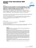
The goal of any HTS campaign is to identify active compounds (“hits”) and
choose the best candidates for lead optimization. This is achieved through a multitude
of steps (Fig. 2). After an assay has been developed and validated, entire chemical
libraries (hundreds of thousands to millions of compounds) are screened against the
target of interest. Primary screening usually involves single measurements of the
activity of each small-molecule compound. These single data points of unknown
3
samples are compared to positive and negative control samples to determine which
compounds are active against the biological target. A robust assay with a Z-factor
between 0.5 and 1 is conducive to single point testing. An assay with a sub-optimal
Z-factor between 0 and 0.5 would require multiple data points for each compound to
ensure reproducibility of the assay readout.
Figure 2: Illustration of steps involved in the initial drug discovery process
1.3 Hit to lead phase
The hit rate from a primary screen can vary between 0.1 and 1% (Eisenthal
and Danson 2002) depending on the target, the assay format and the cut-off used to
decide if a compound is considered ‘active’ or not. After selecting hits from
compounds tested in the primary screen, the next step is to confirm the activity of
these hits. Establishing a dose-response relationship is an important step in hit
confirmation. It routinely involves a secondary screen in which a range of compound
concentrations usually prepared by serial dilution are tested in an assay to assess the
concentration or dose dependence of the assay's readout. Typically, this dose4
response is expressed as the 50% inhibitory concentration (IC50) in enzyme-, protein-,
antibody-based assays; or as the half maximal effective concentration (EC50) in cellbased experiments. Compounds that display potency in a dose-dependent manner are
chosen for further analysis.
1.3.1 Selectivity
Lead candidates should ideally interfere with only the chosen target, and not
other, related targets. Selectivity toward a drug target decreases the risk of off-target
toxicity that might occur in the clinical trial stage. Screens for selectivity usually
include drug targets of the same protein or receptor family, for example, panels of G
protein-coupled receptors (Swanson and Beasley 2010) or kinases (Fabian et al. 2005;
Goldstein et al. 2008; Karaman et al. 2008). In cases where selectivity between
subtypes is important, screens might include a panel of homologous enzymes,
different protein complexes, or heterooligomers. Selectivity screens enable profiling
of the action of a confirmed hit on a defined spectrum of biological target classes.
Ideally, only those compounds which are highly selective towards the target of
interest will progress to the next stage.
1.3.2 Evaluation of potential lead candidates
It has been studied that more often than not, marketed drugs are similar to the
leads from which they originate (Proudfoot 2002). Therefore it is of utmost
importance to choose the best hits to promote to lead status. The most desirable
binding characteristics a ‘lead’ like compound should have are: non-covalent, high
affinity ligand binding; reversible, competitive binding; and tractability in structure–
activity relationship (SAR) of a series of structural analogues of the binder (Rishton
2003). Furthermore, it has been well established that potency alone is a false predictor
of ‘lead’ likeness (Wunberg et al. 2006) and that an ideal lead molecule must exhibit
a balance of potency, selectivity, and favourable physicochemical properties.
5
Therefore, merely re-confirming target inhibition is an inadequate measure of
the quality of a hit as it doesn’t necessarily ensure that the compound satisfies the
required criteria. Many compounds can appear to possess ‘lead’ like characteristics in
a HTS assay. However, false positives can result from multiple mechanisms,
including: non-specific hydrophobic binding, poor solubility (protein or substrate
precipitation), reactive functional groups, low purity, assay interference, aggregationbased enzyme inhibition and experimental errors (Keseru and Makara 2006). Some
concerns, such as false positivity due to reactive functional groups, can be addressed
by triaging of hit lists by medicinal chemists and elimination of compounds with
undesirable chemical structures (Rishton 1997).
Other concerns such as assay interference require more intensive probing.
Therefore hits are subjected to a battery of follow-up assays or counter screens to
identify those that don’t exhibit the intended biological interaction or falsely appear
active due to confounding factors. The number and stringency of counter screens can
vary widely and depend on the drug target. The next section provides an overview of
some of the ways a compound can appear active in a biochemical assay without
possessing any biological activity and strategies to identify these false positives.
1.4 Sources of false positives in high throughput screening
1.4.1 Interference in assay readout
Current HTS technologies are largely based on sensitive light based detection
methods, such as fluorescence or luminescence, to quantify the effect of a compound
on a target enzyme, receptor or signalling pathway (Inglese et al. 2007). These assay
types are preferred because of their high sensitivity, flexibility across multiple
homogeneous formats, ease of miniaturization, and applicability across a wide range
of targets. However, they are highly sensitive to spectral artifacts (Shapiro et al.
6
2009). For instance, false negatives can occur due to light scattering, coloured, or
fluorescent compounds that contribute to the net fluorescence signal. Small-molecule
compounds are able to interfere with the fluorometric readout in many cases. The
most straightforward interference results from spectral overlap between screening
compounds and the assay system in optical and fluorescence assay formats (Gribbon
and Sewing 2003). Compounds may falsely be identified as inhibitors if they absorb
light at the detection wavelength of the fluorogenic substrate. In such cases, the net
fluorescence signal measured in the assay will be attenuated by the compound to be
tested. As a result, a reduction of the fluorescence signal is detected even in the
absence of any interaction of the compound with the enzyme (Liu et al. 1999; Birdsall
et al. 1983).
A recent study profiling the fluorescence spectral properties (Simeonov et al.
2008) of about 70,000 compounds (PubChem Assay IDs – 587-594,709) found that
2–5% of the compounds in the library fluoresced in the blue spectral region (~350500 nm) and that for several fluorescence-based assays involving excitation in the
blue spectral region, up to 50% of the hits identified in the screen were actually
fluorescently active. The study further reported that when excited at red-shifted
wavelengths (~600 nm); only 0.004–0.01% of the library fluoresced, indicating that
use of red-shifted fluorophores is one way to reduce this mode of generation of false
positives. Other methods to counter spectral interference are: inclusion of a pre-read
after compound addition but prior to fluorophore addition to the reaction; inclusion of
a time delay after excitation of fluorophore (time-resolved); use of a ratiometric
fluorescence output; and use of an alternative assay to confirm the activity (Thorne et
al. 2010).
7
1.4.2 Inhibition of detection system
Assay set-ups that employ enzyme-coupling systems are another example of
a complex system that may suffer from detection interference. Many enzymes form
reaction products that are not amenable to direct detection in an in vitro biochemical
assay. To obtain a convenient spectral readout, the target enzyme’s activity may be
monitored by coupling its product to the reaction of an additional enzyme or auxiliary
enzymes. The coupling reaction utilizes the target enzyme reaction product to
produce a colorimetric (e.g., lactate dehydrogenase-coupled NADPH oxidation to
detect pyruvate formation) or fluorescent (e.g., horseradish peroxidase-coupled
fluorescent dye oxidation to detect H2O2 formation) or luminescent (e.g., luciferasecoupled detection of ATP production by kinases) signal. However, the coupled
enzyme itself may be susceptible to inhibition by small molecules. For example, a
profiling effort of a 70,000 compound library (PubChem Assay ID - 411) determined
that at least 3% of the library inhibited firefly luciferase activity in a concentration
dependent manner (Auld et al. 2008) demonstrating that HTS hit lists may contain a
large number of compounds that inhibit the coupled enzyme rather than the target
enzyme.
Direct assays can be carried out to test if apparent compound activity is due
to inhibition of the coupling enzyme. Inhibitors of the coupling system can also be
eliminated by counter screening hits using the same coupling system, but with a
different target enzyme that produces the same reaction product as the original target
enzyme (Seethala and Zhang 2009). If the other enzyme is related to the original
target enzyme or from the same family, selectivity considerations can be addressed at
the same time. Any compound that is positive in this counter screen may then be
eliminated from consideration regardless of whether it inhibits the coupling enzyme
or the undesired counter screening enzyme. Another method to distinguish between
assay format-dependent inhibition and target-specific inhibition is to re-test the
8
activity of hits in an orthogonal assay, i.e. an assay that has a different readout
compared to the format used in the original screening methodology (e.g., use of
fluorescence readout as opposed to absorbance).
1.4.3 Aggregation-based enzymatic inhibition in biochemical assays
Compound aggregation, through self association of organic molecules in
aqueous media, was recently discovered to be one of the main causes for false
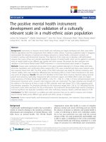
positives in HTS (McGovern et al. 2002). The study by Brian Shoichet’s group
reported that above a certain concentration some small-molecule compounds selfassociate to form aggregate particles. These particles, at 30–400 nm in size, strongly
scattered light detectable by dynamic light scattering and could be visualized by
transmission electron microscopy (Fig. 3).
9
Figure 3: Aggregating compounds visualized by transmission electron
microscopy (McGovern et al. 2002). A to C- 100 µM tetraiodophenolphthalein in
20 mM Tris; D- 50 µM Congo Red in 20 mM Tris; E- 625 µM ANS in 20 mM
Tris. Bar = 100 nm. ANS – negative control. Reprinted with permission from “A
common mechanism underlying promiscuous inhibitors from virtual and highthroughput screening” by McGovern et al. in J Med Chem 2002;45(8):17121722. Copyright 2002 by American Chemical Society.
These ‘aggregators’ that were initially identified as inhibitors of enzyme
targets such as dihydrofolate reductase, thymidylate synthase, insulin receptor,
tyrosine kinases, etc; were also found to inhibit several unrelated model enzymes
such as β-Lactamase, β-Galactosidase and chymotrypsin. Decreased inhibition in the
presence of bovine serum albumin suggested a non-specific mechanism of action and
implied that inhibition by these molecules could be attenuated in the presence of
excess protein. The compounds also showed sensitivity to the molar ratio of inhibitor
to enzyme. Increasing the concentration of the model enzymes by 10-fold
10
significantly decreased the inhibition potency (increased the IC50) of these
‘aggregators’ but not of classical, well-behaved inhibitors. To investigate if an
aggregate-based inhibition model could explain the lack of specificity of many kinase
inhibitors; 15 widely used known kinase inhibitors were analyzed for traits of nonspecific behaviour (McGovern and Shoichet 2003). It was found that more than half
of the kinase inhibitors also inhibited unrelated model enzymes, displayed sensitivity
to enzyme concentration and formed aggregates of 100-1000 nm diameter as
observed by dynamic light scattering. Due to their propensity to inhibit a panel of
unrelated enzymes, inhibitors that act via an aggregation-based inhibition are often
called ‘promiscuous’ inhibitors.
On the basis of the pilot studies, it was proposed that aggregate-forming
compounds may be common in pharmaceutical screening libraries; and that such nonspecific inhibitors could artificially inflate hit rates in screening for new drug leads.
Since these compounds act non-competitively, show little relation between structure
and activity (flat SAR), and have poor specificity, their elimination from hit lists
could potentially save a great deal of effort that would otherwise be spent in trying to
optimize their apparent activity (Borchardt et al. 2004). Therefore, Shoichet et al.
have studied these aggregate-forming inhibitors in great detail and provided a better
understanding of how they work; how frequently they occur in screening libraries;
and techniques that can be used to detect aggregate-based inhibition; as described
below in this section.
In an effort to understand the mechanism of aggregation-based inhibition,
Shoichet’s group studied the interaction of aggregate-forming inhibitors with model
proteins like β-Lactamase. By using centrifugation and gel electrophoresis-based
approaches, it was found that inhibition occurred via the direct binding of enzyme to
aggregate (McGovern et al. 2003). β-Lactamase mutants with increased or decreased
11
thermodynamic stability relative to wild-type enzyme were equally inhibited by
aggregate-forming compounds, suggesting that denaturation by unfolding was not the
primary mechanism of action of aggregate-forming inhibitors. However, visualization
by electron microscopy revealed that enzyme did associate with the surface of
aggregated
molecules.
Interestingly,
β-Lactamase
inhibition
by
compound
aggregation was found to be reversible by non-ionic detergents such as Triton X-100
(McGovern et al. 2003; Ryan et al. 2003). Since the enzyme was thought to be
sequestered by the aggregated compounds, it was inferred that the presence of
detergents either prevented formation of aggregates or interfered in the binding of
enzymes by aggregated compounds.
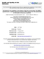
Recently, the stoichiometry of binding of enzyme to aggregates was
elucidated to be as high as 10,000 enzyme molecules per aggregate particle (Coan
and Shoichet 2008). Given the size of the aggregates and the stoichiometry of
binding, the aggregation model suggests that all sequestered enzyme can be
accommodated on the surface on the aggregate (Fig. 4). This deviation from the
classical 1:1 enzyme to inhibitor stoichiometry also explains another phenomenon
generally associated with aggregate forming inhibitors, namely steep dose-response
curves (Shoichet 2006;Feng et al. 2007). In the case of a classical, single-site
inhibitor, inhibition usually increases from 10% to 90% over a large (81-fold)
concentration range, whereas for compounds displaying steep dose-response curves
the same increase in inhibition is observed within a 10-fold range of compound
concentration. Since aggregate-forming inhibitors are known to form aggregates only
above a certain concentration, usually in the micromolar range (Coan and Shoichet
2008), many aggregate-forming compounds are found to have steep dose-response
curves with high Hill coefficients.
12
Shoichet and co-workers recently suggested that partial unfolding of the
protein occurs upon aggregate binding (Coan et al. 2009). They examined changes in
solvent accessibility of the β-Lactamase enzyme upon binding to an aggregateforming inhibitor using hydrogen-deuterium mass spectrometry and noted that
binding to aggregate particles increased deuterium exchange by the enzyme. This
global increase in proton accessibility upon aggregate binding suggested a model
consistent with partial denaturation of the protein (Fig. 4). This mechanism was
confirmed by the observation that enzyme-aggregate complexes were more
susceptible
to
tryptic
proteolysis
compared
to
free
enzyme
molecules.
Figure 4: (A) Model of aggregate and enzyme binding. Reprinted with
permission from “Stoichiometry and physical chemistry of promiscuous
aggregate-based inhibitors” by Coan and Shoichet in J Am Chem Soc
2008;130(29):9606-9612. Copyright 2008 by American Chemical Society. (B)
Mechanism of action of small-molecule aggregators – binding to the aggregate
promotes a partial unfolding event. Reprinted with permission from
“Promiscuous aggregate-based inhibitors promote enzyme unfolding” by Coan
et al. in J Med Chem 2009;52(7):2067-2075. Copyright 2009 by American
Chemical Society.
Subsequent to the initial studies on aggregate-forming inhibitors of βLactamase, aggregate-forming false positives have been discovered among inhibitors
13
of
kinesin
motor
proteins
(Reddie
et
al.
2006),
phospho-
mannomutase/phosphoglucomutase (Liu et al. 2004), and reverse transcriptase
(Frenkel et al. 2005); establishing the incidence of this spurious mode of inhibition
among inhibitors of various enzymes.
In an effort to estimate the prevalence of detergent-sensitive inhibition for a
typical HTS involving a biochemical assay, investigators have tested various smallmolecule libraries for enzyme inhibition sensitive to Triton X-100 using β-Lactamase
as a model enzyme. In a 96-well format assay, it was found that 19% of the 1030
‘drug-like’ compounds tested demonstrated detergent-dependent inhibition when
screened against β-Lactamase at 30 µM (Feng et al. 2005). For a library of ~ 70,000
compounds (PubChem Assay Ids- 584, 585), screened in a 1536-well assay format,
95% of the actives identified in the screen against β-Lactamase were Triton X-100
sensitive (Feng et al. 2007; Babaoglu et al. 2008). A screen of 200,000 compounds
against the cysteine protease cruzain (PubChem Assay ID- 2249) revealed that
approximately 1.9% of the library or 90% of the actives were detergent-sensitive
inhibitors (Jadhav et al. 2010), indicating that the prevalence of this type of assay
interference is neither library-specific nor limited to a particular type of enzyme, as
cruzain and β-Lactamase are structurally and functionally different. Another study on
cruzain inhibitors reported divergent modes of inhibition (competitive or aggregationbased) dependent on assay conditions, within a homologous structure-activity series,
demonstrating that aggregate-based inhibition could be responsible for multiple logs
of apparent(interpretable) SAR (Ferreira et al. 2009).
Recent studies have provided evidence that small-molecule aggregation
exists in more biological contexts and is not just an artifact of in vitro high throughput
biochemical assays. A study investigating the behaviour of aggregates in high protein
concentrations found that aggregates appear to be more stable in ‘in vivo’ like
14
conditions where serum protein is present in abundance (Coan and Shoichet 2007).
Another study illustrating the ability of chemical aggregators to block amyloid fiber
formation by yeast prion proteins and prevent infection of yeast cells by Sup35 prions
(Feng et al. 2008) also points to the fact that aggregates have potentially widespread
effects in biological systems of varying complexity.
Given the fact that many drug-like molecules and some known drugs (Seidler
et al. 2003) are capable of forming colloidal aggregates there has been speculation
that aggregation may affect the bioavailability of drugs within the body. To address
this concern, researchers tested Biopharmaceutics Classification System (BCS) class
II and class IV drugs for aggregate formation in a buffer mimicking conditions in the
small intestine (Doak et al. 2010). It was found that six of these drugs formed colloids
at concentrations equal to or lower than the concentrations reached in the gut,
suggesting that aggregation may have an effect on the absorption and in vivo
distribution of these drugs.
In a nutshell, screening hit lists appear to be inundated by aggregate-forming
inhibitors. These hits are deceptive as the inhibition is reproducible (i.e., these
compounds will consistently inhibit the target under the same experimental
conditions) and dose-dependent. However, their mode of activity is undesirable; and
the lack of sensitivity of their biological activity to structural changes (flat SAR)
makes them poor starting points for medicinal chemistry.
1.4.3.1 Detection of aggregation-based inhibition
This section provides an overview of the different methods currently is use
for detection of aggregation-based inhibition; and their advantages and limitations.
Some methods of aggregation detection rely on characteristics of aggregate-based
inhibition such as steep dose-response curves; sensitivity to detergent, enzyme
15