Directed differentiation of human embryonic stem cells into haematopoietic and definitive endodermal lineages
Bạn đang xem bản rút gọn của tài liệu. Xem và tải ngay bản đầy đủ của tài liệu tại đây (6.96 MB, 296 trang )
DIRECTED DIFFERENTIATION OF
HUMAN EMBRYONIC STEM CELLS
INTO
HAEMATOPOIETIC AND DEFINITIVE ENDODERMAL
LINEAGES
ABRAHAM SUMAN MARY
(M.Sc MICROBIOLOGY, UNIV. OF MUMBAI, INDIA)
A THESIS SUBMITTED FOR THE DEGREE OF
MASTER OF SCIENCE
DEPARTMENT OF BIOCHEMISTRY
NATIONAL UNIVERSITY OF SINGAPORE
2009
ACKNOWLEDGEMENTS
In all things I give YOU glory! You have always led me through amazing paths and
given me gifts that I don’t deserve. I thank you Lord for all the blessings you
constantly shower on me. Everything is possible with God!
I thank Dr. Alan Colman for being my guide and helping me to initiate the work
contained in this dissertation. His support and encouragement have been invaluable.
Thank you, Alan for your support through the years.
Dr. Norris Ray Dunn took me under his wing and guided me through this endeavour.
He has been a true mentor, always willing to teach, and I have learned a lot from him.
Ray, thank you for showing me the way and helping me reach this juncture.
A significant part of the research was conducted at ES Cell International Pte Ltd to
whom I would like to express my sincere gratitude. Triona, Jacqui, Robert, Michael,
Bruce, Chirag, Suzan and so many others have played important roles and encouraged
me at all times.
Critical portions of this work were done at the Institute of Medical Biology, A*Star. I
would like to register my appreciation for the support and help provided by many
people in IMB especially, members of the Ray Dunn lab, Mike Jones lab and Alan
Colman lab.
Kee Yew, thank you for giving me your precious time and helping me with some of
the most important data in this dissertation.
I would like to thank the staff at the Department of Biochemistry and Yong Loo Lin
School of Medicine at the National University of Singapore for providing
administrative support.
Through the years many colleagues and friends have expressed their concern, spoken
encouraging words and pushed me to get to the finish. I thank you all. Varsha, Akila,
Raj, Suzan and Thava, Chaaya, Ajith and Surinder– you have seen my struggles,
listened to my problems and never hesitated to help. Thank you.
My teachers through the years have influenced me in so many fantastic ways. I
remember you all and thank God that you were in my life. Ms. Susan, Mrs.
Chinnamma Kovoor, Mrs. Phadnis, Mrs. Shantini Nair, Mrs. Vaidya, Mrs. V.P.Kale
and George– thank you.
None of this would have been possible but for the support system my family has been.
Thank you, Appa and Mummy for all the love and laughter and for inspiring me to
follow my heart. Sumitha, Aji and Tamanna, and Susan, thank you for the joy you
bring to my life and my world. Pappa and Mamma, thank you for your love and
unconditional support. My grandparents and the rest of my extended family have been
encouraging me whole-heartedly and I sincerely thank all of them.
Ajit, you are my strength and joy. You have been waiting patiently, working so hard
not to distract me and making me smile even when things were tough. Thank you for
being by my side, egging me on and never losing faith in me. You’re simply the best!
i
TABLE OF CONTENTS
Summary…………………………………………………………………………..….v
List of tables………………………………………………………………………....vii
List of figures…………………………………………………………………….....viii
List of symbols………………………………………………………………………..x
Chapter 1: General Introduction
1.1. Embryonic stem cells..............................................................................................1
1.2. Gastrulation– formation of mesoderm and endoderm in the embryo .....................8
1.3. Regenerative medicine and embryonic stem cells ................................................10
1.3.1. Diabetes– a candidate disease for cell therapy .........................................10
1.3.2. Transplantation tolerance of hESC-derived cell therapy ..........................10
1.4. Haematopoiesis .....................................................................................................18
1.4.1. Haematopietic development in the mouse..................................................18
1.4.2. Haematopoiesis in the human embryo.......................................................25
1.4.3. Haematopoietic differentiation from mESCs .............................................26
1.4.4. Haematopoietic differentiation from hESCs..............................................29
1.5. Definitive endoderm formation in the vertebrate embryo ....................................34
1.5.1. Differentiation of mESCs to endodermal derivatives ................................40
1.5.2. Endodermal differentiation from hESCs....................................................45
Chapter 2: Materials and Methods
2.1. Cell culture............................................................................................................52
2.1.1. Human embryonic stem cell culture ..........................................................52
2.1.2. Stromal feeder cells....................................................................................53
2.2. Differentiation protocols.......................................................................................53
2.2.1. Haematopoietic differentiation: Co-culture with stromal cell lines ..........53
2.2.2. Haematopoietic differentiation: Use of cytokines......................................54
2.2.3. Endodermal differentiation: 3D Matrigel protocol ...................................55
2.3. CFU assay .............................................................................................................57
2.4. Flow Cytometry ....................................................................................................58
2.5. Immunocytochemistry ..........................................................................................58
2.6. Differential staining ..............................................................................................59
ii
2.7. RNA extraction .....................................................................................................60
2.8. Quantitative RT-PCR............................................................................................61
2.9. Western blotting....................................................................................................61
2.10. Microarray...........................................................................................................63
2.11. Whole Mount In Situ Hybridisation (WISH)......................................................63
2.11.1. Cloning of genes ......................................................................................63
2.11.2. Riboprobe synthesis .................................................................................64
2.11.3 Whole-mount In Situ Hybridisation (WISH) .............................................65
Chapter 3: Haematopoietic Differentiation
Introduction.………………………………………………………………………….67
Results………………………………………………………………………………..70
3.1. hESC-derived embryoid bodies (EBs) give rise to haematopoietic-like cells
when co-cultured with stromal cell lines………………………………………..70
3.2. hESCs form haematopoietic-like cells when differentiated in presence of prohaematopoietic- cytokines ....................................................................................76
3.3. hESCs maintained on human feeder cells are amenable to haematopoietic
differentiation........................................................................................................80
3.3.1. hESCs maintained on CCD919 cells .........................................................82
3.3.2. hESCs maintained on Ortec143 cells ........................................................85
Conclusion and Discussion…………………………...…….......………………........92
Chapter 4: Endodermal Differentiation
Introduction.………………………………………………………………………….96
Results………………………………………………………………………………..98
4.1. Formation of definitive endoderm within embryoid bodies derived from
hESCs ……………………………………………………………………………98
4.1.1. Bmp4 enhances the endoderm-inducing potential of Activin A in
Matrigel.......................................................................................................99
4.1.2. Matrigel affects the extent, not the outcome of differentiation ................102
4.1.3. Cells expressing FOXA2 and SOX17 are of definitive endodermal origin
as visceral endoderm is suppressed during differentiation .....................107
4.2. Detailed analysis of the role played by Bmp4 in the formation of definitive
iii
endoderm in vitro...............................................................................................110
4.2.1. Activin A signaling leads to the expression of known target genes during
differentiation............................................................................................112
4.2.3. Bmp4 signaling and its downstream effects.............................................118
4.2.4. Bmp4 does not generate cell types associated with its pleiotropic activities
............................................................................................................................123
4.2.5. No evidence of formation of alternate lineages during DE differentiation
............................................................................................................................128
4.2.6. Gene Expression Analysis of Differentiation Using Microarray
Technology................................................................................................131
Conclusion and Discussion…………………………………………………………153
Chapter 5– Future Direction
Introduction...……………………………………………………………………….159
5.1. Haematopoietic Differentiation ..........................................................................160
5.2. Endoderm Differentiation ...................................................................................161
Bibliography……….…………………………………………………………...…..164
Appendices……………………………………………...………………………….187
iv
SUMMARY
Human embryonic stem cells (hESCs) are derived from the inner cell mass of a
5-day old blastocyst. hESCs possess the cardinal properties of unlimited self-renewal
and pluripotency which enable them to give rise to the approximately 220 different
cell types that comprise the human body. Theoretically, harnessing this property of
hESCs could provide an inexhaustible source of cell therapy material for diseases like
diabetes, Parkinson’s disease, etc.
In order to assess the usefulness of hESCs in regenerative medicine, I
investigated the ability of two cell lines– hES2 and hES3– to generate derivatives of
mesoderm and endoderm in vitro. Differentiation into these particular lineages was of
interest to me as hESC-derived β-like cells could be used as cell therapy for Type I
Diabetes and haematopoietic cells from the same source could possibly be used to
induce transplantation tolerance in a host receiving the allogeneic hESC-derived cell
therapy graft. To this end, I evaluated published strategies that used stromal cell
support or cytokines to differentiate hESCs or hESC-EBs into haematopoietic-like
cells. Differentiation either on a cell layer of OP9 stroma or in the presence of
cytokines like SCF, IL-4, TPO and Flt3L generated haematopoietic cells from both
hES2 and hES3 EBs. The haematopoietic identity of these cells was established by the
expression of relevant markers like CD45, CD14, CD34, CD83 and CD86 and the
formation of colony forming units in methylcellulose cultures. The combination and
concentrations of the cytokines used or the stroma itself seemed to bias the
differentiation towards a granulocytic fate as no erythroid cells were formed at any
stage. This finding might also indicate the restricted differentiation potential of hES2
and hES3. Though not efficient, the differentiation achieved in this study provides
proof-of-principle that these 2 cells lines can be directed to a haematopoietic fate.
v
A simultaneous investigation of the endodermal potential of these cells resulted
in the development of a three-dimensional differentiation strategy in which hESC-EBs
embedded in Matrigel were exposed to Activin A and Bmp4 to generate definitive
endoderm. Differentiation progressed in a developmentally relevant sequence with the
formation of TBRA-expressing primitive streak-like cells followed by FOXA2- and
SOX17-expressing endodermal cells. These cells differentiated further in the presence
of growth factors that promote pancreatic development and maturation to generate
PDX1+ pancreatic progenitors which gave rise to insulin-secreting β-like cells albeit at
a low efficiency. The unexpected combinatorial effect of Activin A and Bmp4 on the
formation of endoderm was investigated in detail using molecular techniques that
dissected the individual role of these factors in the differentiation. However, no clear
mechanism of action was evident from these studies. A global view of the
differentiation was obtained using microarray technology which revealed expression
of novel genes and novel expression patterns of known genes in this system.
Expression analysis of a few selected genes in the early mouse embryo showed
hitherto uncharacterized expression domains some of which may be relevant to
endoderm formation. The significance of these genes in the specification of endoderm
will be addressed in future studies employing other model systems like Xenopus and
Zebrafish.
vi
LIST OF TABLES
Table 1.1. Marker genes expressed during the differentiation of mouse and human ES
cells to Definitive Endoderm.
Table 3.1. Various stromal feeders used, their features and outcome of differentiation.
Table 3.2. Cytokines used in haematopoietic differentiation of hESC-derived EBs
and their known functions.
Table 4.1. Genes expressed during gastrulation in the mouse and/ or associated with
the formation of DE.
Table 4.2. Preliminary list of genes for detailed analysis.
Table 4.4. Summary of WISH genes expression domains in the mouse embryo.
Table 4.5. Novel genes involved in endoderm differentiation of hESCs.
Table 5.1. Available information on knock-out phenotypes in the mouse.
vii
LIST OF FIGURES
Figure 1.1. Embryonic origin of human embryonic stem cells and their in vitro
characterisation.
Figure 1.2. Lineage tree of embryo-derived cells and cell lines.
Figure 1.3. Gastrulation and specification of the germ layers.
Figure 1.4. Pancreas and diabetes.
Figure 1.5. Haematopoietic development.
Figure 1.6. Pluripotent hESCs for cell replacement therapy.
Figure 1.7. Haematopoiesis in the mouse embryo.
Figure 1.8. A model for hemangioblast development.
Figure 1.9. Haematopoiesis in the human embryo.
Figure 1.10. Strategies for haematopoietic differentiation from ES cells.
Figure 1.11. Development of endoderm in the vertebrate embryo.
Figure 1.12. Nodal pathway.
Figure 3.1. Summary of various protocols tested.
Figure 3.2. hESC-EBs co-cultured with OP9 stromal cells give rise to haematopoietic
colony forming units.
Figure 3.3. hESC-EBs treated with cytokines give rise to haematopoietic-like cells in
culture.
Figure 3.4. hESC-EBs differentiated in presence of cytokines generate haematopoietic
colony forming units in Methocult.
Figure 3.5. hESC grown on CCD919 human feeders differentiate in presence of
cytokines to generate haematopoietic colonies.
Figure 3.6. hESCs cells grown on Ortec143 differentiated in presence of cytokines to
give rise to haematopoietic-like cells.
Figure 3.7. hESCs grown on Ortec feeders differentiate in presence of cytokines to
generate haematopoietic colonies.
Figure 4.1. Three dimensional differentiation in the presence of Activin A and Bmp4.
viii
Figure 4.2. Activin A and Bmp4 induce upregulation of endodermal markers and
simultaneous downregulation of pluripotency markers.
Figure 4.3. Activin A and Bmp4 together are more effective in inducing endodermal
differentiation than either growth factor alone.
Figure 4.4. Absence of Matrigel adversely affects the extent of endodermal
differentiation.
Figure 4.5. Markers of visceral endoderm are suppressed during the definitive
endoderm formation phase of differentiation.
Figure 4.6. Proposed model for synergistic activity of Activin A and Bmp4.
Figure 4.7. Genes characteristic of Nodal/ Activin A signaling.
Figure 4.8. Phosphorylation of Smad2/3 in response to the Activin A signal.
Figure 4.9. Genes expressed in response to Bmp4 signaling.
Figure 4.10. Phosphorylation status of Smad1/5/8 in response to the Bmp4 signal.
Figure 4.11. Primordial Germ Cell markers are expressed transiently at extremely low
levels.
Figure 4.12. Trophoblast markers are expressed at extremely low levels, mainly in
response to Bmp4.
Figure 4.13. Differentiation does not induce significant mesodermal gene expression.
Figure 4.14. No significant expression of neuronal markers during differentiation.
Figure 4.15. Samples loaded on Illumina BeadChip for microarray analysis.
Figure 4.16. Heat map shows global changes in gene expression corresponding to
growth factor treatment.
Figure 4.17. Genes expressed in conditions that form DE were chosen for further
analysis
Figure 4.18. Quantitative PCR in mouse embryo samples for genes shortlisted from
microarray.
Figure 4.19. Whole mount in-situ hybridisation in mouse embryos.
Figure 4.20. Expression of genes characterised by WISH during in vitro
differentiation of hESCs.
ix
LIST OF SYMBOLS
α – Alpha
β – Beta
Δ – Delta
δ – Delta
μ – Mu
° – Degree
x
CHAPTER 1: GENERAL INTRODUCTION
1.1. Embryonic stem cells
The isolation of mouse and human embryonic stem cells (ES cells) heralded a
new era in regenerative medicine, raising hopes for effective cellular therapies to treat
conditions like diabetes and heart disease. Working with the simple goal of replacing
defective, diseased or lost cell types, embryonic stem cell-based therapy promised the
repair of damaged tissue/s or organs of the human body. ES cells are derived from the
inner cell mass (ICM) of the blastocyst-stage pre-implantation embryo which gives
rise to the approximately 220 specialised cell types in the human body. ES cells
possess the cardinal properties of self-renewal and pluripotency which enable them to
give rise to all the cells that comprise the vertebrate body. The first successful
isolation and study of pluripotent ES cells was accomplished in the mouse in 1981
(Evans and Kaufman 1981; Martin 1981). The ease with which mouse embryonic
stem cells (mESCs) can be derived and manipulated has made them an ideal model
system for the study of developmental biology. In vitro differentiation of mESCs has
successfully given rise to cells of the various germ layers and provided valuable
insights into the events in early development of the embryo like hemangioblast
development (Keller 2005; Keller 1995). mESCs retain their pluripotency despite
extended in vitro culture and generate all three germ layers and the germ line when reintroduced into mouse blastocysts (Bradley et al. 1984).
ES cell research achieved another milestone in 1994 when Bongso et al. (1994)
reported the isolation and culture of ICM cells with stem cell-like morphology from
human blastocysts (Fig 1.1A) though cultures failed beyond 2 passages. The first
long-term culture (4-5 months) of human embryonic stem cells (hESCs) was
accomplished using mouse fibroblast feeder layers by Thomson et al. (1998) (Fig
1.1B). These cells show characteristic expression of pluripotency markers like the
1
transcription factor OCT4/ POU5F1 1 and the cell surface antigen Tra1-60 (Fig 1.1 DE). Unlike mESCs the pluripotent nature of hESCs cannot be demonstrated through
chimera formation as there are obvious ethical concerns in generating mosaic human
embryos that require development in utero. Therefore, in vitro differentiation into the
three embryonic germ layers– ectoderm, mesoderm and endoderm– and in vivo
teratoma formation assays are used to substantiate pluripotency of hESCs. For in vitro
culture of hESCs, mouse or human primary “feeder” monolayers are still popular
though substrates like Matrigel are also widely used. In addition to the traditional
method of mechanical dissection, enzymes like Trypsin and Collagenase IV have
been successfully used for passaging hESCs. Culture media also play an important
role in maintenance of the undifferentiated state. For example, unlike mouse ES cells,
hESCs do not require LIF (leukemia inhibitory factor) or Bmp4 (bone morphogenetic
protein 4) for maintenance of self-renewing, undifferentiated cultures. A combination
of Activin A/ Nodal and Fgf2 has been shown to be sufficient to maintain hESCs in
the pluripotent state even in the absence of feeders, fetal bovine serum or Matrigel
(Xiao et al. 2006; Vallier et al. 2005; James et al. 2005; Beattie et al. 2005).
If mouse ES cells are grown in suspension cultures on low-attachment surfaces
in the absence of feeder support, they form aggregates of differentiating cells called
embryoid bodies or EBs (Fig 1.1C) (Reubinoff et al. 2000; Doetschman et al. 1985;
Evans 1981). Various precursors representing the three germ layers including
haematopoietic and endothelial progenitors emerge as these EBs spontaneously
differentiate (Keller 1995).
1
Gene names are italicized. Human genes- all capital letters eg. OCT4. Mouse genes- first letter capital
eg. Oct4.
2
A
T
D
B
Oct3/4
hESC colony
10X
E
Tra 1-60
5X
C
hESC-EB
10X
5X
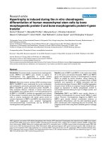
Figure 1.1. Embryonic origin of human embryonic stem cells and their in vitro
characterisation. (A) 5-day human blastocyst with inner cell mass (ICM), blastocoel
cavity (C) and trophectoderm (T). Image from the Advanced Fertility Centre, Chicago
(B) A single hESC colony,
here hES3, maintained on a mitotically inactivated mouse embryonic fibroblast
(MEF) monolayer. Typically a hESC colony grown under these conditions has the
dense, white ‘central button’ surrounded by a thinner halo of cells with a crisp border.
(C) hESC-derived embryoid bodies (EBs) in suspension culture. (D, E)
Immunostaining performed on hESC colonies for pluripotency markers. Nuclear
staining for transcription factor OCT4 (D) and cell surface staining for Tra 1-60 (E)
show that more than 90% of the cells in all colonies stain positive for these two
markers.
3
Detailed investigation of the differentiating ES/ EB system suggests that it
recapitulates to a limited degree the early events of embryonic development (Dvash
and Benvenisty 2004; Dvash et al. 2004; Rust et al. 2006). EBs derived from hESCs
organise themselves in a manner reminiscent of the early post-implantation mouse
embryo, with features like an outer jacket of extraembyonic (visceral) endoderm (Rust
et al. 2006). These similarities prompted the use of the EB system as a model to
stimulate in vitro the early events of mammalian axis specification and germ layer
patterning. Several methods of EB formation– in hanging drops, in low-attachment
plates, in 3D matrices (synthetic and natural) and the use of various growth factors in
all or some of these methods– are commonly used to induce differentiation.
Though they were thought to be equivalent to the ICM, it was suggested that ES
cells are cell culture artefacts as they adapt well to in vitro growth conditions and
show properties not usually associated with the embryo such as dependence on
exogenous cytokines/ growth factors (Buehr et al. 2003; Smith 2001; Rossant 2001).
Later studies provided evidence that ES cells likely bear closer resemblance to
embryonic germ (EG) cells as several germ cell markers like Dppa3 (Stella) were
expressed in ES cells (Zwaka and Thomson 2005). Derivation of pluripotent cell lines
from the mouse epiblast, called EpiSCs, brought to light similarities between these
cells and hESCs (Tesar et al. 2007; Brons et al. 2007). EpiSCs and hESCs have the
ability to give rise to trophectoderm in the presence of Bmp4 which mESCs do not
possess (Xu et al. 2002; Beddington and Robertson 1989). Another similarity between
these two cell types is the requirement for Activin A/ Nodal signaling to maintain
pluripotency, a property that has been previously demonstrated for hESCs (Vallier et
al. 2005). Inhibition of Activin signaling resulted in rapid downregulation of
pluripotency genes in both cell types. This may reflect the embryonic stage to which
4
hESCs are equivalent, since Activin/ Nodal signaling is known to be required for
maintenance of pluripotency in the epiblast of the post-implantation embryo (Brennan
et al. 2001). The importance of Activin/ Nodal signaling in the maintenance of hESC
pluripotency has been re-iterated in recent studies detailing the derivation and
maintenance of induced pluripotent stem (iPS) cells (Takahashi et al. 2007; Takahashi
and Yamanaka 2006). iPS cells are generated from mouse and human adult fibroblasts
by nuclear reprogramming using a few critical transcription factors like SOX2,
OCT3/4, KLF4 and C-MYC. Human iPS cells were found to be similar to hESCs in
several aspects including morphology, growth kinetics, cell-surface antigen profile
and gene expression. In addition it has been shown that iPS cells can differentiate into
the three germ layers in vivo and form teratomas identical to hESCs. A family tree of
the various embryonic and extraembryonic lineages summarises these relationships
(Fig 1.2). The lineage tree emphasizes that as the biology of ES cells continues to be
unravelled, there is mounting confidence that culture regimes can be developed which
direct pluripotent ES cells toward a desired cell fate that would be therapeutically
useful.
Much progress has been made towards gaining a better understanding of hESC
biology and translating the technology from the bench to the bedside. However, the
hESC lines on which most of these studies were performed might have restricted use
in the clinic, as they have all come in contact with materials or reagents of foreign
origin (Bongso et al. 2008; Hentze et al. 2007). Recently, this presumed roadblock
was deemed acceptable when the Food and Drug Administration (FDA), USA granted
permission for the use of oligodendrocyte cells derived from hESCs for Phase I
clinical trials to treat patients with spinal cord injury. GRNOPC1, oligodendroglial
progenitor cells, were derived from the H7 hESC line (Thomson et al. 1998) and have
5
been demonstrated to support re-myelination and nerve growth stimulation in animal
models of acute spinal cord injury (Kierstead et al. 2005). The current clinical trial
will be an attempt to demonstrate the safety of using these cells in humans though it
has been shown to elicit a poor immune response in the immune-deficient animal
model (Okamura et al. 2007).
The isolation of clinically compliant hESC lines was recently achieved (Crook
et al. 2007). Six hESC lines were derived on clinical grade human fibroblasts, Ortec
143, and maintained in chemically defined medium containing Knockout Serum
Replacement supplemented with basic fibroblast growth factor (bFGF). None of the
reagents used during derivation and expansion were of animal origin and the entire
process was carried out under cGMP (current good manufacturing practice)
guidelines. Even with derivation of qualified lines and defined culture methods, the
recurring challenges of directing the differentiation of hESCs to generate cell types in
numbers sufficient for clinical applications and ensuring acceptance of the transplant
and preventing rejection by the recipient’s immune system remain. Harnessing and
understanding the differentiation potential of hESCs and employing that knowledge to
gain insight into mammalian development is the focus of the thesis. Such studies
require experimental strategies that are guided by the knowledge of how a vertebrate
embryo develops and forms a complex organism. Hence it is important to review key
aspects of the mammalian developmental sequence especially, formation of the three
primary germ layers in the embryo.
6
Figure 1.2. Lineage tree of embryo-derived cells and cell lines. The various stages
of embryonic development from fertilization to E6.5 are represented in this image.
Embryonic Stem (ES) cells are derived from the inner cell mass while epiblast stem
cells (EpiSCs) are of epiblast origin. Human ES cells (hESCs) and mouse EpiSCs
have been found to share several characteristics which imply that hESCs might
actually be derivatives of epiblast-stage embryos. The extraembryonic endoderm is
the source of XEN cells while TS cells represent the extraembryonic ectoderm
lineage. Schematic used with permission from Tesar et al. 2007, Nature.
7
1.2. Gastrulation– formation of mesoderm and endoderm in the embryo
Gastrulation is defined by a series of complex morphogenetic events in
combination with cell proliferation and differentiation that generate the three
embryonic germ layers and establish a vertebrate body plan (Arnold and Robertson
2009; Tam and Loebel 2007; Rossant and Tam 2004). In the mouse embryo
gastrulation is initiated by the recruitment of epiblast cells to the primitive streak
around E6.5 (Fig 1.3). There, epiblast cells undergo an epithelial to mesenchymal
transition (EMT) as they ingress through the primitive streak, emerging as definitive
endoderm (DE) and the mesoderm (Tam and Beddington 1992; Lawson et al. 1991).
Mesoderm is formed as an epithelial sheet that expands from either side of the
primitive streak (Tam and Behringer 1997). Extensive studies on cells of the cardiac
mesoderm showed that the timing of ingression through the streak and the position of
these cells in the epiblast determines their lineage fate (Tam and Behringer 1997; Tam
and Zhou 1996; Lawson et al. 1991). The newly formed motile mesoderm migrates
laterally between the outer visceral endoderm (VE) layer and the epiblast, while the
definitive endoderm moves to the outer surface of the embryo by displacing the
visceral endoderm proximally (Lawson et al. 1986). However, recent work from
Kwon et al. (2008) suggests that the DE is formed by intercalation of epiblast cells
with the underlying VE and not by complete displacement of the visceral layer. This
work is discussed in more detail in section 1.5. Understanding the complex events that
characterise gastrulation is critical for the creation of experimental strategies to
generate relevant cell types for therapeutic use (discussed below).
8
A
B
C
Figure 1.3. Gastrulation and specification of the germ layers. (A) Gastrulation in
the human embryo results in the specification of the three germ layers. During this
process, prospective endodermal and mesodermal cells ingress through the primitive
streak (arrows) to form definitive endoderm and mesoderm respectively. Image used
with permission from Dias et al. 2004 Neurosurgical Focus. (B) Gastrulation in
mouse embryo occurs at E6.5 and forms the three germ layers. Image used with
permission from Tam et al. 2007 Nature Reviews Genetics. (C) Cellular organisation
of the mouse embryo after the process of gastrulation is complete. Image used with
permission from Arnold et al. 2009 Nature Reviews Molecular Cell Biology.
9
1.3. Regenerative medicine and embryonic stem cells
1.3.1. Diabetes– a candidate disease for cell therapy
Autoimmune destruction of insulin-secreting pancreatic β-cells within the Islets
of Langerhans causes Type I diabetes which makes up about 5-10% of all diagnosed
cases (Fig 1.4). Clinical islet transplantation using cadaveric islets is to date, the most
successful cell-based therapy that has been used to treat this condition (Shapiro et al.
2006; Robertson 2004). However, the demand for such islets far exceeds the actual
supply especially, since the modern procedure called the Edmonton protocol utilises
approximately 10,000 islet ‘units’ per kilogram of bodyweight. Therefore, alternative
sources of β-cells need to be identified and hESCs are an appealing source.
Pluripotent hESCs retain the capability to differentiate into cells representing all three
embryonic germ layers (Keller 2005). By directing the differentiation of hESCs to
generate functional beta (β) cells, one hopes to create an inexhaustible supply of these
cells for the treatment of Type I Diabetes. This has led to immense interest in the
differentiation of hESCs into endodermal derivatives. One part of this thesis (Chapter
4) describes my contribution to the development of in vitro β cell differentiation
protocols, with particular emphasis on the formation of the definitive endoderm, the
parental lineage of the pancreas.
1.3.2. Transplantation tolerance of hESC-derived cell therapy
As research efforts intensify towards deriving transplantable cell therapy
material like insulin-secreting β-like cells from hESCs, issues pertaining to graft
acceptance/ rejection must be addressed. Rejection of hESC-derived cell populations
is a significant concern as their immunological signature is indisputably foreign
10
(Draper and Andrews 2002; Drukker et al. 2002). Interestingly, several studies have
shown that undifferentiated hESCs and their differentiated progeny may in fact be
immune-privileged or can be transplanted specifically into immune-privileged sites
like the spleen (Li et al. 2004; Drukker et al. 2006). Transplantation into areas like the
spleen are under consideration largely due to the low expression of Major
Histocompatibility Complex (MHC) class I molecules on the surface of hESCs and
the resultant low immunostimulatory capacity of these cells. Though hESCderivatives show increased expression of MHC class I, this does not alter the immune
response.
Recently, it was demonstrated that mESCs and their derivatives with similar
MHC I signatures can induce a potent immunological reaction even with a single
difference between the donor and host Minor Histocompatibility antigen (mH)
profiles (Robertson et al. 2007). However, these authors found that the inherent
immune-privileged status of mESCs could be harnessed with minimal intervention to
induce tolerance and prevent rejection. Highlighting the differences between the
mouse and human systems, a very recent study shows that hESCs and their
derivatives might not be as immune-privileged as previously thought and are capable
of triggering a severe immune response in a xenogeneic host like the mouse
(Swijnenburg et al. 2008). In this study, hESCs transduced with a double fusion
reporter gene consisting of firefly luciferase and enhanced GFP were tracked in vivo
using bioluminescent imaging. Severe infiltration of the graft 5 days after
transplantation with immune cells and detectable levels of anti-hESC antibodies in the
recipient serum together demonstrate active rejection of the graft. However, this
reaction could be mitigated with the use of immunosuppressive drugs like tacrolimus
(binds calcineurin and thereby inhibits T-cell signaling ) and sirolimus (blocks
11
activation of T- and B- cells by inhibiting interleukin-2 responsiveness) that
prolonged
hESC
graft
survival
up
to
28
days.
The
disadvantage
of
immunosuppression is the undesirable side-effects that it triggers including
nephrotoxicity, liver disease, increased risk of infections and a compromised immune
system. Though much progress has been made, it is clear that more studies are
required before any of the above strategies can be put to clinical use. Nevertheless,
one step forward is the recently approved clinical trial for oligodendrocyte precursor
cells derived from hESCs. The outcome of this safety study is eagerly anticipated as
longevity of the graft within humans will pave the way for effective cell therapy.
If the inherent immune-privileged status of hESCs is inadequate to aid
transplantation, one strategy is to induce tolerance with the use of hESC-derived
haematopoietic cells (Drukker and Benvenisty 2004). Haematopoietic stem cells
(HSC) are mesodermal derivatives that serve as progenitors to all cells that circulate
in the peripheral blood and differentiate into several myeloid or lymphoid lineages
during development (Fig 1.5). Theoretically, haematopoietic cells derived from the
same exact source as the therapeutic graft, for example, a given pluripotential hESC
line, could tolerise the recipient towards the incoming transplant material irrespective
of its cellular nature (Kaufman and Thomson 2002). Tolerance could either be
induced (1) through mixed haematopoietic chimerism or (2) through tolerogenic
dendritic cells (DCs) (Fig 1.6) (Drukker and Benvenisty 2004; Fairchild et al. 2004).
Mixed haematopoietic chimerism refers to the use of haematopoietic progenitor
cells to establish a resident donor population in the host. This grants donor-specific
tolerance to the host and allows any other material from the same donor to be
accepted with out any adverse reaction. Clinical examples of this phenomenon in
humans have been reported (Alexander et al. 2008; Kawai et al. 2008).
12
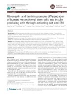
Figure 1.4. Pancreas and diabetes. The pancreas consists of Acinar cells which
perform its exocrine functions and clusters of cells called Islets of Langerhans which
perform its endocrine functions. Acinar cells secrete digestive enzymes like trypsin
and chymotrypsin into the small intestine. Islets of Langerhans secrete various
hormones into blood from its four main cell types which are (1) alpha (α) cells that
secrete glucagon, (2) beta (β) cells that secrete insulin, (3) Delta (δ) cells that secrete
somatostatin and (4) PP cells that secrete pancreatic polypeptide. The beta cells sense
glucose levels in the blood and secrete Insulin to allow uptake of this important
nutrient. Decreased production of Insulin leads to hyperglycemia and all the
symptoms associated with the metabolic disease Type I Diabetes. Schematic diagram
adapted
from
the
NIH
Stem
Cells
Information
Resource
at
/>
13