Effects of glucocorticoids on sulfotransferase 1a (SULT1A) activities and the efflux of sulfate conjugates
Bạn đang xem bản rút gọn của tài liệu. Xem và tải ngay bản đầy đủ của tài liệu tại đây (1.37 MB, 107 trang )
EFFECTS OF XENOBIOTICS ON SULFOTRANSFERASE 1A
(SULT1A) ACTIVITIES AND THE EFFLUX OF SULFATED
CONJUGATES
SHERRY NGO YAN YAN
NATIONAL UNIVERSITY OF SINGAPORE
2003
EFFECTS OF XENOBIOTICS ON SULFOTRANSFERASE 1A
(SULT1A) ACTIVITIES AND THE EFFLUX OF SULFATED
CONJUGATES
SHERRY NGO YAN YAN
(BSc Biochemistry, Massey University, New Zealand)
A THESIS SUBMITTED FOR THE
DEGREE OF MASTER OF SCIENCE IN BIOCHEMISTRY
DEPARTMENT OF BIOCHEMISTRY
2003
Acknowledgement
Many thanks to my fellow colleagues and labmates who have had to bear with my
seemingly endless frustrations from all the unsuccessful experiments I had encountered in
the course of completing this project. I am truly grateful for their endless support and
encouragement they have given me throughout the course of my study.
I also extend my sincere thanks to Prof Sit Kim Ping and Ms Lim Beng Gek for
the Hep G2 cells, the use of the HPLC instrument and for providing guidance on certain
technical aspects of my experiments.
I also sincerely thank my supervisor, Dr Theresa Tan whom without, I would not
have been able to successfully complete my project for this degree. I am grateful for all
her guidance and advice she has given me throughout these past years.
Last but not least, this project was made possible with Grant R183-000-059-213,
which was funded by the National Medical Research Council (NMRC) of Singapore.
Table of Contents
Contents
Page no.
1.
Introduction
1.1
Drug metabolism
1.2
Sulfation
1.2.1 Sulfation: An Overview
1.2.2 PAPS Synthetase
1.2.3 Cytosolic Sulfotransferases
1.3
The Cytosolic SULT Superfamily
1.3.1 An Overview
1.3.2 SULT1 Family
1.3.3 SULT2 Family
1.4
Gene Expression And Regulation Of Cytosolic SULTs
1.5
Hepatic Vectorial Transport
1.5.1 An Overview
1.5.2 Hepatic Xenobiotic Uptake Transporters
1.5.3 Hepatic Xenobiotic Efflux Transporters
1.6
Effects Ff Glucocorticoids On Cytosolic SULTs And
Xenobiotic Transporters
1
1
2
2
4
7
12
12
13
16
18
20
20
22
24
27
2.
Objective And Scope Of This Work
29
3.
Materials and Methods
3.1
Materials
3.2
Methods
3.2.1 Cell Culture Of Hep G2
3.2.2 Treatment Of Hep G2 With Glucocorticoids
3.2.3 Cell Viability
3.2.4 Assay of SULT1A1 and SULT1A3 Activities
In Hep G2
3.2.5 Efflux Assays Of Sulfated Conjugates Of
Dopamine And -Nitrophenol
3.2.6 Reverse-phase High-performance Liquid
Chromatography (RP-HPLC) Detection and
Separation Of The Sulfated Conjugates Of
Dopamine And -Nitrophenol From Na235SO4
3.2.7 Assay Of PAPSS Activity (PAPS
Generation Assay)
3.2.8 Statistical Analysis
3.2.9 RNA Isolation And Reverse-transcription
(RT)-PCR Of SULT1A Isoforms And
30
30
31
31
31
33
33
34
35
36
36
37
Contents
Page no.
Xenobiotic Transporters
3.2.10 RT-PCR of SULT1A3 Followed By
Chemiluminescence Detection
38
Results
4.1
Cell Counting And Cell Viability Of Hep G2
4.2
RP-HPLC Chromatograms From SULT1A Assays
4.3
SULT1A Assay: Time-Dependent Sulfation By
SULT1A1 And SULT1A3 In Hep G2
4.4
SULT1A Assay: Effect Of DX And PN On SULT1A1
And SULT1A3 Activities
4.5
RT-PCR Detection: Effect Of DX And PN On
SULT1A3 mRNA Expression
4.6
PAPS Generation Assay: Effect of DX and PN On
SULT1A1 And SULT1A3 Activities
4.7
Efflux Assay: Effect of DX on Xenobiotic
Transporters
4.8
RT-PCR Detection: Effect Of DX On Xenobiotic
Transporters
4.9
Software Analysis: Putative Promoter Elements Of
The SULT1A3 Gene
40
40
40
44
Discussion
5.1
Effects Of Glucocorticoids On Sulfation In Hep G2
Cells
5.2
DX Differentially Induces SULT1A3 But Not
SULT1A1 Activity
5.3
Effects of DX On Efflux Of Sulfated Conjugates In
Hep G2 Cells
5.4
Effects of DX On Detoxification Via Sulfation
Pathway
68
68
6.
Conclusion
75
7.
Future works
76
8.
References
78
9.
Abbreviations
98
4.
5.
45
48
51
52
53
56
70
72
74
List of Figures
Contents
Figure 1.1
Figure 1.2
Figure 1.3
Figure 1.4
Figure 1.5
Figure 2.1
Figure 4.1
Figure 4.2
Figure 4.3
Figure 4.4
Figure 4.5
Figure 4.6
Figure 4.7
Figure 4.8
Figure 4.9
Figure 4.10
Figure 4.11
Figure 4.12
Figure 4.13
Page no.
Human PAPSS1 and PAPSS2
(next page) The highly conserved Region I and IV amino
acid SULT signature sequences
Proposed reaction mechanism of sulfuryl transfer
catalyzed by SULTs
The human SULT enzyme family
Hepatic vectorial transport
Schematic outline of the scope of this work
Separation of -nitrophenyl-35sulfate from sodium-sulfate
(Na235SO4)
Separation of dopamine-35sulfate from Na235SO4
Separation of PAP35S from Na235SO4
Time-dependent (A) -nitrophenyl-ST and (B) dopamineST activities in Hep G2
(A) SULT1A1 and (B) SULT1A3 activities in Hep G2
following three days of DX treatment
(A) SULT1A1 and (B) SULT1A3 activities in Hep G2
following three days of PN treatment
(A) SULT1A1 and (B) SULT1A3 activities in preconditioned Hep G2 cells prior to three days of DX
treatment
(A) SULT1A1 and (B) SULT1A3 activities in preconditioned Hep G2 cells prior to three days of PN
treatment
RT-PCR of SULT1A3 and -actin in Hep G2 cells
following three days of GC treatment
Panel A: SULT1A3 mRNA levels in Hep G2 following
three days of GC treatment
PAPS generation by PAPSS in Hep G2 following three
days of DX and PN treatment
Efflux of (A) -nitrophenyl-35sulfate and (B) dopamine35
sulfate in Hep G2 following three days of DX treatment
RT-PCR of various isoforms of MRP xenobiotic
5
9
12
16
21
29
41
42
43
44
45
46
47
47
48
49
51
52
54
Contents
Figure 4.14
Figure 4.15
Figure 4.16
Figure 4.17
Figure 4.18
Figure 5.1
Figure 5.2
Page no.
transporters from total RNA extract of DX-treated Hep G2
cells
RT-PCR of various isoforms of OATP transporters
from total RNA extract of DX-treated Hep G2 cells
SULT1A3 cDNA sequence (GenBank Accession Number:
U20499)
Annotations of the human SULT1A3 genomic sequence
(GenBank Accession Number: NT_042812)
BLAST result from alignment of proximal 5’UTR of
SULT1A3 cDNA onto the ~6.3 kb region proximal to the
translational start site on SULT1A3 genomic sequence
Putative regulatory factors and elements of the human
SULT1A3 gene
(A) Prednisolone and (B) Dexamethasone
Potential response elements in the 5’-untranslated region
of human MRP1
53
57
62
65
67
69
74
List of Tables
Contents
Table 1.1
Table 1.2
Table 1.3
Table 3.1
Table 3.2
Table 3.3
Table 4.1
Table 4.2
Table 4.3
Table 4.4
Page no.
Phase II conjugation reactions
Characteristics of human PAPSS1 and PAPSS2
Names of the corresponding SULTs that are listed in
Figure 1.2, based on the new nomenclature and their
GenBank Accession Numbers
Buffer compositions of PBS and HBSS
Solvent composition for the separation of dopamineand -nitrophenyl-sulfate from Na235SO4
Primer sequences of various isoforms of SULT 1A,
MRP, OATP and the control, -actin for RT-PCR
Typical cell concentration and viability of Hep G2
following three days of GC treatment
Intensities of SULT1A3 blot following
chemiluminescence detection of RT-PCR products
mRNA levels of the various transporters in DX-treated
Hep G2 cells
Consenus sequences of SP1, AP1, AP2, CAAT and
GRE used by MatInspector software
2
6
10
32
35
38
40
50
55
67
Summary
Sulfation by sulfotransferases (SULTs) is pharmacologically important for
detoxification of endogenous compounds and xenobiotics. Glucocorticoid (GC)
regulatory elements have been identified for rat SULT1A1. In this study, the effects of
dexamethasone (DX) and prednisolone (PN) on human SULT1A and 3’phosphoadenosine 5’-phosphosulfate synthetase (PAPSS) activities, and DX on mRNA
expression of xenobiotic transporters were explored using Hep G2 cells.
PAPSS activities were unaltered by both DX and PN. While SULT1A1 activity
was unaltered by DX and PN, 10-7M DX increased SULT1A3 activity by 80% which
correlated to the increase in mRNA levels of 1.8 folds. Software analysis of the 5’
flanking region of human SULT1A3 gene showed the presence of a consensus binding
site for the GC receptor. Such a site was not present for SULT1A1.
MRP and OATP isoforms were generally DX-inducible. MRP3 mRNA
expression was down-regulated, whereas a biphasic response was observed for MRP2.
Efflux of -nitrophenyl-sulfate was down-regulated by DX by nearly 50%; probably due
to increased uptake, possibly by OATP proteins and/or reduced export. Dopamine-sulfate
was up-regulated by 150% at 10-7 M DX; probably a result of increased efflux in addition
to the increased SULT1A3 activity.
1.
Introduction
1.1
Drug Metabolism
Drug metabolism essentially comprises Phase I (functionalization reactions),
Phase II (conjugative reactions) and Phase III (involving protein transporters for drug
excretion). Phase I reactions generally include oxidation, reduction, hydrolysis, hydration
although there exists other rarer reactions such as isomerization and dimerization,
transamidation, decarboxylation, etc. (Kauffman, 1990).
Phase II conjugations are carried out by a diverse group of enzymes acting on
numerous types of compounds. The conjugation processes generally lead to bioinactivation of the drugs or xenobiotics to form water-soluble products that can be readily
excreted through bile or urine. As such, Phase II reactions are said to be the true
“detoxification” pathways since they generate the final inactive, excretable products of a
drug or xenobiotic. Conjugation reactions that comprise the Phase II detoxification
pathways, the enzymes involved and the types of drugs conjugated are as listed in Table
1.1 (Kaufman, 1990).
Phase III transport systems will be discussed in Section 1.5.
Conjugation reaction
Enzyme
Functional group
Glucuronidation
UDP-glucuronyltransferase
-OH, -COOH, -NH2,
-SH
Glycosidation
UDP-glycosyltransferase
-OH, -COOH, -SH
Sulfation
Sulfotransferase
-NH2, -SO2NH2, -OH
Methylation
Methyltransferase
-OH, -NH2
Acetylation
Acetyltransferase
-NH2, -SO2NH2, -OH
Amino acid conjugation
Glutathione conjugation
-COOH
Glutathione-S-transferase
Epoxide, Organic halide
Fatty acid conjugation
-OH
Condensation
Various
Table 1.1
Phase II conjugation reactions (Kauffman, 1990)
1.2
Sulfation
1.2.1
Sulfation: An Overview
Sulfation plays a role in homeostasis and regulation of many important
endogenous chemicals such as catecholamines, steroids as well as other macromolecules
(Coughtrie et al, 1998). In addition, it serves as one of the detoxification pathways for the
various xenobiotics, although occasionally it results in the activation of the xenobiotic to
a reactive electrophile (Buhl A et al, 1990; Falany, 1991; Glatt, 1997).
The energy-requiring, sulfation process is catalysed by the substrate-specific
sulfotransferases, using 3’-phosphoadenosine 5’-phosphosulfate (PAPS) and ATP as
cosubstrates for the sulfation reaction. Sulfation reactions utilize PAPS as the sulfate
donor. PAPS is made in the cytosol as a two-step enzymatic process (Robbins and
Lipmann, 1958). Firstly, ATP sulfurylase catalyzes the formation of adenosine-5’phosphosulfate (APS) from inorganic SO4 in the presence of ATP. Subsequently, APS
kinase catalyzes the formation of PAPS from the phosphorylation of APS (using ATP as
the phosphate donor). The primary source of sulfur is free SO42-, which is transported into
the cytosol by a variety of transporter or symporter molecules (Falany, 1997a; Falany,
1997b; Weinshilboum et al, 1997; Kullak-Ublick et al, 2000).
For post-translational protein modification via sulfation, PAPS is delivered to the
Golgi network with the aid of the PAPS translocase, where the secreted proteins can be
sulfated by the substrate-specific membrane sulfotransferases (Mandon et al, 1994;
Ozeran et al, 1996; Schwarrtz et al, 1998). For metabolism of endogenous compounds or
detoxification of xenobiotics, the PAPS is utilized in the cytosol by the cytosolic
sulfotransferases (Klassen et al, 1997).
Sulfotransferases (SULTs) exist as cytosolic and membrane-bound enzymes.
Cytosolic SULTs catalyze the sulfation of endogenous and exogenous small-molecule
substrates like steroids, hormones, neurotransmitters and xenobiotics, including
therapeutic drugs, in animals. In plants, similar reactions occur with flavonols (Coughtrie
et al, 1998). Membrane-bound SULTs typically catalyze the sulfation of macromolecules,
such as proteoglycans, glycosaminoglycans, polysaccharides and tyrosyl residues within
proteins (Huttner, 1982; Hashimoto et al, 1992).
1.2.2 PAPS Synthetase
ATP sulfurylase and APS kinase constitute the sulfate activation pathway in both
higher and lower organisms. In simpler organisms (bacteria, yeasts, algae, protozoa), they
exist as two separate and relatively small polypeptides (Klassen and Boles, 1997;
Farooqui, 1980). However, in higher organisms including mammals, they exist as a single
bifunctional enzyme, called the PAPS synthetase (PAPSS) (Lyle et al, 1994). Two
different isoforms of PAPSS, PAPSS1 and PAPSS2, are known to exist in humans, mice
and the marine worm Ureches caupo (Li et al, 1995; Rosenthal et al, 1995; Besset et al,
2000). The human PAPSS1 and PAPSS2 proteins show 76.5% amino acid sequence
identity (Kurima et al, 1998; ul Haque et al, 1998).
Historically, PAPS synthesis is assumed to occur exclusively in the cytosol. In
fact, cytosol and Golgi apparatus are the only sites of PAPS utilization by known
sulfotransferases (Falany CN, 1997a; Falany CN, 1997b; Bowman and Bertozzi, 1999).
However, it has been reported that human PAPSS1 is a nuclear protein, in contrast to
PAPSS2 which is cytosolic. Besset et al demonstrated that the APS kinase domain targets
PAPSS1 to the nucleus in a number of mammalian cell lines (Besset et al, 2000). In
addition, nuclear targeting of PAPSS1 in yeast functionally complements ATPsulfurylase and APS-kinase-deficient strains (Besset et al, 2000). Furthermore, ectopic
PAPSS2 expression in mammalian cells dramatically localized the cytosolic PAPSS2 to
the nucleus, when coexpressed with PAPSS1 (Xu et al, 2000).
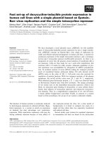
Human PAPSS1 and PAPSS2 are very similar in structure. Figure 1.1 shows that
both genes are made up of 12 exons, but exon 1 (the first splice junction) contains an
additional codon in PAPSS2. All exon-intron splice sites for the two genes are virtually
identical. Introns of PAPSS1 vary from 1.6 kb to 21.9 kb whereas the introns of PAPSS2
are generally shorter than those of PAPSS1. Table 1.2 summarizes the characteristics of
human PAPSS1 and PAPSS2 genes. The 5’-flanking region of PAPSS1 did not contain
any TATA or CAAT sequences. The transcriptional start site did not contain an Initiator
(Inr) sequence. However, a TATA box was located at 21 bp upstream of the transcription
initiation site for PAPSS2 (Xu et al, 2000).
Figure 1.1
Human PAPSS1 and PAPSS2 (Xu et al, 2000)
PAPSS1
PAPSS2
GenBank Accession: AF097710-AF097721
Chromosome band 4q24
108 kb
12 exons
2.7 kb mRNA transcript
No TATA box/motif
GenBank Accession: AF160503-AF160509
Chromosome band 10q23-24
>37 kb
12 exons
4.2 kb mRNA transcript
TATA motif at -21 bp from transcriptional
start site
Several putative Sp1 binding sites at 5’flanking region
Slice junctions conform to ‘GT-AG’ rule
Highly expressed in liver and adrenal gland
Several putative Sp1 binding sites at 5’flanking region
Slice junctions conform to ‘GT-AG’ rule
Low expression in liver, skeletal muscle and
adrenal gland
Table 1.2
Characteristics of human PAPSS1 and PAPSS2
Schwartz et al showed that the rat PAPS synthetase uses a channeling mechanism
to transfer APS from the sulfurylase to the kinase active site. The defect in PAPS
production observed in brachymorphic mice was primarily due to the decreased ability to
channel APS, hence, the inability to generate PAPS efficiently (Schwartz et al, 1998).
Similar observations were made by Hastbacka et al and Lyle et al, who reported the
brachymorphic mouse phenotype attributed to defects in the ATP sulfurylase/ APS kinase
protein (Hastbacka et al, 1994; Lyle et al, 1995). More importantly, a variant sequence
within the human PAPSS2 orthologue has been associated with spondiloepi-metaphyseal
dysplasia, an inherited syndrome in humans, phenotypically similar to the brachymorphic
phenotype in mice (ul Haque et al, 1998). This clearly signifies the role of PAPS
synthetase in the generation of PAPS for sulfation.
1.2.3
Cytosolic Sulfotransferases
The sulfotransferases (SULTs) constitute a diverse range of enzymes that make
up an emerging superfamily. Historically, the reactions catalyzed by these low-capacity
enzymes have been termed “sulfation”, although chemically, they are more accurately
described as sulfonation. Sulfonation/sulfation by the sulfotransferases involves the
transfer of sulfonate group from PAPS to the acceptor substrate (e.g. endogenous
compound, neurotransmitter or xenobiotic) to form either a sulfate or sulfamate conjugate
(Weinshilboum et al, 1997).
Cytosolic sulfotransferases usually are found as hetero- and homodimers, with
monomer molecular weight ranging from 30 to 36 kDa (Falany, 1991). However, in some
plants and mammals, they can exist as catalytically active monomers (Takikawa et al,
1986).
SULTs are single / globular proteins with characteristic five-stranded parallel
-sheets. The -sheet constitutes the PAPS-binding site and the core active site of the
enzyme. Consequently, both these sites are highly conserved in cytosolic and membranebound SULTs. As such, all sulfotransferases are categorized as members of a single gene
superfamily. The membrane-bound SULTs are attached to the membranes of the Golgi
network at the amino-terminal end (Negishi et al, 2001).
Protein sequence alignments of cytosolic sulfotransferases of different species
identified various regions of sequences that were highly conserved (Marsolais and Varin,
1995; Weinshilboum et al, 1997). As shown in Figure 1.2, two of those regions are
located relatively near the termini of the protein sequence; one being near the amino
terminus (Region I) and the other near the carboxy terminus (Region IV).
Through the cloning of SULT cDNAs, the consensus sequence that has been
identified in Region I is YPKSGTxW and in Region IV is RKGxxGDWKNxFT, where
“x” represents any amino acid. The motif of Region IV is similar to the glycine-rich
phosphate-binding loop (the “P-loop”), present in some ATP and GTP-binding proteins.
Consequently, it is hypothesized that the portion of Region IV that contains the sequence
GxxGxxK might be a homologue for the glycine-rich region, followed by a conserved
lysine, present in some P-loop motifs (Komatsu et al, 1994). Through site-directed
mutagenesis experiments in guinea pigs, the conserved G and K in this region were
shown to be essential for enzymatic activity and the binding of 35S-PAPS as a
photoaffinity ligand for the enzyme (Komatsu et al, 1994). Furthermore, similar studies
with SULTs in plants had led to the conclusion that the invariant lysine within Region I
might be important for the stabilization of an intermediate formed during the sulfonation
reaction (Marsolais and Varin, 1995).
Figure 1.2
(next page) The highly conserved Region I and IV amino acid SULT
signature sequences (Weinshilboum et al, 1997)
“Position” refers to amino acid number with the protein sequences for regions I
and IV. “x” represents any amino acid. Black columns denote amino acids with >
93% identity with residues at that position. Black columns with an asterisk
denote > 93% identity within groups of amino acids having similar polarity.
White boxes represent non-identical residue. Arrows indicate invariant amino
acids.
9
9
The enzyme names used in Figure 1.2 were cDNA names based on the old
nomenclature. The new nomenclature for the corresponding SULT cDNA/enzymes is
as listed in Table 1.3.
Old Name
rPST
mPST
hTSPST1
hTSPST2
mfPST
hTLPST
bPST
r1B1ST
rAAFST
bEST
hEST
gpEST
mEST
rEST
rEST-6
rSMP2
rHSST1
rHSST2
rHSST3
mfHSST
hDHEAST
gpHSST1
gpHSST2
fcFST3
fbFST3
fcFST4
atST
Table 1.3
New Name
Rat SULT1A1
Mouse SULT1A1
Human SULT1A1
Human SULT1A2
Monkey SULT1A1
Human SULT1A3
Bovine SULT1A1
Rat SULT1B1
Rat SULT1C1
Bovine SULT1E1
Human SULT1E1
Guinea pig SULT1E1
Mouse SULT1E1
Rat SULT1E1
Rat SULT1E1
Rat SULT2A1
Rat SULT2A1
Rat SULT2B1
Rat SULT2B1
Monkey SULT2A1
Human SULT2A1
Guinea pig SULT2A1
Guinea pig SULT2B1
Flaveria chloraefolia (plant) SULT-like flavonol
Flaveria bidentis (plant) SULT-like flavonol
Flaveria chloraefolia (plant) SULT-like flavonol
Arabidopsis thaliana (plant) SULT-like flavonol
Accession No.
X52883
L02331
L19999
X78282
D85514
L19956
U35253
U38419
L22339
X56395
U08098
U09552
S78182
S76489
S76490
J02643
M31363
M33329
D14989
D85521
U08024
U06871
U35115
M84135
U10275
M84136
Z46823
Names of the corresponding SULTs that are listed in Figure 1.2, based
on the new nomenclature and their GenBank Accession Numbers
SULTs were traditionally named after the substrates they catalyze. However,
this form of naming system is often misleading and confusing because different
SULTs show overlapping substrate specificities. As such, a systematic
nomenclature, similar to that used for classifying the cytochrome P450
enzymes, is in use but not yet finalized for the SULTs. For this new
nomenclature, members of each family is indicated by a number after “SULT”;
and members of each subfamily is indicated by a letter after each subfamily
number.
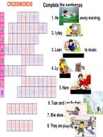
More recently, using the human estrogen SULT (hEST) which is responsible for
sulfation of estrogens, Negishi et al demonstrated that the conserved lysine (K47) within
Region I and another highly conserved serine (S137 in hEST or S138 in mouse EST) are
essential not only for PAPS-binding site, but also for catalysis. Figure 1.3 shows the sidechain nitrogen of the conserved lysine forms an H-bond with an O-atom of the 5’phosphate group in the PAPS molecule. The hydroxyl side-chain of the conserved serine
interacts with an O-atom in the 3’-phosphate. X-ray crystallography of hEST also showed
that the side-chain of the conserved Ser137 interacted with the side-chain of the
conserved K47. As a result of the interaction, the side-chain nitrogen of the conserved
lysine is repelled from the bridging oxygen of the PAPS molecule. It was also observed
that the serine decreased PAPS hydrolysis when the substrate was absent from the active
site. However, mutation of the serine residue markedly increased PAPS hydrolysis. This
led to the conclusion that the conserved serine may serve to regulate the sulfuryl transfer
process by interacting with the catalytic lysine (Negishi et al, 2001).
In addition, using x-ray crystals of mouse EST (mEST)-PAP-vanadate, Negishi et
al also demonstrated that the conserved histidine at position 108 (H107 in human) and the
conserved lysine at position 48 (K47 in human) appeared to be catalytic residues. He
reported that mutation of His107 of the hEST to asparagine rendered the enzyme
incapable of hydrolyzing PAPS nor catalyzing the sulfation reaction (Negishi et al, 2001).
Figure 1.3
Proposed reaction mechanism of sulfuryl transfer catalyzed by SULTs
(Negishi et al, 2001)
Residue numbers are taken from hEST.
1.3
The Cytosolic SULT Superfamily
1.3.1
An Overview
Presently, at least 44 cytosolic sulfotransferases have been identified in
mammals, ranging from rats and mice to dogs, rabbits, cows, guinea pigs and monkeys.
In humans, at least 11 different sulfotransferases have been identified (Nagata and
Yamazoe, 2000). These enzymes are classified into three sub-families based on their
amino acid sequence identity and substrate specificity (Yamazoe et al, 1994;
Weinshilboum et al, 1997). Members of the sub-family SULT1 preferentially sulfate
phenols (including estrogens and iodothyronines) and catechols (including
catecholamines). SULT2 family members mainly sulfate steroids, sterols and other
alcohols (Yamazoe et al, 1994; Strott, 1996; Weinshilboum et al, 1997). In general,
amino acid sequence comparisons between members of each family yield at least 45%
similarity. However, members of subfamilies show at least 60% amino acid sequence
identity (Nagata and Yamazoe, 2000).
1.3.2
SULT1 Family
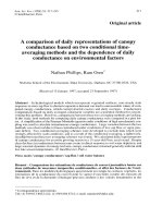
As shown in Figure 1.4, the human SULT1 family, presently known to be
the largest family, comprises of SULT1A, SULT1B, SULT1C and SULT1E enzymes.
The human SULT1A subfamily has three members namely, SULT1A1, SULT1A2 and
SULT1A3. These three genes differ by less than 10% at amino acid level (Figure 1.4) and
are physically mapped to a small chromosomal region 16p.
SULT1A1 preferentially sulfates simple phenols. Classical phenolic substrates are
-nitrophenol and -napthol, although weaker activities have been observed with
sulfation of catechols, hydroxyarylamines, and iodothyronines. SULT1A1 also sulfates
the common xenobiotics, acetaminophen and minoxidil (Reiter et al,1982; Young et al,
1988; Falany and Kerl, 1990; Duanmu et al, 2000; Honma et al, 2001). SULT1A2
sulfates simple phenols and catechols, albeit at a lower catalytic activity and shows a
higher Km value when compared to SULT1A1 (Dooley, 1998a). SULT1A3 has been
observed to preferentially catalyze the sulfation of catecholamines; the classical substrate
being dopamine. It has only a limited activity for -nitrophenol (Veronese et al, 1994;
Honma et al, 2001). Other substrates for SULT1A3 include tyramine, 5hydroxytryptamine, salbutamol, isoprenaline and dobutamine (Honma et al, 2001).
Human SULT1A1 and SULT1A2 are mapped to the chromosomal position
16p12.1-p11.2 and are approximately 45 kb apart (Her, 1996; Gaedigk, 1997). SULT1A3
is localized 100 kb away (Dooley, 1998b). SULT1A1 is expressed in many tissues but is
abundant in the human liver, brain, kidney and platelets. However, SULT1A2 apparently
is expressed only in the adult human liver as well as in the fetal liver and spleen.
SULT1A3 is also expressed in the fetal liver and brain, with lower levels in the lung and
kidney (Dooley et al, 2000; Nagata and Yamazoe, 2000).
To date, only one form of SULT1B (SULT1B1) has been identified in human.
SULT1B1, localized on chromosome band 4q13, is highly expressed in the liver (Dooley
et al, 2000; Meinl and Glatt, 2001). Although not much is known about this isoform,
SULT1B1 has been shown to catalyze sulfation of 3,3’,5’-triiodothyronine and nitrophenol in human and rat livers, at a much lower affinity. These substrates are
sulfated by members of the SULT1A family at a much higher affinity (Dunn et al, 2000;
Tsoi et al, 2001).
SULT1C was first isolated from rat as an N-hydroxy-2-acetylaminofluorenespecific sulfotransferase (Nagata et al, 2000). Since then, two human SULTs have since
been identified through the EST database, namely SULT1C1 and SULT1C2 (Her et al,
1997). Although not much is known about these enzymes presently, SULT1C1 and
SULT1C2 share about 63% identical at the amino acid level as shown in Figure 1.4.
SULT1C2 is thought to be a “dead” enzyme because it shows little or no activity towards
any standard substrates; probably due to an amino acid change in the active site of the
enzyme (Coughtrie and Johnston, 2001). SULT1C is localized to the human chromosome
band 2q11.1-11.2 (Her et al, 1997).
Only one form of human SULT1E has been identified so far, and it is named
SULT1E1. SULT1E1, found on chromosome band 4q13.1, is known as a typical estrogen
SULT, with the Km value for -estradiol being the lowest among the human SULTs.
Experimental data suggests that estrogen sulfation is the main physiological role of
SULT1E1 in humans (Nagata and Yamazoe, 2000).
SULT
1A1
SULT
1A2
SULT
1A3
SULT
1B1
SULT
1C1
SULT
1C2
SULT
1E1
SULT
2A1
SULT
2B1a
SULT
2B1b
SULT
4A1
SULT
1A1
.
SULT
1A2
SULT
1A3
SULT
1B1
SULT
1C1
SULT
1C2
SULT
1E1
SULT
2A1
SULT
2B1a
SULT
2B1b
SULT
4A1
96
93
53
53
51
50
34
33
33
32
.
90
57
53
51
49
34
33
33
32
.
52
53
50
48
34
34
34
34
.
53
52
54
36
34
34
32
.
62
48
34
31
31
33
.
47
35
32
32
31
.
34
32
32
33
.
48
43
27
.
97
29
.
29
.
Figure 1.4
The human SULT enzyme family (Weinshilboum et al, 1997)
Amino acid similarities between members of the SULT superfamily.
SULT4 represents a novel SULT which has yet to be characterized.
1.3.3
SULT2 Family
Relative to the SULT1 family, limited studies have been done with the
characterization of members of the SULT2 family. Currently, SULT2 family comprises
SULT2A and SULT2B. More than one form of SULT2A have been isolated from rodents
and they exhibit different substrate specificity on the sulfation of hydroxysteroids