Exploration of the functional significance of mig 2 in human cancer cell susceptibility to cytotoxic agents and cell growth control a pilot study
Bạn đang xem bản rút gọn của tài liệu. Xem và tải ngay bản đầy đủ của tài liệu tại đây (1.6 MB, 123 trang )
EXPLORATION OF THE FUNCTIONAL SIGNIFICANCE
OF MIG-2 IN HUMAN CANCER CELL SUSCEPTIBILITY
TO CYTOTOXIC AGENTS AND CELL GROWTH
CONTROL: A PILOT STUDY
LIU KUN
(M.B.B.S., CHINA MU)
A THESIS SUBMITTED FOR
THE DEGREE OF MASTER OF SCIENCE
DEPARTMENT OF PHYSIOLOGY
NATIONAL UNIVERSITY OF SINGAPORE
2003
ACKNOWLEDGEMENTS
In submitting this thesis, I would like to express my deepest appreciation to my
supervisor, Dr. Shen Shali, for her patient instructions and constant encouragement
throughout this course of research. Her kind personality and invaluable suggestions will
always be in my memory.
My deepest appreciation goes to the members of our laboratory: Angela and Lay
Hoon, for their assistance, company and friendship. I would also like to extend my
gratitude to all the staff and students in the Department of Physiology, who make
Physiology a wonderful team and me proud of being part of it.
My special thanks go to Zhang Xin and Sun Yu. Thank you for being there in my
time of need. You are my best friends!
Thanks to Ayub for all the happiness; to my parents for the unconditional love
they always give to me in my life; to my dearest sister and brother-in-law for their care
and kindly support. I would never be here without you all.
Finally I acknowledge the National University of Singapore for their award of
Research Scholarship to enable me to complete my master’s study.
i
TABLE OF CONTENTS
Acknowledgement
i
Summary
vii
List of Tables
ix
List of Figures
x
List of Illustrations
xii
Chapter 1 Introduction and literature review
1
1.1
Overview of mig-2
1
1.1.1
Identification of mig-2
1
1.1.2
IE Gene
1
1.1.2.1 Classification of mitogen inducible genes
1
1.1.2.2 Some critical proteins encoded by IE genes
2
1.1.2.3 Mig-2 belongs to IE genes
4
1.2
Potential significance of mig-2 gene --- our hypothesis
5
1.2.1
Possible involvement of mig-2 in anticancer drug resistance
5
1.2.1.1 Overview of drug resistance in cancer chemotherapy
5
1.2.1.2 Genetic factors in anticancer drug resistance
6
1.2.1.3 Identification of a novel gene in mouse
8
1.2.1.4 Preliminary study on mig-2
9
1.2.2
11
Possible role of mig-2 in cell growth control
1.2.2.1 Overview of cell cycle and growth control
11
ii
1.2.2.2 Mig-2 involved in mitogenic signaling cell cycle control
12
1.3
Recent studies on mig-2
15
1.3.1
Mig-2 gene involved in cell adhesion
15
1.3.2
Integrin signaling pathway in cell growth control
16
1.3.3
Apoptosis related to integrin---Anoikis
18
1.3.4
Homologues of mig-2
19
1.3.5
PH domain and FERM domain
20
1.4
Strategies to explore mig-2 gene function
21
Chapter 2 Objectives
23
2.1
23
Objectives of the study
Chapter 3 Materials and methods
25
3.1
Sequence analyses and statistics
25
3.2
Cell lines and cell culture
25
3.3
RNA isolation
25
3.4
One-Step RT-PCR
26
3.5
cDNA synthesis
27
3.6
PCR amplification of mig-2 full-length cDNA
28
3.7
Cloning mig-2
30
3.7.1
Purification of PCR product
30
3.7.2
Cloning mig-2 into pcDNA3.1 (+)
30
3.7.3
Transformation
31
iii
3.7.3.1 Principle of bacterial transformation in gene cloning
31
3.7.3.2 Procedure of transformation by heat shock
34
3.7.4
Plasmid miniprep and screening
34
3.8
Automated sequencing
36
3.9
Transfection
38
3.9.1
Principle of cationic lipid mediated transfection
38
3.9.2
Transient transfection
39
3.9.2.1 Optimization for transfection efficiency
39
3.9.2.2 Procedure of optimized transfection
39
3.9.3
41
Stable transfection
3.9.3.1 Principle of stable transfection
41
3.9.3.2 Determination of Geneticin (G418) concentration
41
3.9.3.3 Selection of stably transfected cell lines
44
3.10
Proliferation and cytotoxicity assays
44
3.10.1
MTT assay
44
3.10.1.1 Principle of MTT assay
44
3.10.1.2 Optimization of the parameters of MTT assay
44
3.10.1.3 Procedure of optimized MTT assay
45
3.10.2
45
Drug treatment
3.10.2.1 Optimization for drug concentration and duration
46
3.10.2.2 Optimized anticancer drug treatment
46
3.11
Cell growth
46
3.11.1
Cell growth curve
46
iv
3.11.2
Colony formation
47
3.12
Apoptosis detection
47
3.12.1
Flow cytometry
47
3.12.2
TUNEL assay
47
3.12.2.1 Principle of TUNEL assay
47
3.12.2.2 Procedure of TUNEL assay
49
3.13
Serum starvation and stimulation
50
3.14
Cellular localization of mig-2
50
3.15
Antisense technology
53
3.15.1
Principle of antisense technology
53
3.15.2
Antisense oligonucleotide
53
3.15.3
Antisense plasmid
56
3.15.3.1 Cloning mig-2 antisense into pcDNA3.1 (+)
56
3.15.3.2 Transfection of antisense plasmid and selection
56
Chapter 4 Results
57
4.1
Sequence analyses and bioinformatics
57
4.1.1
Mig-2 in human genome
57
4.1.2
Mig-2 cDNA sequence
57
4.1.3
Mig-2 protein sequence
57
4.2
Mig-2 gene expression in human cancer cell lines
66
4.3
Cloning mig-2
66
4.3.1
PCR amplification of the full-length cDNA of mig-2
66
v
4.3.2
Cloning mig-2 into pcDNA3.1 (+)
66
4.4
Optimizations Data
69
4.4.1
MTT assay
69
4.4.2
Drug treatment
73
4.4.3
Transfection efficiency
73
4.5
Sensitivity of mig-2 transfected HT29 cells to different
anticancer agents
76
4.5.1
Drug treatment on transiently transfected HT29
76
4.5.2
Establishment of stably transfected HT29
76
4.5.3
Drug treatment on stably transfected HT29
76
4.6
Antiproliferative effect of mig-2
81
4.6.1
Growth curve of stable cell lines
81
4.6.2
Colony formation
81
4.7
Apoptotic effect of mig-2
84
4.7.1
Flow cytometry
84
4.7.2
TUNEL assay
84
4.8
Cellular localization of mig-2 protein
84
4.9
Expression of mig-2 is serum-dependent
88
4. 10
Down-regulation of mig-2 gene by antisense technology
88
Chapter 5 Discussion
91
Chapter 6 Conclusions
102
References
103
vi
SUMMARY
In our previous study, we identified a novel gene in mouse that was capable of
reversing the acquired drug resistance in murine tumor cells against a panel of anticancer
drugs including etoposide, vincristine, cisplatin and tamoxifen. Interestingly, BLAST
search revealed a 60% homology between the novel mouse gene and a human gene,
mitogen-inducible gene-2 (mig-2). This finding raised an intriguing question: whether or
not mig-2 could exhibit similar function, i.e. reversing drug resistance, in human cancer
cells. Mig-2 was identified in 1994 as an immediate early gene in mitogen-mediated
signal transduction in human fibroblasts cells WI-38. To date its biological functions,
however, remain poorly understood.
Two recent publications disclosed some investigations on mig-2. In one study, mig-2
was found to be one of the components in cell extracellular matrix adhesion complex,
participating in cell shape modulation. In the other study, two novel mig-2 homologous
genes were identified. One was found overexpressed in human lung and colon
carcinomas whereas the other was found silenced in patients with Kindler syndrome, an
autosomal recessive skin disorder. These studies, together with its identification process,
further aroused the question on mig-2 gene function. We therefore initiated a pilot study
aiming to explore the functional significance of mig-2. Our hypothesis was that mig-2
might enhance cancer cell susceptibility against anticancer drugs. The study aimed to test
our hypothesis by exploring the potential functions of gene mig-2 in human cancer cells.
The human colon cancer HT-29 cell line, a mig-2-null cell line as examined
experimentally, was chosen for transfection studies to investigate mig-2 gene function.
vii
Our data showed that re-expression of mig-2 in HT29 cells exerted antiproliferative
effects and induced apoptosis. The growth inhibitory effect of mig-2 was confirmed by
colony formation and cell growth assay. Mig-2 mediated apoptosis was determined by
flow cytometry and TUNEL assay. However, MTT assay failed to demonstrate any
significant enhancement of the killing effect of anticancer drugs on HT29 cells
transfected with mig-2. Furthermore, mig-2-GFP fusion protein revealed that mig-2 was
predominantly localized in the cytoplasm of both HT29 and MCF-7 cells. The expression
of endogenous mig-2 was serum-dependent.
In conclusion, mig-2 gene may play an important role in the regulation of human cell
proliferation; however the underlying mechanism is yet studied.
viii
LIST OF TABLES
Table 1-1
Some of the cell cycle control genes in the GeneSever
14
Table 3-1
One-step RT-PCR
26
Table 3-2
Reverse transcription
27
Table 3-3
PCR amplification of mig-2 cDNA
28
Table 3-4
Restriction enzyme digestion with BamH I and Xho I
31
Table 3-5
Ligation of mig-2 into plasmid
31
Table 3-6
Plasmid DNA PCR screening
35
Table 3-7
Preparation of TdT incubation buffer
49
Table 3-8
PCR amplification of mig-2 ORF
51
Table 4-1
The BLAST hits of mig-2 protein in human
63
Table 4-2
The top BLAST hits of mig-2 protein in 11 organisms
64
ix
LIST OF FIGURES
Figure 1-1
Sensitivity against different drugs in 4 cell lines
10
Figure 3-1
mRNA secondary structure prediction using web servers
55
Figure 4-1
One-step RT-PCR of mig-2 expression screening
in 4 human cancer cell lines
67
Figure 4-2
Total RNA extracted from MCF-7 cells
67
Figure 4-3
PCR generating of full-length mig-2 cDNA
68
Figure 4-4
Plasmid DNA screening by PCR
70
Figure 4-5
Plasmid DNA digested by EcoR I
71
Figure 4-6
Optimization of incubation of MTT
72
Figure 4-7
Optimization of cell number
72
Figure 4-8
Optimizations of drug concentrations in HT29
74
Figure 4-9
Optimization of transfection efficiency
75
Figure 4-10
Expression of mig-2 detected by One-Step RT-PCR
in transient transfected cells
77
Figure 4-11
Sensitivity of transient transfected HT-29 cells
to etoposide, 5-FU and staurosporine
78
Figure 4-12
One-Step RT-PCR to detect mig-2 expression in
stably transfected cells
79
Figure 4-13
Sensitivity of stably transfected HT-29 cells
to etoposide, 5-FU and staurosporine
80
Figure 4-14
Growth curves of mig-2/plasmid stably transfected HT29
82
Figure 4-15
Colony formation assay on mig-2 transfected HT29 cells
83
Figure 4-16
Apoptotic effect of mig-2 gene transfection in HT29 cells
by flow cytometry
85
x
Figure 4-17
TUNEL assay on the apoptotic effect of mig-2 transfection
86
Figure 4-18
Intracellular localization of mig-2 protein
87
Figure 4-19
Expression of mig-2 is dependent on serum stimulation
89
Figure 4-20
One-Step RT-PCR to detect mig-2 gene expression
in antisense treated cells
90
xi
LIST OF ILLUSTRATIONS
Illustration 3-1
The procedure of generating full-length mig-2 cDNA
from mRNA
Illustration 3-2
Structure of constructed pcDNA 3.1 (+) with mig-2
29
cDNA insertion
32
Illustration 3-3
Gene cloning by bacterial transformation
33
Illustration 3-4
Principle of BigDye sequencing
37
Illustration 3-5
Principle of cationic lipid medicated transfection
38
Illustration 3-6
Procedure of cationic lipid mediated transfection
40
Illustration 3-7
Principle of establishing stable cell line
42
Illustration 3-8
The determination of optimal concentration for Geneticin
43
Illustration 3-9
Principle of TUNEL labeling with fluorescence
48
Illustration 3-10
The construction of mig-2 ORF into pEGFP-N2
52
Illustration 3-11
Principle of antisense technology
54
Illustration 4-1
Gene map of mig-2
58
Illustration 4-2
Detailed map of mig-2 gene in human genome
59
Illustration 4-3
The full length cDNA sequence of mig-2 and its
coding amino acid sequence
61
Illustration 4-4
Schematic representation of primary structure of mig-2
protein
62
Illustration 4-5
Alignment of mig-2 protein with its homologies
65
xii
CHAPTER1
1.1
1.1.1
INTRODUCTION AND LITERATURE REVIEW
Overview of mig-2
Identification of mig-2
Mitogen inducible gene-2 (mig-2) was initially identified in human diploid fibroblasts
cell line WI-38 (Wick et al., 1994). The WI-38 cells were starved in serum free medium
for 72 hours (h) to establish quiescent cells system. The quiescent cells were stimulated
when adding serum back into the culture medium. Differential cDNA library screening
was performed to identify serum inducible genes during G0
S progression. Besides 11
known genes found, 4 novel genes were identified in their study, which were designated
as mig-1, mig-2, mig-3 and mig-4 respectively. Interestingly, one of the novel genes,
mig-2, was reported possessing a 60% homology with an open reading frame (ORF) in
the non-coding strand of the human transcription factor HTF-4 (Zhang et al., 1991), a
helix-loop-helix transcription factor. This finding raised intriguing questions on mig-2
gene. The biological functions of mig-2 gene, however, have remained unknown since its
identification.
1.1.2
IE Gene
1.1.2.1 Classification of mitogen inducible genes
The expression of mitogen inducible genes is induced in a cell cycle-dependent
manner. These genes express in a limited manner in quiescent cells, however when
quiescent cells are induced to reenter the cell cycle by addition of appropriate mitogenic
stimuli, the products of mitogen inducible genes (mRNA and/or proteins) accumulate in
cytoplasm at various times as the cells progress through the cell cycle.
1
The mitogen inducible genes are divided into 3 different categories: Immediately early
(IE) primary genes, delayed early (DE) secondary genes and late genes.
A) IE genes are defined by three criteria: 1. Their transcripts, which are at low or
undetectable levels in quiescent cells, rapidly accumulate to detectable levels within one
hour of growth-factor stimulation. 2. Their expression is independent of de novo protein
synthesis. 3. Their transcriptional activation is mediated by growth-factor stimulation.
According to these criteria, more than 60 IE genes have been identified in 3T3 fibroblasts
(Lim et al., 1989). Many IE genes are encoding transcription factors such as c-Fos, c-Jun,
NF-κB, etc.
B) DE genes are expressed approximately 3-8 h after mitogen stimulation and
before the onset of DNA synthesis. Many of these DE genes require de novo protein
synthesis for their expression, suggesting that they are regulated by IE genes. However, at
present there are few known examples of DE genes that are direct targets of specific IE
genes. Although a number of DE genes have been identified, undoubtedly many others
are yet to be described.
C) Expression of late genes reaches peak level during S phase. It has been shown
that in normal cell cycle progression the late genes are subjected to a clear cell cycle
regulation.
1.1.2.2 Some critical proteins encoded by IE genes
Characterization of the proteins encoded by IE genes is crucial for understanding the
genetic program activated by growth factors. The hypothesis that IE proteins mediate the
downstream cellular responses to growth factors has been supported by the discovery that
a number of them are known or likely transcription factors, oncoproteins, and cytokines.
2
Moreover, expression of antisense RNAs, in some cases, results in inhibition of cell
proliferation (Heikkila et al., 1987, Holt et al., 1988).
The following list a few critical and intensive characterized proteins encoded by IE
genes, which could shed some light on the functional exploration on the uncharacterized
IE genes like mig-2.
A) Fos and Jun Family Members: considerable exciting findings have surrounded
the Fos and Jun protooncogenes, particularly since the discovery that they encode
proteins that can form heterodimers and constitute the major components in the
transcription factor AP-1 (Vogt and Bos, 1990). C-Fos is one of the first protooncogenes
that is rapidly activated by growth factors (Müller et al., 1984). Most of other Fos-related
proteins are encoded by IE genes as well, including Fos B (Zerial et al., 1989), Fosrelated antigen 1 (Fra-1; Cohen and Curan, 1988), and Fra-2 (Nishina et al., 1990). Both
c-Jun and a related gene, Jun B are IE genes. Another member of the Jun family, Jun D,
is expressed continuously and is not inducible by growth factors (Hirai et al., 1989).
B) Rel and NF-κB: the protooncogene rel is an IE gene activated by serum (Bull
et al., 1989), and its product can function in regulating transcription (Bull et al., 1990).
Rel is homologous to the DNA-binding subunit (p50) of NF-κB (Ghosh et al., 1990), a
transcription factor that binds the NF-κB site in the immunoglobulin enhancer (Lenardo
and Baltimore, 1989). NF-κB plays a significant role in the inducible expression of a
large number of genes, including cytokines, cytokines receptors, major histocompatibility
antigens, and genes in a number of viruses.
C) Myc: the c-Myc protooncogene is among the first genes to be recognized as an
IE gene (Kelly et al., 1983). Overexpression of c-Myc or microinjection of c-Myc protein
3
into 3T3 cells partially alleviates the cell’s requirement for exogenous growth factors for
DNA synthesis (Kaczmarek et al., 1985). Also, Myc antisense oligonucleotides inhibit G1
cells from progressing into S phase (Heikkila et al., 1987; Holt et al., 1988) indicating
that the function of Myc is important for cell growth.
D) Serum-Response Factor: the p67SRF serum-response factor appears to play a
role in the transcriptional activation of c-Fos and a number of other IE genes. The
molecular cloning of p67SRF makes possible the characterization of its pattern of
expression and the discovery that it is encoded by an IE gene (Norman et al., 1988).
p67SRF is a phosphoprotein of 508 amino acids, and it most likely binds DNA as a
dimmer. Its DNA binding domain is homologous to that of the yeast transcription factor
MCM1, which regulates the expression of genes involved in mating and is required for
the maintenance of minichromosomes (Passmore et al., 1989).
1.1.2.3 Mig-2 belongs to IE genes
Wick et al. showed that the induction of mig-2 was independent on the protein
synthesis de novo because mig-2’s expression could be induced in the presence of protein
synthesis inhibitors such as puromycin and emetine. The peak level of expression for
mig-2 was 7-20 fold compared to the expression in quiescent cells. The time for reaching
peak was approximately 4 h. The induction time for mig-2 was less than 8 h (Wick et al.,
1994). According to these findings they concluded that mig-2 belonged to IE genes group
in the mitogen stimulated signal transduction. Considering the complexity and
importance of proteins encoded by IE genes as we described above, its potential
significance needs to be investigated further.
4
1.2
Potential significance of mig-2 gene --- our hypothesis
Mig-2 gene has remained uncharacterized since its identification in 1994. However, it
shows potential significance in anticancer drug resistance and cell growth control. Based
on our previous data, we made the hypothesis that mitogen inducible gene-2 might
enhance cancer cell susceptibility against anticancer drugs and initiated a pilot study on
mig-2 function.
1.2.1
Possible involvement of mig-2 in anticancer drug resistance
1.2.1.1 Overview of drug resistance in cancer chemotherapy
The term “Chemotherapy” was given by Paul Ehrlich in the early 20th century during
his searching for a chemical that would cure syphilis. In late 1940s, Gillman and his
colleagues treated a patient with lymphosarcoma using nitrogen mustard, whose
anticancer action led to a sharp rise of interest in cancer chemotherapy program and a
large scale of screening program for the discovery of new anticancer drugs. Nowadays,
there are approximately 60 drugs available for the treatment of various types of cancer.
They are derived from a variety of sources and act on many different biochemical
processes within the cells. The majority of them are developed empirically. In recent
years our knowledge of the regulation of growth and death in cancer cells has expanded
enormously. This leads to a better understanding of how these chemotherapeutic agents
work and change the face of drug development.
The success of chemotherapy in the treatment of some malignant diseases was
dramatic. For example, before 1980 the five-year disease-free survival in childhood acute
lymphoblastic leukemia was 39%, by the end of 1990s, this had increased to 63% (Guy
and Caroline, 2001). However, in the common epithelial malignant diseases of adult life
5
such as carcinomas of breast, colon, lung and liver, etc., the impact of chemotherapy was
disappointing. The major obstacle to an effective chemotherapy is the development of
resistance to anticancer drugs in cancer cells.
Anticancer drug resistance is defined as a state of insensitivity or decreased sensitivity
of a population of cancer cells to drugs that ordinarily cause cell death. Two types of
resistance have been defined on the basis of the ability of tumor cells to either escape
from drug effects spontaneously (innate resistance) or after a delay (acquired resistance).
The phenomenon of drug resistance was recognized very early in the history of cancer
chemotherapy. About 50 years have passed since the initial publication of this
phenomenon (Burchenal et al., 1950). To date numerous mechanisms of anticancer drug
resistance have been identified and studied using principally human cancer cell lines. All
of them are associated with one or more levels of the action of a drug in a cell. It is a
general principle that a drug must first be transported into a cell and then may need some
intracellular activation, and finally binds to the target to exert its cell-killing effects.
Recently the downstream regulation of cell death has been studied intensively such as
apoptosis and cell cycle control, which may play a critical role in the drug killing.
1.2.1.2 Genetic factors in anticancer drug resistance
A wide variety of genetic factors have been implicated as determinants of anticancer
drug resistance including oncogenes, tumor suppressor genes, cell cycle checkpoints,
intracellular signaling cascades, mechanisms of apoptosis, etc. Many mechanisms of
drug resistance are described in which drug-target interaction is modified. These
mechanisms, basically regarded as upstream events, include: transporting drug into cells,
drug activations, interactions between drugs and targets, and finally intervention of cell
6
repair processes. However there is at least one step downstream to make the actions of a
drug successful. That is the process of programmed cell death or so-called apoptosis of
cancer cells. It has been demonstrated that most anticancer agents exert their killing
effects by inducing cancer cell to undergo apoptosis. Thus any factor that prevents cells
from apoptosis may confer resistance to the cancer cells against a broad panel of
anticancer drugs. Apoptosis is increasingly recognized as a key mechanism for drug
resistance.
There is considerable evidence that an important early event in apoptosis involves p53
(Greenblatt et al., 1994). P53 protein binds to specific areas on the DNA and functions as
a transcription factor that is capable of initiating a number of complex sequences,
including apoptosis. Studies have shown that the status of p53 correlates with the
sensitivity of cells to undergo apoptosis. Homozygous cells for wild-type p53 are much
more sensitive to a number of different anticancer agents as well as radiation than those
homozygous for mutated or nonfunctional p53 (Lowe et al., 1993 and Xia et al., 1995),
and heterozygous cells for p53 appear to have intermediate-level sensitivity.
P53 is not the only initiator of apoptosis in the cells. Certain oncogenes, for example
c-myc, produce positive signals for cell proliferation and may actually sensitize the cells
to undergo apoptosis at a low level of cell damage (Martin SJ and Green DR, 1995). On
the other hand, many signaling pathways exist between a number of cell-surface
receptors and Bcl-2 family proteins, for example interleukin-3 receptor ligation leads to
the phosphorylation of Bad, which prevents Bad from binding to Bcl-XL thus free Bcl-XL
to block apoptosis (Zha et al, 1996). Within the framework of signaling pathways
7
between cell death and survival, there are multiple points at which the activation of
apoptosis can be abrogated and subsequent resistance may occur.
In addition to the relatively well-studied area of apoptosis, a number of other modes of
cell death have been characterized. These include mitotic catastrophe which is associated
with damage to the mitotic spindle, occurs at the G2/M checkpoint, and may have special
relevance to the action of spindle poison such as the taxanes and vinca alkaloids (Sané et
al., 1999 and Dumontet et al., 1999). Another important mechanism is necrosis, which
differs from apoptosis in a number of ways: for example apoptosis is the controlled autodigestion by activation of endogenous proteases resulting in cell shrinkage, membrane
blebbing and nuclear condensation, whereas necrosis is an acute inflammatory and
pathological cell death typified by cell swelling and lysis. Many variants of typical
apoptosis are described as well, which can occur if the caspase system is impaired or
nonfunctional. It suffices to say that the study of various death programs will have
enormous impact on our understanding of resistant state as well as how various
anticancer drugs act.
1.2.1.3 Identification of a novel gene in mouse
In our previous study, we identified a novel gene in mouse murine lymphoma cell line
EL4. The gene was found to be capable of reversing the acquired drug resistance of the
cells against a series of anticancer drugs including etoposide, vincristine, cisplatin, and
tamoxifen. The P value was less than 0.01 in all cases (Shen, et al., 1999). Interestingly,
BLAST search revealed that this gene shared a high sequence homology (60%) at both
nucleotide and protein level with the human mig-2 gene. This finding led us to ask if
8
mig-2 gene was able to enhance the killing effect of anticancer drugs on human cancer
cells.
1.2.1.4 Preliminary study on mig-2
In order to establish our hypothesis on the relationship between mig-2 expression and
cell sensitivity to anticancer drugs, we carried out some preliminary study. Firstly, we
examined the expression level of mig-2 gene in 4 human cancer cell lines including
MCF-7, Colo205, HT29 and HCT116. We observed that mig-2 gene was highly
expressed in MCF-7 and HCT116 but was not expressed in HT29 or Colo205.
Subsequently we investigated the sensitivity of these four cell lines to a series of
commonly used anticancer drugs based on the information provided by the
Developmental Therapeutics program NCI/NIH (DTP). The drugs we chose were
etoposide, 5-Fluorouracil (5-FU), mitomycin C, vincristine and cisplatin. The cytotoxic
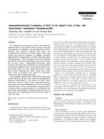
effect was represented by the absolute values of logGI50 in Figure 1-1. GI50 is the drug
concentration that causes 50% cell growth inhibition. Our investigation showed that
MCF-7 and HCT116, which expressed mig-2 gene, were more sensitive (higher logGI50
absolute value) to all the chosen drugs except for vincristine than Colo205 and HT29 that
lost mig-2 expression. In each case the P value was less than 0.05.
This preliminary study, together with the homology between mig-2 and the novel gene
identified in mouse, strongly supported our hypothesis on the possible role of mig-2 in
anticancer drug resistance.
9
MCF7
A
HCT
116
CoLo
205
HT29
<--GAPDH
<--Mig-2
etoposide
8.5
8
7.5
7
6.5
6
5.5
5
4.5
4
doxorubicin
5-FU
mitomycin C
vincristine
9
T2
LO
C
O
H
20
6
11
H
C
T-
C
M
5
cisplatin
F7
|logGI50|
B
|logGI50| of different drugs in the 4 cell lines
FIGURE1-1 Sensitivity against different drugs in 4 cell lines. A) One-Step RT-PCR
showing the expression status of mig-2 in the 4 cell lines. B) The absolute value of
logGI50 of anticancer drugs against these 4 cell lines, representing cell susceptibility to
the above anticancer drugs.
10
1.2.2
Possible role of mig-2 in cell growth control
1.2.2.1 Overview of cell cycle and growth control
The mammalian cell cycle is the focus of a large number of studies at both genetic and
molecular level. The cell cycle can be described as the period between the formation of
the daughter cell, by division of a mother cell, and the subsequent time in which the cell
divides to form two more daughter cells (Mitchison, 1971). This period was initially
divided into two parts called interphase and mitosis. With the advent of radiographic and
cytophotometric techniques, interphase was further divided into four phases. The
pioneering studies of Howard and Pelc in 1951 were the first to use radiographic to
determine when DNA was being actively synthesized (Howard and Pelc, 1951, 1953).
These studies, along with additional microscopic observations, provided a means to
introduce the concept of the cell cycle. They divided the cycle into four stages: (1) the
presynthetic gap or gap1 (G1); (2) DNA synthesis (S phase), the time of bulk
incorporation of radio-labeled DNA precursor and subsequent DNA replication; (3) the
post-synthetic gap or gap 2(G2), the interval between the end of S and the initiation of
mitosis; and (4) mitosis, the period during which the chromosomes condense, and
cytokinesis is completed (Howard and Pelc, 1953).
The cell cycle described above was accepted for those cells that were actively
dividing. However, it soon became obvious that in an adult animal, not all cells were
actively dividing at all time. Thus a further modification was introduced into this system.
With the addition of those non-dividing cells, the diagram of the mammalian cell cycle
had to be modified to take into account the fact that some cells continually divide, others
leave the cycle but remain the ability to reenter it, and still others leave and never
11
proliferate again (Wier and Scott, 1986). Thus the modified model of cell cycle included
actively proliferating cells, which cycle through G1, S, G2 and M; non-dividing cells or
terminally differentiated (TD) cells and the non-dividing or quiescent (G0) cells.
1.2.2.2 Mig-2 involved in mitogenic signaling cell cycle control
Because mig-2 was identified in the progression of G0
S phase upon serum
stimulation (Wick et al, 1994), it was regarded as a gene involved in the mitogenmediated cell cycle regulation and possibly stimulating cells to proliferate. It was noted
that most of anticancer drugs were primarily effective against rapidly dividing cells
(Tomida and Tsuruo, 1999), which further supported our hypothesis that mig-2 might
enhance the cell sensitivity in cancer chemotherapy.
To initiate the mitogenic response, growth factors interact with, and consequently
activate, specific membrane-bound receptors. Receptor activation, in turn, stimulates the
formation of various signaling molecules, which transfers the mitogenic signal from cell
membrane to the internal part. Regarding the cell cycle regulation, it is known that
mitogenic signaling by growth factors activates the Ras-Raf-MEK-ERK signal
transduction cascade (Schaeffer and Weber, 1999). Ras proteins are located at the inner
face of the plasma membrane where they serve as relay switches to transmit extracellular
signal-mediated stimuli to cytoplasmic signaling cascades (Boguski and McCormick,
1993). Ras proteins function as GDP/GTP-regulated switches that cycle between an
active GTP-bound state and an inactive GDP-bound state. Mitogenic signals stimulate a
transient formation of active GTP-bound Ras and activated Ras in turn interacts with
downstream effector targets. The intensively characterized Ras effectors are the Raf
serine/threonine kinases (Campbell et al., 1998; Shields et al., 2000). Activated Raf
12