MOLECULAR AND FUNCTIONAL CHARACTERISATION OF THE ROLE OF HYDROGEN SULPHIDE IN SEXUAL MEDICINE
Bạn đang xem bản rút gọn của tài liệu. Xem và tải ngay bản đầy đủ của tài liệu tại đây (2.19 MB, 151 trang )
MOLECULAR AND FUNCTIONAL CHARACTERIZATION OF
THE ROLE OF HYDROGEN SULPHIDE IN SEXUAL MEDICINE
ROESWITA LEONO LIAW
B.Sc. (Hons), National University of Singapore
A THESIS SUBMITTED FOR THE DEGREE OF
MASTER OF SCIENCE
DEPARTMENT OF OBSTETRICS & GYNAECOLOGY
NATIONAL UNIVERSITY OF SINGAPORE
2011
i
ii
ACKNOWLEDGEMENTS
First and foremost, I would like to express my heartfelt gratitude to my project supervisor,
Professor Ganesan P Adaikan for the great opportunity to work on this interesting project and
also for his invaluable advice, patient guidance and encouragement throughout the course of
this project.
I would also like to thank Dr Balasubramanian Srilatha, my co-supervisor, for her helpful
input and constructive suggestions which were instrumental to the development of the project.
My sincere thanks also go out to Dr Jun Meng for the friendship, advice, support, bantering of
ideas along the way as well as for sharing his expertise and setting aside time for consultation
on troubleshooting problems with regards to real time PCR and western blot. I would also like
to thank Miss Maryam Jameelah, past member of this lab who has done a good job in
maintaining an orderly lab environment and also for providing assistance.
I would like to extend my appreciation to both the academic and non-academic staff of the
Department of Obstetrics & Gynaecology, NUS for the kind help they rendered along the
way.
I would also like to express my gratitude to the National University of Singapore for granting
me the graduate research scholarship and hence allowing me to pursue my interest in
research. The project was made possible under the NMRC grant (R-174-000-104-213)
awarded to my supervisors.
i
Last but not least, I would like to express my heartfelt appreciation to my parents and family
members. Without their strong support and loving encouragements, this project would not
have reached fruition.
ii
TABLE OF CONTENTS
ACKNOWLEDGEMENTS...................................................................................................... i
TABLE OF CONTENTS........................................................................................................ iii
SUMMARY ............................................................................................................................. vi
LIST OF FIGURES .............................................................................................................. viii
LIST OF TABLES ................................................................................................................... x
LIST OF ABBREVIATIONS ................................................................................................ xi
1.
INTRODUCTION............................................................................................................ 1
1.1 Penile structure and innervation ....................................................................................... 1
1.2 Erectile dysfunction ......................................................................................................... 3
1.2.1 Pathophysiology of erectile dysfunction ................................................................... 4
1.2.2 Management of erectile dysfunction ......................................................................... 5
1.3 Gasotransmitters .............................................................................................................. 7
1.3.1 Hydrogen sulphide .................................................................................................... 8
1.3.1.1 Overview of H2S ................................................................................................ 8
1.3.1.2 Biosynthesis of H2S............................................................................................ 9
1.3.1.3 Metabolism of H2S ........................................................................................... 11
1.3.1.4 Roles of H2S in erectile function ...................................................................... 12
1.3.2 Nitric oxide ............................................................................................................. 16
1.3.2.1 Overview of NO ............................................................................................... 16
1.3.2.2 Biosynthesis of NO .......................................................................................... 16
1.3.2.3 Metabolism of NO ........................................................................................... 19
1.3.2.4 Roles of NO in erectile function ...................................................................... 20
1.3.2.5 RhoA/Rho-kinase in contractile mechanism ................................................... 21
1.3.3 Cross talk between H2S and NO ............................................................................. 22
2.
RESEARCH INTEREST AND OBJECTIVES .......................................................... 25
3.
MATERIALS AND METHODS .................................................................................. 26
3.1 Materials ........................................................................................................................ 26
3.1.1 Drugs ....................................................................................................................... 26
3.1.2 Chemicals ................................................................................................................ 26
3.2 Experimental Methods ................................................................................................... 28
3.2.1 Cell culture .............................................................................................................. 29
3.2.1.1 Media preparation ............................................................................................ 29
iii
3.2.1.2 Isolation of rat erectile tissue ........................................................................... 29
3.2.1.3 Primary culture of rat corpus cavernosum smooth muscle .............................. 29
3.2.1.4 Trypan blue exclusion assay ............................................................................ 31
3.2.2 Experimental protocol to investigate the involvement of second messenger cGMP
and cAMP in H2S action .................................................................................................. 31
3.2.2.1 Measurement of cGMP and cAMP concentration ........................................... 32
3.2.3 Experimental protocol to investigate effects of H2S on erectile function in vivo ... 33
3.2.3.1 Measurement of intracavernosal pressure ........................................................ 34
3.2.4 Experimental protocol to investigate effects of H2S on biochemical parameters in
vivo ................................................................................................................................... 37
3.2.4.1 Measurement of H2S production (CBS/CSE activity) in corpus cavernosum . 37
3.2.4.2 Measurement of plasma H2S concentration ..................................................... 38
3.2.4.3 Measurement of NO concentration in plasma and corpus cavernosum ........... 38
3.2.5 Experimental protocol to investigate effects of H2S on expression of targeted
mRNAs in vitro ................................................................................................................ 39
3.2.5.1 Extraction of total RNA from rat corpus cavernosum ..................................... 39
3.2.6 Reverse transcription of RNA to cDNA ................................................................. 41
3.2.7 Real Time (Quantitative) RT-PCR.......................................................................... 41
3.2.8 Experimental protocol to investigate the effects of H2S on expression of target
proteins in vitro ................................................................................................................ 44
3.2.8.1 Protein extraction from rat corpus cavernosum tissue ..................................... 44
3.2.8.2 Isolation of cytoplasmic and total membrane protein ...................................... 44
3.2.8.3 Western blot ..................................................................................................... 45
3.2.9 Experimental protocol to investigate the involvement of testosterone in H2S’
effects ............................................................................................................................... 46
3.2.9.1 Castration procedure in rat model .................................................................... 47
3.2.9.2 Measurement of testosterone concentration ..................................................... 47
3.2.10 Statistical analysis ................................................................................................. 48
4.
RESULTS ....................................................................................................................... 49
4.1 Effects of treatments in vivo........................................................................................... 49
4.2 Effects of treatments on NO level in plasma and corpus cavernosum in vivo ............... 51
4.3 Effects of treatments on H2S level in plasma and H2S production in corpus cavernosum
in vivo ................................................................................................................................... 53
4.4 Effects of NaHS on cGMP and cAMP level in vitro ..................................................... 54
4.5 RNA samples ................................................................................................................. 56
4.6 Gene expression of eNOS .............................................................................................. 56
iv
4.7 Gene and protein expression of sGCα1 and sGCβ1 ....................................................... 57
4.8 RhoA/Rho-Kinase pathway ........................................................................................... 63
4.8.1 Gene expression of RhoA, ROCK I and ROCK II ................................................. 63
4.8.2 Protein expression of RhoA and ROCK II .............................................................. 66
4.9 Effects of testosterone .................................................................................................... 70
4.10 Summary of results ...................................................................................................... 73
5.
DISCUSSION ................................................................................................................. 74
5.1 Effects of H2S on erectile response ................................................................................ 74
5.2 Relationship between H2S, NO and erectile function .................................................... 75
5.3 Effects of H2S on the cGMP and cAMP second messenger system .............................. 80
5.4 Effect of H2S on eNOS .................................................................................................. 84
5.5 Effects of H2S on sGC ................................................................................................... 85
5.6 Effects of H2S on RhoA/Rho-Kinase pathway .............................................................. 89
5.7 Effects of testosterone .................................................................................................... 93
6.
CONCLUSION............................................................................................................... 96
7.
BIBLIOGRAPHY .......................................................................................................... 98
v
SUMMARY
Hydrogen sulphide (H2S) is an endogenously produced gasotransmitter with a similar role as
nitric oxide (NO) which has long been recognised as an important mediator in erectile
physiology. Several studies have investigated the role of H2S in erectile function and H2S was
found to exert definitive pro-erectile effects. The aim of this thesis is to elucidate the
contribution of H2S to erectile response and shed some light on the mechanism(s) involved,
including any possible cross talk between H2S and NO.
It was observed that NaHS, a H2S-donor, significantly improved the magnitude of erectile
response to cavernous nerve electrical stimulation in rats. This improvement was associated
not only with an increase in the systemic H2S concentration and H2S biosynthesis in the
corpus cavernosum (CC) of these rats but also with increased production of NO in both
plasma and CC. The cross talk between H2S and NO was evident in this tissue. Further in
vitro studies revealed that H2S increased endothelial nitric oxide synthase (eNOS) mRNA
expression and cyclic guanosine monophosphate (cGMP) level. Moreover, H2S also exerted
an effect on the NO pathway downstream of NOS, namely increasing the expression of both
the active and inactive forms of soluble guanylyl cyclase (sGC) β1 and stimulating the
translocation of sGCα1 from the cytosol to the membrane. Overall, H2S seems to play a
‘supportive’ role with respect to NO pathway in erectile physiology, amplifying NO
signalling through dual action of increasing NO production and sensitizing the sGC towards
NO. In addition, studies using castrated animals demonstrated that testosterone is not a
requirement for the pro-erectile effect of H2S; however, testosterone is clearly implicated in
this cross talk. High testosterone level seems to favour the cross talk, with H2S boosting NO
production in this condition while low testosterone seems to cause H2S to ‘switch’ to an NOindependent mechanism for its pro-erectile effect. Interestingly, H2S seems to act as a backup
when the NO pathway is compromised. Under condition of high NO (observed in animals
treated with sildenafil), normal H2S level and production were observed, while under
vi
condition of low NO (observed in animals treated with NO synthase inhibitor L-NAME), high
H2S level was observed. Thus, shortage of NO can trigger the production of H2S, which can in
turn stimulate the production of NO. The finding from this study that exogenous H2S seems to
stimulate endogenous H2S production also shed some light on the possible auto-regulation of
H2S through positive feedback.
The pro-erectile effect of H2S was also likely to result from its attenuating effect on the
RhoA/Rho-Kinase contractile pathway. In this system, H2S was shown to downregulate the
level of RhoA and Rho Kinase II (ROCK II) proteins which may have direct implication on
corporal smooth muscle tone.
In summary, findings from this thesis work show that H2S plays an important physiological
role in erectile function. It is likely to exert its pro-erectile effects through multiple
mechanisms of action including a complex cross talk with NO as well as modulation of the
contractile, anti-erectile pathway.
vii
LIST OF FIGURES
Figure 1.1
The anatomy and mechanism of penile erection……………….......
3
Figure 1.2
Enzymatic production of H2S............................................................
10
Figure 1.3
Non-enzymatic endogenous production of H2S................................
11
Figure 1.4
H2S metabolism.................................................................................
12
Figure 1.5
H2S as an inhibitor of superoxide formation.....................................
15
Figure 1.6
Synthesis of NO from L-arginine......................................................
18
Figure 1.7
Relaxation of penile smooth muscle via the NO/cGMP pathway.. ...
21
Figure 3.1
Schematic diagram of the colorimetric competitive EIA for cGMP
measurement.....................................................................................
33
Figure 3.2
Schematic representation of experimental protocol for in vivo study
34
Figure 3.3a
Animal preparation and the pelvic plexus........................................
36
Figure 3.3b
Perineal anatomy of the rat...............................................................
36
Figure 4.1
Effects of treatments on magnitude of erectile response to electrical
stimulation…………………………………………………………
50
Effects of chronic in vivo treatments of sildenafil, NaHS, L-NAME
and PAG on nitric oxide concentration in (A) plasma and (B) corpus
cavernosum.......................................................................................
52
Effects of chronic in vivo treatments of sildenafil, NaHS, L-NAME
and PAG on(A) hydrogen sulphide concentration in plasma and (B)
hydrogen sulphide production in corpus cavernosum................... ..
54
Effects of 30 minutes incubation of NaHS at indicated dosage
on cGMP concentration in primary culture of rat corpus cavernosum
at passage 1-3……………………………………………………....
55
Effects of 30 minutes incubation of NaHS at indicated dosage
on cAMP concentration in primary culture of rat corpus cavernosum
at passage 1-3……………………………………………………. .
55
Relative expressions of eNOS mRNA in rat CC after NaHS treatment
at different time points as assessed by real time PCR....................
57
Relative expression of sGCα1 mRNA in rat CC as assessed by
real time PCR...................................................................................
58
sGCα1 protein expression in rat corpus cavernosum (TMP) in
control and NaHS treated group......................................................
59
Figure 4.2
Figure 4.3
Figure 4.4
Figure 4.5
Figure 4.6
Figure 4.7
Figure 4.8a
viii
Figure 4.8b
sGCα1 protein expression in rat corpus cavernosum (cytosolic fraction)
in control and NaHS treated group.................................................
60
Figure 4.9
Relative expression of sGCβ1 mRNA in rat CC as assessed by
real time PCR...................................................................................
61
Temporal expression of sGCβ1 protein in rat corpus cavernosum
(total tissue lysate)...........................................................................
61
sGCβ1 protein expression in rat corpus cavernosum (TMP) in
control and NaHS treated group......................................................
62
Figure 4.10
Figure 4.11a
Figure 4.11b
sGCβ1 protein expression in rat corpus cavernosum (cytosolic fraction)
in control and NaHS treated group............................................... .
63
Figure 4.12
Relative expression of RhoA mRNA in rat CC as assessed by
real time PCR..................................................................................
65
Relative expression of ROCK II mRNA in rat CC as assessed by
real time PCR................................................................................
65
RhoA protein expression in rat corpus cavernosum (TMP) in
control and NaHS treated group.....................................................
67
RhoA protein expression in rat corpus cavernosum (cytosolic fraction)
in control and NaHS treated group...............................................
68
ROCK II protein expression in rat corpus cavernosum (TMP) in
control and NaHS treated group....................................................
69
Figure 4.13
Figure 4.14a
Figure 4.14b
Figure 4.15a
Figure 4.15b
ROCK II protein expression in rat corpus cavernosum (cytosolic fraction)
in control and NaHS treated group................................................
70
Figure 4.16
Effects of castration and treatment on plasma testosterone
total level.......................................................................................
71
Effects of NaHS and testosterone treatment on the magnitude of
erectile response (ICP/MAP) in normal and castrated rats...........
72
Effects of NaHS and testosterone treatment on plasma NO
concentration...............................................................................
72
Relaxant and anti contractile effects of H2S................................
96
Figure 4.17
Figure 4.18
Figure 6.1
ix
LIST OF TABLES
Table 1
List of reagents, chemicals and kits used........................................
28
Table 2
In vitro treatment of rat CC primary culture for cGMP and
cAMP measurement........................................................................
31
Table 3a
Real time RT-PCR mixture.............................................................
42
Table 3b
Primer sequences for each gene of interest, including eNOS,
sGCα1, sGCβ1, ROCK I, ROCK II and β-Actin......................... .
42
Antibody information (primary and secondary) and the
conditions used in western blot for sGCα1, sGCβ1, RhoA
and ROCK II.................................................................................. ..
46
Table 4
x
LIST OF ABBREVIATIONS
5-HT
5-hydroxytryptamine
AAT
aspartate (cysteine) aminotransferase
AAV
adeno-associated virus
AC
adenylyl cyclase
AOAA
aminooxyacetic acid
AS
argininosuccinate
ASL
argininosuccinate lyase
Asp
L-aspartate
ASS
argininosuccinate synthase
BH4
tetrahydrobiopterin
cAMP
cyclic adenosine monophosphate
CBS
cystathionine β-synthase
CC
corpus cavernosum
CDO
cysteine deoxygenase
cGK
cGMP-dependent protein kinase
cGMP
cyclic guanosine monophosphate
CO
carbon monoxide
CSD
cysteine sulphinate decarboxylase
CSE
cystathionine γ-lyase
DAG
diacylglycerol
DMEM
Dulbecco’s modified Eagle’s medium
EC
enzyme commison number
ED
erectile dysfunction
EDRF
endothelium-derived relaxing factor
EIA
enzyme immunoassay
ELISA
enzyme-linked immunosorbent assay
xi
eNOS
endothelial nitric oxide synthase
ET-1
endothelin-1
FAD
flavin adenine dinucleotide
FMN
flavin mononucleotide
GDP
guanosine diphosphate
GPCR
G protein-coupled receptor
GTP
guanosine triphosphate
H2S
hydrogen sulphide
HRP
horseradish peroxidase
HSP
heat shock-related protein
hVSMCs
human vascular smooth muscle cells
IBMX
3-Isobutyl-1-methylxanthine
ICP
intracavernosal pressure
iNOS
inducible nitric oxide synthase
IP3
inositol triphosphate
K2HPO4
potassium phosphate dibasic trihydrate
KH2PO4
potassium dihydrogen phosphate
KHPO4
potassium hydrogen phosphate
L-NAME
Nω-Nitro-L-arginine methyl ester hydrochloride
LPS
lipopolysaccharide
MAP
mean arterial pressure
MBS
myosin binding subunit
MLC
myosin light chain
MLCK
myosin light chain kinase
MLCP
myosin light chain phosphatase
MPST
3-mercaptopyruvate sulphurtransferase
mRNA
messenger ribonucleic acid
NADPH
nicotinamide adenine dinucleotide phosphate
xii
NaHS
sodium hydrosulphide hydrate
NANC
non-adrenergic non-cholinergic
nNOS
neuronal nitric oxide synthase
NO
nitric oxide
NO2-
nitrite
NO2
nitrogen dioxide
NO3
nitrogen trioxide
NO3-
nitrate
NOS
nitric oxide synthase
NTC
no template control
ODQ
1H-[1,2,4]oxadiazolo[4,3-a]quinoxalin-1-one
ONOO
peroxynitrite
OP
open probability
PAG
DL-propargylglycine
PDE
phosphodiesterase
PE
phenylephrine
PGE1
prostaglandin E1
PKA
protein kinase A
PKB
protein kinase B
PKG
protein kinase G
RhoGDI
rho-guanine dissociation inhibitor
RhoGEFs
guanine nucleotide exchange factors
ROCK
rho kinase
RQ
relative quantitative value
rRNA
ribosomal ribonucleic acid
RSNO
s-nitrosothiols
SEM
standard error of mean
sGC
soluble guanylyl cyclase
xiii
SNP
sodium nitroprusside
SO
sulphite oxidase
SOD
superoxide dismutase
STZ
streptozotocin
T1/2
half-life
TMP
total cellular membrane protein
TSMT
thiol S-methyltransferase
TST
thiosulphate:cyanide sulphurtransferase
VIP
vasoactive intestinal polypeptide
Zn
zinc
xiv
1. INTRODUCTION
1.1 Penile structure and innervation
The erectile tissue is comprised of two functional compartments namely the paired corpora
cavernosa and corpus spongiosum. The corpora cavernosa consist of smooth muscle fibers
intertwined in the extracellular matrix of collagen and elastin; they are surrounded by multiple
interconnecting sinusoidal spaces called lacunae and eventually by a thick fibroelastic sheath,
the tunica albuginea (Figure 1.1) (Lue, 2000). Arterial blood flow to the corpus cavernosum
(CC) is provided by the cavernosal arteries through branches of multiple resistance helicine
arteries which lead directly into the lacunae. Venous outflow from the corpus cavernosum is
provided by subtunical venous plexus which drains blood from the lacunae into emissary
veins that pierce through the tunica albuginea and eventually into the deep dorsal vein (Banya
et al., 1989; Porst and Sharlip, 2006). When the smooth muscles of the helicine arteries are
relaxed, blood inflow to the lacunar spaces increases. Relaxation of the smooth muscle of the
trabeculae then dilates the lacunae, allowing for the expansion of the erectile tissue against the
tunica albuginea which in the process, compresses the subtunical venules against the tunica
(the stretching of the tunica also compresses the emissary veins), restricting the venous
outflow. Penile erection is achieved through this combined increase in arterial inflow and
reduction in venous outflow; a process referred to as the veno-occlusive mechanism (Saenz de
Tejada et al., 1991). Full erection phase is achieved when the increase in intracavernous
pressure (to around 100 mmHg from 10-15 mmHg in the flaccid state) lifted the penile body
from its dependent position to an erect state. This is followed by the rigid erection phase
where the pressure becomes suprasystolic (>120 mmHg) with the contraction of the perineal
(ischiocavernosus) muscles (Dean and Lue, 2005).
The penis is innervated by both somatic (dorsal) and autonomic nerve fibers (Lue, 2000). In
the pelvis, they merge to form cavernous nerves. The somatic nerves supply the penis with
1
sensory fibers and are therefore primarily responsible for penile sensation. They also supply
the perineal skeletal muscles with motor fibers to facilitate the contraction of the pelvic floor
smooth muscle which would help to increase the corporeal body pressure and subsequently
help to achieve maximum rigidity and ejaculation (Kandeel et al., 2001). The autonomic
nerve supplies are comprised of parasympathetic and sympathetic branches, which are
involved in the initiation and inhibition of erection respectively (Steers, 1994). The
parasympathetic nerve fibers divide into two different nerve terminals upon entering the CC:
1) cholinergic (acetylcholine) nerve terminals at endothelial cells and 2) non-adrenergic, noncholinergic (NANC) nerves ending at cavernosal smooth muscles (Adaikan et al., 1991).
Erection inducing/stimulatory neurotransmitters include those from central nervous system
such as dopamine (via D2 receptors) (Andersson, 2001), melanocortins (via melanocortin
receptors) (Martin et al., 2002), serotonin (via 5-HT receptor 2C (Stancampiano et al., 1994;
Millan et al., 1997)), glutamate (Zahran et al., 2000), EP peptides (hexarelin peptide
analogues), vasoactive intestinal polypeptide (VIP) (Ottesen et al., 1984; Adaikan et al.,
1986); neurotransmitters from the peripheral nervous system such as acetylcholine
(Andersson, 2001), and NANC such as nitric oxide (NO) (Burnett et al., 1992; Burnett, 2002).
The sympathetic nervous system mediates corporal vasoconstriction and smooth muscle
contraction and therefore, has a role in maintaining penis in flaccid state as well as in
mediating detumescence after orgasm. The sympathetic nerve fibers innervate cavernous
smooth muscle (stimulating α1 adrenoceptors) and cavernous vessels (stimulating mostly α2
adrenoceptors in penile and cavernous arteries (Andersson and Wagner, 1995) and mostly β2
adrenoceptors in helicine arteries (Saenz de Tejada et al., 1996)).
Generally, penile erection is associated with relaxation of the corporal smooth muscle and
flaccidity with contraction. The relaxation of the smooth muscle in the penile vasculature is
also as important in erectile physiology as cavernosal smooth muscle relaxation. A balance
exists between smooth muscle contraction and relaxation and this is regulated for the most
part through a complex interplay of autonomic neurotransmitters.
2
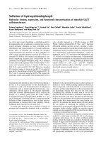
Figure 1.1 The anatomy and mechanism of penile erection. The cavernous (autonomic)
nerves regulate penile blood flow during detumescence and erection while the dorsal
(somatic) nerves are mainly responsible for penile sensation. The mechanisms of erection and
flaccidity are shown in the inserts (Lue, 2000).
1.2 Erectile dysfunction
Erectile dysfunction (ED) is defined as the persistent inability to generate enough corporal
body pressure necessary for vaginal penetration and/or the failure to maintain this level of
rigidity in the penis until ejaculation for satisfactory sexual performance (Lizza and Rosen,
1999). It is a major health concern not only because it can significantly affect the quality of
life but also because of its relatively high prevalence; the combined prevalence of ED
(including mild moderate and complete) was estimated to be approximately 52% in men aged
3
between 40 to 70 years (Feldman et al., 1994). It is also strongly associated with age and can
be correlated with hypertension and heart disease (Feldman et al., 1994). In fact, ED has been
found to be a likely indicator of systemic vascular disease and may serve as an early warning
for cardiovascular events such as myocardial infarct or stroke (Speel et al., 2003; Thompson
et al., 2005; Montorsi et al., 2003b). The risk of ED was found to be 26/1000 every year and
this incidence increases with age, hypertension, heart disease and diabetes (Johannes et al.,
2000). In the local context, ED is found to be common amongst Singaporean men; the
prevalence for ED is 42% in forty-year old men and is as high as 77% in sixty-year old men
(Tan et al., 2003).
1.2.1 Pathophysiology of erectile dysfunction
Erectile physiology is an intricate interplay of vascular, neurologic and endocrine factors,
making ED a multifactorial disorder that can be difficult to treat. The dysfunction can be
psychogenic (performance anxiety related) or organic (e.g. as a result of hypertension,
diabetes, hypercholesterolemia, etc). It can also be caused by pharmacological agents (such as
anticholinergic, psychotropic, or antihypertensive medications) (Finger et al., 1997;
Crenshaw, 1996). Medications may get implicated in the development or exacerbation of ED
in several ways: by inhibiting the central/peripheral nervous system, disturbing the
hypothalamic-pituitary-gonadal axis, including androgen production and metabolism, altering
the normal haemodynamics of hypogastric-cavernous arterial beds or by disturbing the
control of the corporal vasomotor system (Goldstein and Krane, 1983). There has also been
evidence that smoking is associated with vascular pathology (including atherosclerosis in the
penile arteries) and may be a major risk factor for ED (Mannino et al., 1994).
Organic cause of ED may be systemic such as endocrinal, vascular, neurological or local in
nature. Systemic diseases such as diabetes mellitus (Feldman et al., 1994; McCulloch et al.,
1980; Hidalgo-Tamola and Chitaley, 2009), renal failure (Palmer, 1999), cancer (Andersen,
4
1985; Cull, 1992) and chronic liver disease (Kew, 1988; Burra et al., 2010) have been
associated with ED. One of the most common forms of ED is related to vascular
insufficiency, which includes arterial and venous insufficiency (Mulcahy, 2006). In arterial
insufficiency, arterial supply is disrupted, usually from atherosclerosis or hypertension,
resulting in poor penile perfusion. Venous insufficiency or leakage refers to inadequate
trapping of blood in the corpora which may be caused by intrinsic abnormality in the smooth
muscle, incomplete smooth muscle relaxation, or primary veno-occlusive dysfunction
(Mulcahy, 2006). Chronic central nervous system disorders (e.g. Alzheimer’s or Parkinson’s
disease, stroke), spinal cord injuries (trauma), or diabetes mellitus (Lue, 2000) may also affect
the erectile pathway, reflexogenic erections and/or erectile response to psychogenic stimuli
(Smith and Bodner, 1993; Courtois et al., 1993). Similarly, local penile disorders such as
Peyronie’s disease (Hellstrom and Bivalacqua, 2000; Lopez and Jarow, 1993; Ralph et al.,
1996), phimosis (Alexander, 1993; Morgentaler, 1999), priapism (El-Bahnasawy et al., 2002),
or any congenital penile malformations/anomalies (Matter et al., 1998) may interfere with
normal erectile function resulting in ED.
1.2.2 Management of erectile dysfunction
There are several ways in which ED can be managed. These include psychological and
behavioural counseling, drug therapy, the use of non-surgical devices (e.g. vacuum pump and
constrictive ring), or surgery (e.g. repair of penile abnormality, penile prosthesis implantation,
arterial revascularization or venous ligation) (Kandeel et al., 2001). The choice of treatment
should be considered based on the etiology behind the dysfunction. Generally, patients
presenting with ED that is secondary to an underlying disease should be treated for the
primary pathology, e.g. diabetic men with better glycemic control have been found to have
lower odds ratio for ED (Fedele et al., 1998). Some drug therapies that have been used with
varying success, are reproductive hormones (androgen replacement in hypogonadal men
presenting with ED) (Arver et al., 1996; Mulhall, 2004), α2-adrenoceptor antagonist
5
(yohimbine) (Ernst and Pittler, 1998), centrally-acting drugs such as dopaminergic agonist
(apomorphine) (Altwein and Keuler, 2001), and long-acting opiate antagonist (naltrexone)
(Brennemann et al., 1993). Besides systemic medications, local vasoactive agents can also be
administered through direct intracavernosal injection (for example papaverine, phentolamine
(Dinsmore, 1990), prostaglandin E1 (PGE1, alprostadil) (Virag and Adaikan, 1987), and VIP
(Adaikan et al., 1986)), or transurethral application (e.g. alprostadil) (Montorsi et al., 2003a).
There are 11 known families of phosphodiesterase (PDE) enzyme systems, comprising of at
least 60 distinct species, each differing in its kinetic properties, substrate specificity and tissue
distribution (Bischoff, 2004). Phosphodiesterase type 5 (PDE-5) is the predominant cGMP
metabolizing enzyme in penile arteries and CC, but it is also localised in lungs, platelet and
vascular smooth muscle cells. Sildenafil, a classical PDE-5 inhibitor, approved in March 1998
and its successors have emerged as the first line of treatment and still are the most widely
prescribed oral therapy for ED (Montorsi et al., 2003a; Al-Shaiji and Brock, 2009), mainly
because of their ease of use, efficacy and relatively low incidence of adverse effects (Fazio
and Brock, 2004). Sildenafil is also known to be highly selective for PDE-5, compared to
other PDEs (Bischoff, 2004). However despite its general efficacy, there remains a
subpopulation of patients with ED (about 30-40%) who are resistant to this treatment
regimen, necessitating a search for alternative approaches (Hatzimouratidis and Hatzichristou,
2005). Sildenafil works by inhibiting PDE-5, the enzyme that breaks down 3’5’-cyclic
guanosine monophosphate (cGMP) - an important mediator in erectile physiology involved in
smooth muscle relaxation - to 5’-GMP (Corbin and Francis, 1999), effectively increasing the
cGMP level and thereby amplifying the cavernosal smooth muscle relaxation occurring after
sexual arousal (Montorsi et al., 2003a). This means that the erectogenic effect of sildenafil
relies very much on prior release of NO following sexual stimulation (the binding of NO to
soluble guanylyl cyclase (sGC) increases the activity of the enzyme which would
subsequently convert GTP to cGMP and increase the cGMP level (Ignarro, 2000)) and/or
6
possibly, the available cGMP pool in the body. Failure of sildenafil therapy that is observed in
some patients may be attributed partly to insufficient production of NO (Rajfer et al., 2002).
Agents whose mechanism of action is independent of the NO/cGMP production may prove to
be useful in pathological cases of ED where the NO/cGMP pathway is compromised.
1.3 Gasotransmitters
The neurotransmission in erectile physiology involves both sympathetic and parasympathetic
pathways of the pelvic region. The sympathetic, anti-erectile neurotransmitter in human
penile tissue is noradrenergic causing contraction of the CC muscle (Adaikan and Karim,
1981; Giuliano et al., 1993); this transmitter is the main agent helping to keep the penis in
rugose state. The parasympathetic neurotransmitter of erection to the cavernosum is not
cholinergic (that is, not releasing acetylcholine, as it is in some other systems in the body) or
adrenergic (that is not releasing noradrenaline). This type of neurotransmission was
discovered and coined as NANC by Burnstock (Burnstock et al., 1964; Burnstock 1972) and
subsequently was termed ‘nitrergic’ by Rand in 1992 (Rand 1992). The existence of NANC in
rat anococcygeus and bovine retractor penis muscle was first reported by Gillespie (Gillespie
1972) and by Klinge and Sjostrand (Klinge and Sjostrand, 1974). Similarly, the identification
of NANC neurotransmission in the human CC was first reported by Adaikan and Karim
(Adaikan and Karim, 1978; Adaikan, 1979) and this neurotransmitter was confirmed to be
nitrergic, releasing NO (Adaikan et al., 1991).
Cellular signaling is usually initiated by the binding of factors or neurotransmitters to
receptors on the plasma membrane. The resulting interaction between ligand and receptor
generates intracellular second messengers which then relay the extracellular signals to
different parts inside the cell, resulting in the modulation of cellular activities. The discovery
of NO as an endothelium-derived relaxing factor (EDRF) in 1987 (Marsh and Marsh, 2000)
represents the identification of cellular signaling mechanism that is receptor-independent. It
7
was observed that NO acted like a classical neurotransmitter, but with a different signaling
mechanism. The term ‘gasotransmitter’ was then conceived to designate this molecule to
distinguish it from classical neurotransmitters (Wang, 2002). Generally, to qualify as
gasotransmitters, the molecules must possess the following characteristics: 1) they must be
endogenously produced; 2) they must be freely permeable to membranes so that their effect(s)
do not need to rely on membrane receptors; 3) their production and metabolism must be
regulated; 4) at physiological concentration, they must have specific and well-defined
function(s); and 5) regardless of whether their effects are mediated by intracellular second
messenger or not, they should have specific molecular and cellular targets (Wang, 2002).
Currently three gasotransmitters have been identified: nitric oxide, hydrogen sulphide (H2S)
and carbon monoxide (CO).
1.3.1 Hydrogen sulphide
1.3.1.1 Overview of H2S
Decades of occupational health and environmental studies have described H2S as a toxic
pollutant that is detrimental to human health. This perspective has undergone a paradigm shift
in recent years with the emergence of evidence for profound physiological effects of H 2S.
Hydrogen sulphide seems to be able to exert a multitude of biological effects, having been
implicated in inflammation (Zanardo et al., 2006), antinociception (Distrutti et al., 2006),
myocardial ischaemia-reperfusion (Elrod et al., 2007), cardiovascular pathology, shock/sepsis
(Mok et al., 2004; Collin et al., 2005), pulmonary hypertension, and diabetes (Łowicka and
Bełtowski, 2007). Essentially, H2S is a lipophilic colorless gas with a ‘rotten-egg’ odor. It is
also a weak acid; it can dissolve in water and dissociates to form HS - and H+ through the
following reaction: H2S ↔ HS- + H+ ↔ S2- + 2H+. The Henderson–Hasselbalch equation
predicts that at the physiological pH of 7.4 and temperature of 37°C, 18.5% of the sulphide
will exist as H2S, with the remaining 81.5% as HS- (Dombkowski et al., 2004). It is still
8
currently unknown which of these molecules (H2S, HS- or S2-) mediate the observed
biological effects of H2S (Whiteman and Moore, 2009).
1.3.1.2 Biosynthesis of H2S
Most of the evidence for the physiological role of hydrogen sulphide is based on the
observation that it is endogenously produced in tissues that are pertinent to its proposed roles
(either as a vasorelaxant or neuromodulator). This means that the methodologies used to
accurately measure this gas, which is both labile and present at relatively low concentration,
must be rigorously assessed in order to avoid potential artifacts. Unfortunately, unlike NO
which can be measured using its stable oxidation products (NO2- and NO3-), H2S has no
known stable or specific end product from its biosynthesis (SO32- and SO42- cannot be used to
measure hydrogen sulphide production as they can also be formed from direct oxidation of Lcysteine with cysteine deoxygenase; refer to Figure 1.2). However, a majority of the studies
(employing different analytical techniques) reported plasma H2S in similar range (25-80 µM
in rat and humans) with few exceptions (Whiteman and Moore, 2009), thereby suggesting that
the measurements are likely to be credible.
Significant amount of H2S is produced in most tissues in mammals including the penile tissue
(Srilatha et al., 2007), with higher production being observed in brain, liver, kidney, and the
cardiovascular system (Doeller et al., 2005; Zhao et al., 2003). The majority of the
endogenous H2S is synthesised from L-cysteine by two pyridoxal-5’-phosphate (vitamin B6)
dependent enzymes, cystathionine β-synthase (CBS, enzyme commission number (EC
4.2.1.22)) and cystathionine γ-lyase (CSE, EC 4.4.1.1) (Figure 1.2). The expression of these
enzymes is tissue-specific; CBS is predominantly found in the central nervous system while
CSE is expressed mainly in the liver, vascular and non vascular smooth muscles (Szabó,
2007). Human penile tissue homogenates express both CBS and CSE mRNA and protein
(d'Emmanuele di Villa Bianca et al., 2009). Another enzyme that can contribute to H2S
9