UNDERSTANDING THE MOLECULAR MECHANISM OF CENTRINS IN TRYPANOSOMA BRUCEI
Bạn đang xem bản rút gọn của tài liệu. Xem và tải ngay bản đầy đủ của tài liệu tại đây (1.83 MB, 125 trang )
UNDERSTANDING THE MOLECULAR MECHANISM OF
CENTRINS IN TRYPANOSOMA BRUCEI
ZHANG YU
B.Sc., Yunnan University
A THESIS SUBMITTED FOR
THE DEGREE OF MASTER OF SCIENCE
DEPARTMENT OF BIOLOGICAL SCIENCES
NATIONAL UNIVERSITY OF SINGAPORE
2012
ACKNOWLEDGEMENT
I would like to express my deepest and most sincere gratitude to my supervisor, Dr.
Cynthia He, for allowing me to join her team to carry on research work in the past years,
her constant support, guidance and encouragement throughout the research work, and
her help in thesis writing. Her innovative insight and logical way of thinking have been
of great value for me, under the influence of which my knowledge was enhanced and
the depth of my scientific thinking was increased quite a lot.
I would sincerely like to express my thanks to Associate Professor J. Sivaraman for
providing valuable suggestions which indeed helped the biophysical studies on centrins
immensely as well as his student K. Thangavelu for assisting me in carrying FPLC and
CD experiment in their lab.
I wish to thank Dr. Wandy Beatty in the Molecular Microbiology Imaging Facility at
Washington University School of Medicine for her assistance with EM analysis.
I would also like to thank all my labmates, both present and past. Their kindness and
friendship enable me to work and study in a lively and active atmosphere. Their
substantial support and valuable suggestions on my official presentations were really
appreciated. Particular thanks are given to Wan Min for her help in in vivo GST
pull-down experiment.
i
I also extend my thanks to my prethesis committee members, A/P Low Boon Chuan,
A/P Liou Yih Cherng and Dr. Maki Murata-Hori for their valuable feedback and advice
during my prethesis presentation.
My sincere appreciation goes to Professor Zhiyuan Gong for recruiting me from China,
enabling me to have such a great opportunity to study in Department of Biological
Sciences, National University of Singapore as a postgraduate student. The experience
of working with so many brilliant people here, which I can never forget, is a great
treasure of my life.
My special appreciation goes to my parents, who h ve een giving me infinite love,
always kept me away from family responsibilities and encouraged me to concentrate on
my study. And I am also grateful to my husband for his self-giving support and care.
Finally, I would like to render my appreciation to National University of Singapore for
providing me the graduate research scholarship during these years.
ii
TABLE OF CONTENTS
ACKNOWLEDGEMENT .............................................................................................. i
TABLE OF CONTENTS ............................................................................................. iii
SUMMARY ............................................................................................................... viii
LIST OF PUBLICATIONS RELATED TO THIS STUDY ......................................... x
LIST OF FIGURES ...................................................................................................... xi
LIST OF TABLES ..................................................................................................... xiii
LIST OF ABBREVIATIONS AND SYMBOLS ....................................................... xiv
Chapter 1
Introduction ............................................................................................... 1
1.1 Trypanosoma brucei ........................................................................................ 1
1.1.1 Trypanosoma brucei, a parasite causing trypanosomiasis .................... 1
1.1.2 Phylogeny ............................................................................................. 2
1.1.3 Cellular anatomy of procyclic T. brucei ............................................... 3
1.1.4 Cell cycle .............................................................................................. 6
1.1.4.1 The major cell cycle events of T. brucei .................................... 6
1.1.4.2 Unusual cell cycle control mechanisms in T. brucei ................. 7
1.2 Centrin.............................................................................................................. 8
1.2.1 EF-hand motif ....................................................................................... 9
1.2.2 Three-dimensional structure of Centrins ............................................ 10
1.2.3 Function of centrins ............................................................................ 13
1.2.3.1 Centrins on contractile structures............................................. 13
iii
1.2.3.2 Centrins on microtubule organizing centers (MTOCs) ........... 14
1.2.3.3 Other cellular functions of centrins.......................................... 15
1.3 TbCentrin2 and TbCentrin4 in T. brucei ....................................................... 16
1.4 Purpose of this study ...................................................................................... 18
Chapter 2
Materials and methods ............................................................................ 20
2.1 Molecular cloning .......................................................................................... 20
2.1.1 Polymerase chain reaction (PCR) ....................................................... 20
2.1.2 DNA gel electrophoresis ..................................................................... 20
2.1.3 Measurement of DNA concentration .................................................. 21
2.1.4 Restriction endonuclease digestion ..................................................... 21
2.1.5 DNA ligation ....................................................................................... 21
2.1.6 Sequencing of DNA ............................................................................ 22
2.1.7 Preparation of heat-shock competent E. coli cell................................ 22
2.1.8 Transformation of E. coli by heat shock ............................................. 23
2.1.9 Isolation of plasmid DNA from E. coli ............................................... 24
2.1.10 Long-term storage of E. coli ............................................................. 24
2.2 Protein methods ............................................................................................. 24
2.2.1 SDS Polyacrylamide Gel electrophoresis (SDS-PAGE) .................... 24
2.2.2 Staining of proteins in SDS-PAGE gels with Coomassie Blue .......... 25
2.2.3 Western blottings ................................................................................ 26
2.2.4 Expression of recombinant proteins in E. coli .................................... 27
2.2.5 His-tagged protein purification ........................................................... 27
iv
2.2.6 GST-tagged protein purification ......................................................... 28
2.2.7 FPLC ................................................................................................... 29
2.2.8 In vitro GST pull-down....................................................................... 29
2.2.9 In vivo GST pull-down ....................................................................... 30
2.2.10 Protein dialysis .................................................................................. 31
2.2.11 Bradford assays ................................................................................. 31
2.2.12 Concentrating protein samples by centrifugation ............................. 32
2.2.13 Circular dichroism (CD) spectroscopy ............................................. 32
2.3 T. brucei ......................................................................................................... 32
2.3.1 Culture of procyclic T. brucei ............................................................. 32
2.3.2. Genomic DNA isolation from T. brucei ............................................ 33
2.3.3 Long-term storage of T. brucei cells ................................................... 34
2.3.4 Transient and stable transfection of procyclic T. brucei ..................... 34
2.3.5 Cloning of stable transformants by serial dilution .............................. 35
2.3.6 RNAi experiment ................................................................................ 35
2.3.7 Immunofluorescence assays of T. brucei ............................................ 36
2.3.8 Sample preparation for immuno cryoEM ........................................... 37
2.4 Yeast two-hybrid screening methods ............................................................. 37
2.4.1. Isolation of mRNA from T. brucei .................................................... 37
2.4.2 Synthesis of first-strand cDNA ........................................................... 38
2.4.3 Amplification of ds cDNA by long distance PCR (LD-PCR) ............ 39
2.4.4 Preparation of yeast competent cells................................................... 39
v
2.4.5 Small-scale yeast transformation ........................................................ 40
2.4.6 Transformation of yeast strain AH109 with ds cDNA and
pGADT7-Rec ............................................................................................... 41
2.4.7 Yeast mating ....................................................................................... 42
2.4.8 Long-term storage of yeast cells ......................................................... 42
Chapter 3
Ca2+-regulated activity of TbCentrin2 and TbCentrin4 .......................... 43
3.1 Brief introduction ........................................................................................... 43
3.2 Results ............................................................................................................ 43
3.2.1 Analysis of the primary structures of TbCentrin2 and TbCentrin4 .... 43
3.2.2 Ca2+-induced electrophoretic mobility shift for TbCentrin2 and
TbCentrin4 ................................................................................................... 45
3.2.3 Analysis of structural changes of TbCentrin2 and TbCentrin4 by
circular dichroism (CD) spectroscopy ......................................................... 47
3.2.4 Ca2+-dependent self-assembly ............................................................ 49
3.2.5 Verification of centrin-centrin interactions by GST pull-down .......... 54
3.3 Discussion ...................................................................................................... 56
Chapter 4
Identification of TbCentrin2- and TbCentrin4-binding partners ............ 61
4.1 Introduction .................................................................................................... 61
4.2 Results ............................................................................................................ 64
4.2.1 Identification of binding partners of TbCentrins by yeast two-hybrid
screening ...................................................................................................... 64
4.2.1.1 Auto activation test of TbCentrin2 and TbCentrin4 ................ 64
vi
4.2.1.2 cDNA library construction ....................................................... 66
4.2.1.3 Yeast two-hybrid screening results using TbCentrin4 as bait
protein .................................................................................................. 68
4.2.1.4 Cellular distribution patterns of SUMO1/Ulp2, beta-adaptin, and
synaptotagmin ...................................................................................... 72
4.2.1.5 Synaptotagmin localized to FAZ-ER ....................................... 74
4.2.1.6 Colocalization between synaptotagmin and TbCentrin4 ......... 77
4.2.1.7 In vitro and in vivo GST pull-down assay to test interaction
between synaptotagmin and TbCentrins .............................................. 79
4.2.2 Search for TbCentrin-binding proteins by homology screening......... 82
4.2.3 Searching proteins containing the motif
[F/W/L]X2W[K/R/H]X21-34[F/W/L]X2W[K/R/H] in T. brucei ............... 87
4.3 Discussion ...................................................................................................... 91
4.3.1 Candidates identified by yeast two-hybrid screening ......................... 91
4.3.2 Candidates identified by the rest two strategies .................................. 93
4.3.3 Continuing on binding-partners identification of TbCentrins ............ 94
Chapter 5
Conclusion and future directions ............................................................ 95
References .................................................................................................................... 98
Appendix: Data of TbCentrin2-RNAi rescue experiment ......................................... 106
vii
SUMMARY
Trypanosoma brucei is the causative agent of sleeping sickness in humans and nagana
in livestock in Africa, posing enormous burden to African healthcare and word wide
economy. In addition to being of great medical and economic importance, the
unicellular eukaryotic parasite with simple anatomy is a model system with advantages
for addressing the fundamental questions on organelle biogenesis and positioning
during the cell cycle. In T. brucei, organelles like basal body, flagellum, Golgi, nucleus,
and kinetoplast (the aggregated mitochondrial DNA) are present in single copies, each
at a characteristic location in the cell. During the cell cycle, all these organelles
duplicate and separate properly before onset of cytokinesis to ensure production of
proliferative daughter cells. Centrins, TbCentrin2 and TbCentrin4, have been
demonstrated to be essential for proper cell cycle progression of T. brucei. In addition
to being localized to basal bodies, TbCentrin2 and TbCentrin4 mark a previously
unknown, bi-lobed structure, which is in close proximity with Golgi apparatus. RNAi
experiment revealed that depletion of TbCentrin2 inhibited duplication of basal bodies,
flagellum, kinetoplast, and Golgi, and subsequent cell division; depletion of
TbCentrin4 has no obvious effect on organelles duplication, but the coordination
between nucleus division and cell division seems to be disturbed. This thesis further
investigated the molecular mechanisms of TbCentrin2 and TbCentrin4 in Trypanosoma
brucei.
viii
Centrins are EF-hand containing proteins that bind Ca2+. They are regulatory proteins
functioning through specific binding partners. The chapter 3 of this thesis confirmed
Ca2+ binding of these two TbCentrins, suggesting the role of these two TbCentrins as
Ca2+ sensors during cell cycle progression. Additionally, while Ca2+-dependent
self-assembly was observed with TbCentrin2, TbCentrin4 did not self-assemble in the
absence or presence of Ca2+. This may partially explain the functional difference of
these two TbCentrins in cell cycle progression as revealed by their different RNAi
phenotypes. The chapter 4 of this thesis describes the efforts in identifying binding
partners of TbCentrin2 and TbCentrin4 in T. brucei. Two proteins, TbPOC5 and
TbFOP, were characterized as putative binding partners of TbCentrins on the basal
bodies. Bi-lobe binding partner(s), however, has/have not been found through these
studies. In the future, while continue to search for bi-lobe centrin binding partner(s), the
functions of the two basal body proteins, TbPOC5 and TbFOP, and the relationship
between either of the two proteins and TbCentrin2/4 shall be further investigated for
comprehensive understanding of the roles of these two TbCentrins on the basal bodies.
ix
LIST OF PUBLICATIONS RELATED TO THIS STUDY
Zhang, Y., and He, C.Y.. Centrins in unicellular organisms: functional diversity and
specialization. Protoplasma. Jul 24, 2011. PMID: 21786168
x
LIST OF FIGURES
Figure 1.1 Schematic representation of major cell cycle events of T. brucei ........ 5
Figure 1.2 Structures of a typical Ca2+-binding EF-hand motif........................... 11
Figure 1.3 Schematic representation of domain organization of centrins ........... 12
Figure 1.4 TbCentrin2 and TbCentrin4 colocalize on basal bodies and bi-lobed
structure, but perform different cellular functions. ...................................... 17
Figure 3.1 Primary structural characteristics of TbCentrin2 and TbCentrin4 ..... 44
Figure 3.2 Gel mobility shift assay of TbCentrin2 and TbCentrin4 .................... 46
Figure 3.3 Circular dichroism spectra of TbCentrin2 (TbCen2) and TbCentrin4
(TbCen4) in the presence and absence of Ca2+ ............................................ 48
Figure 3.4 Purification of TbCentrins directly fused to 6×His ............................ 50
Figure 3.5 Ca2+-induced self-assembly of TbCentrin2 and TbCentrin4 .............. 53
Figure 3.6 Verification of centrin-centrin interactions by GST pull-down assay 55
Figure 4.1 Auto activation test of TbCentrin2 and TbCentrin4 as BD-fusions ... 65
Figure 4.2 Library construction for yeast two-hybrid screening ......................... 67
Figure 4.3 Yeast two-hybrid screening to identify binding partners of TbCentrin4
...................................................................................................................... 70
Figure 4.4 Cellular distribution patterns of SUMO1/Ulp2, beta-adaptin, and
synaptotagmin .............................................................................................. 73
Figure 4.5 Localization of synaptotagmin to the FAZ ......................................... 75
Figure 4.6 Investigation of ultrastructural localization of synaptotagmin by
immuno cryoEM .......................................................................................... 76
Figure 4.7 Overlap between synaptotagmin-YFP and TbCentrin4 (TbCen4) on the
bi-lobed structure ......................................................................................... 78
Figure 4.8 In vitro GST pull-down assay to test the interaction between
TbCentrins and synaptotagmin .................................................................... 80
Figure 4.9 In vivo GST pull-down assay to test the interaction between
synaptotagmin and TbCentrin4 .................................................................... 81
xi
Figure 4.10 Cellular localization of TbPOC5 ...................................................... 85
Figure 4.11 Cellular localization of TbFOP ........................................................ 86
Figure 4.12 Cellular distribution patterns of Tb927.10.8610, Tb927.10.8730 and
Tb11.01.1970 ............................................................................................... 90
xii
LIST OF TABLES
Table 4.1 List of non-redundant proteins identified by yeast two-hybrid screening
...................................................................................................................... 71
Table 4.2 T. brucei homologues of centrin binding proteins identified in other
organisms ..................................................................................................... 84
Table 4.3 List of proteins containing motif:
[F/W/L]X2W[K/R/H]X21-34[F/W/L]X2W[K/R/H] ................................... 89
xiii
LIST OF ABBREVIATIONS AND SYMBOLS
Chemicals and reagents
Ade
adenine
APS
ammonium persulfate
3-AT
3-amino-1,2,4-triazole
BME
β-mercaptoethanol
CaCl2
calcium chloride
CO2
carbon dioxide
DAPI
4', 6-diamidino-2-phenylindole
DMSO
dimethyl sulfoxide
DTT
dithiothreitol
EDTA
ethylenediaminetetraacetic acid
dNTP
deoxyribonucleotide triphosphate
HCl
hydrochloric acid
His
histidine
IPTG
isopropyl-beta-D-thiogalactoside
Kan
kanamycin
KOH
potassium hydroxide
KOAc
potassium acetate
Leu
leucine
LiAc
lithium acetate
MnCl2
manganese chloride
MOPS
3-(N-morpholino)propanesulfonic acid
NaCl
sodium chloride
NaOH
sodium hydroxide
PEG
polyethylene glycol
PMSF
Phenylmethanesulfonyl fluoride
RbCl
Rubidium chloride
xiv
SDS
sodium dodecyl sulphate
SUMO
small ubiquitin-related modifier
TAE
tris-acetate-EDTA
TE
tris-EDTA
TEMED
tetramethylethylenediamine
Tris
tris(hydroxymethyl)-aminomethane
Trp
tryptophan
Units and measurements
∞
infinity
Ω
ohm
µF
microfarads
µl
microliter(s)
µg
microgram(s)
µg/ml
microgram(s) per milliliter
µM
micromolar
g/L
gram(s) per liter
kD/kDa
kiloDaltons
L
liter(s)
min
minute(s)
ml
milliliter(s)
mg/ml
milligram(s) per milliliter
ng
nanogram(s)
nm
nanometer(s)
OD600
optical density at wavelength 600 nm
rpm
revolutions per minute
sec
second(s)
°C
degree celsius
xv
V/cm
volt per centimeter
Others
AD
activating domain
BD
binding domain
BLASTP
basic local search alignment tool (search protein database
using a protein query)
CD
circular dichroism
CDC31
cell division cycle 31
DIC
differential interference contrast
DNA
deoxyribonucleic acid
cDNA
complementary DNA
EM
electron microscopy
ER
endoplasmic reticulum
FAZ
flagellar attachment zone
FPLC
fast protein liquid chromatography
ICL
infraciliary lattice
MTOC
microtubule organizing centre
MtQ
microtubule quartet
MW
molecular weight
NER
nuclear excision repair
NMR
nuclear magnetic resonance
PCR
polymerase chain reaction
RE
restriction enzyme
RNA
ribonucleic acid
mRNA
messenger RNA
RNAi
RNA interference
SPB
spindle pole body
xvi
SSU
small subunit
YFP
yellow fluorescent protein
xvii
Introduction
Chapter 1 Introduction
1.1 Trypanosoma brucei
1.1.1 Trypanosoma brucei, a parasite causing trypanosomiasis
Trypanosoma brucei, a unicellular parasite, is the causative agent of sleeping sickness
in humans and nagana in livestock. The trypanosomiasis caused by T. brucei is mostly
restricted to sub-Saharan Africa, which is the natural habitat for its insect vector, the
tsetse flies (Weller, 2008). While possessing a complex life cycle alternating between
the insect vector and the mammalian host, parasites of two reproductive stages - the
blood stream form stage that causes diseases and the procyclic stage - can be cultivated
in vitro (Cross, 2001). Furthermore, the robust growth of the procyclic stage parasites
in vitro makes them extremely amenable to biochemical and molecular genetic
analyses. There are two types of human sleeping sickness: the chronic disease caused
by T. brucei gambience and the acute disease caused by T. brucei rhodesiense. Both
types of disease are divided into two stages. During the first stage known as the
hemolymphatic stage, the parasite lives in its host lymph and blood. Then, in the second
stage or the meningoencephalitic stage, the parasite breaks the blood brain barrier,
invades and destructs central nervous system. The second stage is characterized by the
symptom of sleeping disorder, hence the n me ‘sleeping sickness’ (Brun et al., 2010;
Jannin and Simarro, 2008). The disease is fatal if it is left untreated. It is estimated by
the World Health Organization that 60 million people are under the threat of sleeping
thickness, posing an enormous burden to African healthcare and word wide economy.
1
Introduction
Furthermore, the subspecies T. brucei brucei is also the causative agent of nagana in
livestock. It infects animals only, but can negatively affect humans through food losses
as a result of live stock disease (Weller, 2008).
1.1.2 Phylogeny
T. brucei belongs to the order kinetoplastidae and family Trypanosomatidae (De Souza,
2001). Members of the order kinetoplastidae are characterized by possessing a
kinetoplast, a disc like aggregation of mitochondrial DNA. Family Trypanosomatidae
is characterized by the presence of a single flagellum (Honigberg, 1963). Because of
the corkscrew-like motion initially observed in some species in this family, like T.
brucei, Greek trypano (borer) soma (body) is used to name this family. Other medically
important species in this family are Trypanosoma cruzi and Leishmania, causing
Ch g ’s disease and Leishmaniasis, respectively (Englund et al., 1982). T. brucei is
among the earliest-branching eukaryotic organisms. It probably embarked on its own
evolutionary branch more than 500 million years ago, prior to its invertebrate and
vertebrate hosts (Cross, 2001), as revealed by a eukaryotic evolutionary tree drawn
according to the small subunit (SSU) ribosomal RNA gene sequences, which have been
used as the standard molecular measure for reconstructing phylogenetic relationships
(Dacks and Doolittle, 2001).
2
Introduction
1.1.3 Cellular anatomy of procyclic T. brucei
Although morphological differences exist between T. brucei at distinct stages of the life
cycle, the procyclic T. brucei acts as a paradigm for the basic architecture of T. brucei
(McKean, 2003). Morphologically, procyclic T. brucei cell has a long and slender
shape (~20µm in length and ~4µm in broadest diameter) with a single flagellum
laterally attached to the cell body in a left-handed helix from close to the posterior end
towards the anterior tip (Figure 1.1 A) (Hoog et al., 2010). The slender cell shape is
maintained by a subpellicular microtubule corset. More than 100 microtubules are
aligned along the long axis of the cell with regular inter-microtubule spacing
(~18-22nm). These microtubules are cross-linked with each other and to the plasma
membrane with their plus (+) end towards posterior and minus (-) end to anterior (Gull,
1999).
Inside the cell, single-copy organelles such as the basal body pair, the Golgi apparatus,
the kinetoplast, the nucleus, and the flagellum are located at fixed positions with
distinct polarity. As schematically represented in Figure 1.1 A, with the nucleus
occupying the centre of the cell, and the kinetoplast near the posterior end, the single
Golgi stack is located between the nucleus and the kinetoplast juxtaposed to the
flagellar pocket, from which the flagellum protrudes out of the cell body (Field et al.,
2000; Sherwin and Gull, 1989; Warren et al., 2004). At the base of the flagellar pocket
is the basal body pair, which is physically linked to the kinetoplast through a tripartite
adhesion complex (Ogbadoyi et al., 2003). The mature basal body seeds a typical 9+2
3
Introduction
axoneme of the flagellum (Sherwin and Gull, 1989). With the exception of the most
anterior part, almost the entire extracellular part of the flagellum is physically attached
to the cell body via the flagellum attachment zone (FAZ), which ends at the tip of the
cell body. On the cytoplasmic side, the FAZ is defined by an electron dense filament
and microtubule quartet (MtQ) with associated endoplasmic reticulum. The MtQ and
associated endoplasmic reticulum are located immediately to the left of the FAZ
filament when viewed from the posterior of end of the cell (Sevova and Bangs, 2009;
Sherwin and Gull, 1989). The microtubules of the MtQ originate near to the basal
bodies, thus having the opposite polarity to the subpellicular microtubules (Lacomble
et al., 2009).
4
Introduction
Anterior
Posterior
(A)
1K1N
(B)
(C)
2K1N
(D)
2K2N
(E)
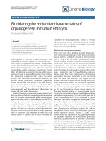
Figure 1.1 Schematic representation of major cell cycle events of T. brucei
Golgi (red dot), nucleus (big blue dot), kinetoplast (small blue dot) flagellum (purple
line) and basal body pair (green), each present at a single copy in an interphase cell; the
single flagellum is attached to the cell body through FAZ (dashed line) (A). When cells
enter the cell cycle, these organelles duplicate and separate in strict order (B, C, and D)
before cytokinesis (E). The duration of cell cycle for procyclic T. brucei cells is ~8.5
hours. T. brucei cell cycle can be divided into three stages: 1K1N, 2K1N and 2K2N
stages, according to the number of nucleus (N) and kinetoplast (K) present in a cell.
New flagellum and FAZ are represented in yellow. Flagellum protrudes out of the cell
body from the flagellum pocket that is not delineated.
5
Introduction
1.1.4 Cell cycle
1.1.4.1 The major cell cycle events of T. brucei
During the cell cycle, the single-copy cellular components must be faithfully duplicated
and properly separated to ensure the continuous reproduction of daughter cells. The
order and timing of cell cycle events have been subjected to extensive investigations
(McKean, 2003). The earliest recognizable morphological events are the duplication of
the basal bodies, the duplication of the Golgi apparatus and outgrowth of a new
flagellum (Figure 1.1 B), which take place concurrent with the kinetoplast DNA
replication. The kinetoplast cycle is different to the nucleus cycle, with kinetoplast
S-phase initiating prior to the onset of nuclear S-phase and the division of kinetoplast
DNA having completed before the onset of nuclear mitosis. According to the number of
nucleus (N) and kinetoplast (K) present in a cell, T. brucei cell cycle is roughly divided
into three stages: 1K1N, 2K1N and 2K2N stages, representing cells containing one
kinetoplast and one nucleus, cells containing duplicated kinetoplasts and one nucleus,
and cells containing duplicated kinetoplasts and duplicated nuclei, respectively (Figure
1.1). In normal conditions, 1K2N cell does not exist since kinetoplast always separates
before separation of nucleus.
The replicated kinetoplast, flagella and Golgi apparatus segregate, powered by the
movement of the basal bodies (Figure 1.1 C). Mitosis then occurs with an intranuclear
spindle formed without disruption of the nuclear envelope (Figure 1.1 D) (Ogbadoyi et
al., 2000). The cytokinesis of T. brucei occurs soon after mitotic chromosomal
6
Introduction
segregation via a unidirectional ingression of a cleavage furrow along the helical axis of
the cell from anterior between the old and new flagella (Figure 1.1 E). It has been
proposed that the structural information required to position the cleavage furrow is
provided by FAZ since it marks a unique seam in the cytoskeleton (Robinson et al.,
1995).
1.1.4.2 Unusual cell cycle control mechanisms in T. brucei
Various cell cycle checkpoints are employed by eukaryotic cells to verify the accuracy
of cell cycle events before progression into the next phase, thus to ensure the fidelity of
cell division. In yeast and mammalian cells, DNA synthesis is monitored by the DNA
replication/damage checkpoints; the mitotic spindle checkpoint ensures chromosome
alignment at the mitotic plate before entry into anaphase; whether the two copies of
DNA are separated sufficiently to initiate cytokinesis is monitored by cytokinesis
checkpoint (Lodish et al., 2000). Although T. brucei shows the typical periodic,
eukaryotic nuclear events, G1, S, G2 and M phases, different cell cycle checkpoints are
present in T. brucei. Besides nuclear DNA, the single copy kinetoplast DNA shows
periodic cell cycle events as well. This is in contrast with most other eukaryotic cells,
which contain multiple mitochondria whose DNA is continuously replicated
throughout the cell cycle (Pica-Mattoccia and Attardi, 1972). It has been observed that
entry into cytokinesis depends on successful kinetoplast replication and segregation
rather than on mitosis. When mitosis is inhibited by rhizoxin or nuclear DNA synthesis
by aphidicolin, cells can continue to divide, producing daughter cells containing one
7