Design and synthesis of novel metal complex protein conjugation agents for applications in bio imaging
Bạn đang xem bản rút gọn của tài liệu. Xem và tải ngay bản đầy đủ của tài liệu tại đây (3.92 MB, 110 trang )
Design an
nd Synthessis of novvel metal complex-p
c
protein coonjugatio
on
agentts for App
plications in bio-im
maging
W
Wang
Tao
o
Supervisoor: A/P Tanja Weiil
N
NATION
AL UNIV
VERSITY
Y OF SINGAPORE
E
2010
Design and Synthesis of novel metal complex-protein conjugation
agents for Applications in bio-imaging
WANG TAO
(BSc, Sichuan University2008 )
A THESIS SUBMITTED FOR THE DEGREE OF
MASTER OF SCIENCE
DEPARTMENT OF CHEMISTRY
NATIONAL UNIVERSITY OF SINGAPORE
2010
Acknowledgements
ACKNOWLEDGEMENTS
I would like to take this opportunity to express my heartfelt gratitude to those who
help me in research and writing thesis.
A special acknowledgement is given to my supervisor, Associate Professor Tanja
Weil, who gives me kind encouragement and useful instructions all through my research.
She is willing to discuss the difficulties encountered in my project and always give
creative suggestions. She is also very kind and considerable, making our group like a
sweet and happy family.
I would like to my sincerely thank my colleagues Dr. Kuan Seah Ling, Wu
Yuzhou, Chen Xi, Goutam Pramanik, Ng Yuen Wah David, They offer me invaluable
advice and appreciate help throughout the project.
I would like to my parents and friends for their consideration and motivation.
Last but not least, I would like to thank the chemistry department of NUS for
giving me the opportunity to undertake this project.
i
Table of Contents
TABLE OF CONTENTS
Acknowledgements
i
Table of Contents
ii
List of Figures
v
List of Schemes
vi
Index of Abbreviations
vii
Abstract
ix
Chapter 1. Introduction
1
1.1 Metal Complex for MRI or PET
1
1.2 The Biological Function of Folic Acid
4
1.3 The Biological Significance of Somatostatin
6
1.4 Chemical Modification of Proteins
9
1.5 Site-specific Intercalation Into Protein Using a Three-carbon Bridge
13
1.5.1 Disulfide Bonds in Therapeutically Relevant Proteins
13
1.5.2 Reduction of Disulfides and Disulfide Site-specific Intercalation
14
1.6 Design of biocompatible metal-complex protein conjugate
Chapter 2. Project Aim and Design
16
18
ii
Table of Contents
Chapter 3. Results and Discussion
20
3.1 Synthesis of 1, 4, 7-tris (tert-butoxycarbonylmethyl)-1, 4, 7, 10tetraazacyclododecane 3 (DO3tBu) (3)
20
3.2 Synthesis of tert-butyl 2, 2’, 2’’-(10-(2-oxo-2-(prop-2-ynylamino) ethyl)
-1, 4, 7, 10-tetraazacyclododecane-1, 4, 7-triyl) triacetate (8)
22
3.3 Synthesis of tert-butyl 2,2'-(4-(2-tert-butoxyallyl)-10-(6-(2,5-dioxo-2,5dihydro-1H-pyrrol-1-yl)hexanoyl)-1,4,7,10-tetraazacyclododecane-1,7diyl)diacetate (10)
23
3.4 Preparation of DOTA-Folate Conjugate
24
3.5 Synthesis of Tailored Linker (16)
26
3.6 Synthesis of Water Soluble Intercalator (21)
27
3.7 Intercalation of Somatostatin
31
Chapter 4. Experimental
33
4.1 General Procedures
33
4.2 Synthesis of 1, 4, 7-tris (tert-butoxycarbonylmethyl)-1, 4, 7, 10tetraazacyclododecane (DO3tBu) (8)
4.3 Synthesis of 2-bromo-N-(prop-2-ynyl) acetamide (6)
34
36
4.4 Synthesis of Tert-butyl 2,2',2''-(10-(2-oxo-2-(prop-2-ynylamino)ethyl)1,4,7,10-tetraazacyclododecane-1,4,7-triyl)triacetate (8)
4.5 Synthesis of 6-maleimideocaproic acid (9)
iii
37
38
Table of Contents
4.6 Synthesis of Tert-butyl 2, 2’-(4-(2-tert-butoxyallyl)
-10-(6-(2, 5-dioxo-2, 5-dihydro-1H-pyrrol-1-yl) hexanoyl)
-1, 4, 7, 10-tetraazacyclododecane-1, 7-diyl) diacetate (10)
40
4.7 Synthesis of Folate-NHS (11)
41
4.8 Synthesis of Folate-DOTA (13)
42
4.9 Synthesis of Folate-DO3tBu (12)
43
4.10 Synthesis of Mannich Salt (14)
46
4.11 Synthesis of Bis-disulfide (15)
47
4.12 Synthesis of Bis-sulfone (16)
48
4.13 Synthesis of Bromoethyl-bis-sulfide (18)
50
4.14 Synthesis of Piperazine-bis-sulfide (23)
52
4.15 Synthesis of Tert-butyl 4-(2-aminoethyl)
piperazine-1-carboxylate (21)
53
4.16 Synthesis of Tert-butyl 4-(2-aminoethyl)
piperazine-1-carboxylate (31)
54
Chapter 5. Conclusion
56
Chapter 6. References
59
Appendix
68
iv
LIST OF FIGURES
Figure
1.1
Page
The structure of 1, 4, 7, 10-tetraazacyclododecane
-1, 4, 7, 10-tetraacetic acid (DOTA) and folic acid
2
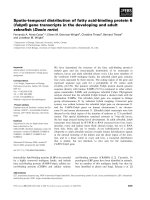
1.2
FR-mediated endocytosis of a folic acid conjugate
5
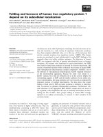
1.3
[18F]-FDG microPET (middle), MR (right), and [66Ga]Ga-DF-Folate
microPET (left) images of mice with subcutaneous folate-receptor-positive
human KB cell tumor xenografts in their intrascapular region
6
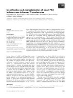
1.4
The structure of somatostatin
7
1.5
(a) Site-specific modification of protein yields homogenity and
(b) Non-specifcification modification of protein results heterogeneity
9
1.6
The bifuncional molecule consisting DOTA complex and folic acid
18
1.7
The bifunctional molecule consisting DOTA complex and folic acid
19
1.8
The bifuncional molecule with maleimide-DOTA complex and
1.9
Protein with free thiol group e.g. BSA
19
The side product in the synthesis of compound 18
29
v
LIST OF SCHEMES
Scheme
2.1
Page
Non-specific modification of a) lysines and
b) cysteines residue on proteins
2.2
11
Mechanism for conjugating a three-carbon
bridge to a native disulfide bond.
2.3
15
Synthesis of tri-tert-butyl
2,2',2''-(1,4,7,10-tetraazacyclododecane-1,4,7-triyl)triacetate (DO3tBu)
2.4
20
Synthesis of tert-butyl 2, 2', 2''-(10-(2-oxo-2-(prop-2-ynylamino) ethyl)
-1, 4, 7, 10-tetraazacyclododecane-1, 4, 7-triyl) triacetate (8)
22
2.5
Synthesis of DOTA-maleimide deriveritives (10)
23
2.6
The first procedure to synthesize Foate-DOTA (13) conjugates
24
2.7
The second procedure to synthesize Foate-DOTA conjugates
25
2.8
Synthesis of bis-disulfone (16) used to
combine metal complex and biomelocule
26
2.9
The second method to synthesized bis-disulfone (16)
27
2.10
Synthesis of water soluble intercalator piperazine bis-sulfone (21)
28
2.11
The second synthetic route towards the water soluble intercalator (21)
29
2.12
The third synthetic route of water soluble intercalator (21)
30
2.13
Intercalation of Somatostatin
31
2.14
Proposed synthetic route for somatostatin DOTA conjugate
58
vi
INDEX OF ABBREVIATIONS
CT
Computed Tomography
d
doublet
DCC
N,N'-dicyclohexylcarbodiimide
DCM
Dichloromethane (Methylene Chloride)
DIEA
N,N-Diisopropylethylamine
DMAP
4-Dimethylaminopyridine
DMF
Dimethylformamide
DMSO
Dimethylsulfoxide
DOTA
1,4,7,10-tetraazacyclododecane-1,4,7,10-tetraacetic acid
EA
Ethyl Acetate
EDC
1-ethyl-3-3(3-dimethylaminopropyl) carbodiimide hydrochloride
ESI
Electron Spray Ionization
Fab
Fast Atom Bombardment
FR
Folate Receptor
HBTU
O-(Benzotriazol-1-yl)-N,N,N′,N′-tetramethyluronium
Hexafluorophosphate
IT-TOF-MS
Ion trap & Time-of-flight Mass Spectrometry
LC-MS
Liquid Chromatography & Mass spectrometry
MIBK
4-Methyl-2-pentanone
MRI
Magnetic Resonance Imaging
PET
Positron Emission Tomography
vii
q
quartet
s
singlet
SPECT
Single-photon-emission Computed Tomography
t
triplet
t-Bu
tertiary butyl
THF
Tetrahydrofuran
TLC
Thin-Layer Chromatography
UV
Ultraviolet
viii
Abstract
Abstract
Targeting particular cells or tissues for imaging e.g. proliferative cells or for
transporting drug molecules plays a vital role in cancer treatment and represents an area
of high scientific interest. In order to contribute to a better detection of proliferative cells,
a sophisticated metal-DOTA imaging agent was designed that is able to specifically
interact with disulfide bridges of proteins by intercalating into accessible disulfide
bridges via two sequential Michael addition-elimination reactions. Such DOTA-protein
conjugates are highly versatile since they can be labeled with
68
Ga for PET or
paramagnetic metals such as Gd(Ⅲ) for MR imaging.
In addition, a folate-metal-DOTA conjugate has been prepared as well as a novel
approach that facilitates somatostatin-metal-DOTA conjugates has been designed for
targeted delivery and first attempts have been undertaken to achieve this challenging goal.
Folic acid or somatostatin-DOTA conjugates require conjugation via a tailored linker.
The synthesis of this linker moiety, the functionalization of the metal-DOTA complex
and the conjugation approach is thoroughly investigated. Based on this strategy, sitedirected labeling of peptides or even larger proteins with a single accessible disulfide
bond such as antibodies becomes feasible. Future work will focus on the in vitro or in
ix
Abstract
vivo evaluation of the Folic acid-DOTA derivative. In addition, a larger number of
gadolinium complexes may be attached to proteins with multiple disulfide bridges which
may yield improved contrast and low detection limit.
x
Chapter 1
Chapter 1.
1.1.
Introduction
Introduction
Metal Complex for MRI or PET
Molecular imaging is one of the most exciting and rapidly growing areas of
science as it enables the characterization and quantification of biological processes at the
cellular and subcellular level in living subjects in an intact manner[1]. It utilizes specific
molecular probes as well as intrinsic tissue characteristics as the source of image contrast,
and offers the opportunity for an improved understanding of integrative biology, earlier
detection and characterization of diseases, and facilitates a better evaluation of
therapeutic treatment[2]. The imaging modalities can be broadly divided into two
categories: anatomical and molecular techniques. Examples of anatomical imaging
technologies include computed tomography (CT) and magnetic resonance imaging (MRI),
which are characterized by high spatial and temporal resolutions. On the other hand,
molecular techniques such as positron-emission tomography (PET) and single-photonemission computed tomography (SPECT) offer excellent sensitivity and often provide
important biochemical information on pathological conditions [3, 4].
1
Chapter 1
Introduction
Figure 1. The structure of 1,4,7,10-tetraazacyclododecane-1,4,7,10-tetraacetic acid
(DOTA) and folic acid
In molecular imaging techniques, the contrast media play an important part in
improving the sensitivity, resolution of images and target specificity at the
molecular/cellular level. However, the toxicity of the contrast agent is a major concern in
its application. For example, lanthanide ions are widely used as MRI contrast agents[5],
radioactive tracers[6], and optical imaging probes[7, 8]. Nonetheless, free lanthanide ions
often exhibit high toxicity in vivo. To circumvent this problem, chelating agents are
extensively used to coordinate to these metal ions and thus minimize their toxicity.
Among various chelating agents, macrocyclic 1,4,7,10-tetraazacyclododecane-1,4,7,10tetraacetic acid (DOTA) (Figure 1) is one of the widely used ligand in molecular imaging
as it outperforms other agents in the ability to form complexes with a large number of
transition and lanthanide metal ions with high thermodynamic stability and kinetic
2
Chapter 1
Introduction
inertness[9]. DOTA complexes, depending on the metal ions, are mainly used in three
areas: magnetic resonance imaging [Gd(Ⅲ), Eu(Ⅲ)], nuclear imaging[111In(Ⅲ), 68Ga(Ⅲ),
64/67
Cu(Ⅱ)], and therapeutic radiopharmaceuticals [90Y(Ⅲ),
177
Lu(Ⅲ)]. Low–molecular-
weight contrast agents for MRI, such as the commercial agent Gd3+-DOTA (DotaremTM)
display low relaxivity and extremely fast excretion rates in vivo. To improve the
relaxivity, Gd(Ⅲ) chelates are conjugated to macromolecules, like proteins[10], micellar
aggregates[11], dendrimers[12], or liposomes[13], thus extending the rotational
correlation lifetime. But most of them are not able to differentiate between “healthy” and
e.g. tumor cells thus preventing cell or tissue-specific molecular imaging. Nonetheless,
remarkable progress has been made in recent years in the development of targeted
contrast agents for diagnostic imaging that allows better differentiation [14, 15]. Various
targeted contrast agents for MRI and PET have been reported which were synthesized via
the
conjugation of metal chelates to various biomolecules, including peptides[16],
proteins[17],
antibodies[18],
oligonucleotides[19]
and
biotin/avidin[20].
These
biomolecules are used as molecular imaging probes which show high binding affinity to
the target receptors, antigens, and nucleic acids being specifically overexpressed in or on
the targeted cells tissues.
3
Chapter 1
1.2.
Introduction
The Biological Function of Folic Acid
Folic acid(Figure 1) is a water-soluble vitamin of the B-complex group and plays
essential roles in numerous bodily functions by participating in the biosynthesis of
nucleic and amino acids [21]. More importantly, it can be utilized for targeted delivery.
Targeted delivery via selective cellular marker improves the efficacy and safety of
the therapeutic and imaging agents. Among cellular surface targets, folate receptors
(FR)-α is most promising and well-investigated in epithelial cancers. The other form of
FR (FR-β) is present in myeloid leukemia and activated macrophages, increasingly
recognized as a cellular target[22]. A variety of molecules including radioimaging agents,
magnetic resonance imaging (MRI) contrast agents, chemotherapeutic agents,
oligonucleotides, proteins, enzyme constructs for prodrug therapy, haptens, liposomes,
nanoparticles and gene therapy vectors have been conjugated to folate for FR-targeted
delivery[22]. FR is significantly upregulated in cancer cells and occurs at very low levels
4
Chaapter 1
Introoduction
in most
m
normaal tissues [223-25]. Follate conjugaates bind FR
F with higgh affinity and are
inteernalized intto tumors viia receptor-m
mediated enndocytosis (Figure
(
2).
e
of a folic acid
a conjugaate[26]
Fiigure 2. FR--mediated endocytosis
The keyy part of tuumor-specifiic imaging is the speciificity of the targeting unit for
mallignant cellls and the capacity of
o the tum
mor-specificc receptor to bind suufficient
quanntities of the
t imagingg agent to achieve high contrastt. In the caase of highh tumor
speccificity , vvery low concentratio
c
on of the
PET imagging agent is needed
d; while
micromolar cooncentration
n is still reequired in MRI[27]. Generally, targeting imaging
i
nts should: (a) show high
h
affinitty (Kd<10−88 M) for thheir cell surrface recepttors, (b)
agen
targget a receptoor that is siignificantlyy upregulateed on canceer cells (>100-fold over normal
cells is preferreed), (c) reveeal rapid cleearance from
m normal (rreceptor neggative) tissuues, and
(d) be uptakenn in sufficcient quantiities by recceptor-exprressing tissuues to alloow high
5
Chapter 1
Introduction
contrast[28]. The folic acid and receptor pair fulfill most of these requirements; hence
they are an attractive ligand/receptor combination for targeted imaging.
Folate-targeted conjugates of radionuclides (Figure 3), like 99mTc[29-31], 111In[32,
33],
66/67/68
Ga[34, 35] and
18
F[36], for SPECT and PET imaging have been developed
and evaluated in preclinical and clinical studies. In addition, a few folate-targeted MRI
contrast agents have also been reported [37, 38].
Figure 3. [18F]-FDG microPET (middle), MR (right), and [66Ga]Ga-DF-Folate microPET
(left) images of mice with subcutaneous folate-receptor-positive human KB cell tumor
xenografts in their intrascapular region[39].
1.3
The Biological Significance of Somatostatin
Somatostatin(Figure 4) is a cyclic tetradecapeptide hormone. It is found in
multiple sites throughout the nervous system, including the cerebral cortex, the brain
6
Chaapter 1
Introoduction
stem
m, the gastrrointestinal tract, and the
t pancreaas. It plays many diverse roles inncluding
inhiibition of enndocrine annd exocrine secretions, modulationn of neurotrransmission
n, motor
and cognitive ffunctions, innhibition off intestinal m
motility, absorption off nutrients annd ions,
vasccular contraactility, and cell prolifeeration[40].
F
Figure
4. Thhe structuree of somatosstatin
Somatoostatin mediiates its bioological effeects throughh interactioon with a faamily of
s
n receptors expressed by a varieety of norm
mal and maalignant
fivee specific somatostatin
tissuues[40]. Som
matostatin receptors
r
haave been iddentified by classical biiochemical binding
7
Chapter 1
Introduction
techniques and in vitro autoradiography on a variety of human tumors, such as pituitary
tumors, endocrine pancreatic tumors, carcinoids, paragangliomas, meningiomas, brain
tumors (astrocytomas), neuroblastomas, and some human breast cancers[41]. Therefore,
somatostatin/somatostatin receptor system is studied intensively in contrast-enhanced
diagnostic imaging and targeted therapy of tumors. The exact mechanism of somatostatin
antineoplastic activity is unknown, but some possibilities are: (1) a direct antiproliferative
effect by blockade of mitogenic growth signal or induction of apoptosis through
interaction with somatostatin receptors; (2) inhibition of secretion of gastrointestinal
hormones thought to be important in tumor growth; and (3) reduction or inhibition of
secretion of growth-promoting hormones and growth factors which stimulate the growth
of cancers[42].
A major progress is made by introducing radiolabelled somatostatin for diagnosis
and treatment of cancers. Somatostatin acts as a bullet to specifically target a maligant
tissue with high affinity through interaction with somatostatin receptors.
8
Chapter 1
1.4.
Introduction
Chemical Modification of Proteins
There is an increasing interest in protein conjugates for diagnosis and therapy.
Even though proteins often display limited pharmacokinetics, low proteolytic stabilities
and the possibility to elicit immune responses, there have been successful attempts of
converting proteins into efficient drug delivery systems or imaging agent [43]. Their
low nanometer sizes, highly defined structures, biodegradability and the presence of a
high number of functional groups available for chemical modifications make them
attractive for applications in targeted drug delivery and bioimaging.
Figure 5. (a) Site-specific modification of protein yields homogenity and (b) nonspecifcification modification of protein results heterogeneity[44]
9
Chapter 1
Introduction
Protein conjugates can be prepared via chemical modification and bioengineering
techniques. Chemical modification approaches can be divided into two major categories;
site-specific and non-specific protein functionalization(Figure 5).
Classical
non-specific
protein
conjugation
techniques
typically
involve
electrophilic reagents targeting the nucleophilic functional groups of lysine (Scheme 1),
cysteine, aspartic acid or glutamic acid side chains, generally providing a heterogeneous
mixture of proteins modified to a different extent and at variable locations in the protein
conjugates[44, 45].
A more specific strategy represents the modification of cysteine residues (Scheme
1) through alkylation with iodoacetamide reagents, disulfide exchange and Michael
addition with maleimides[46-48]. Since free cysteine groups are rare and often
inaccessible, they can be engineered into the protein as point mutations using molecular
biological techniques. Such approaches are usually demanding and expensive and point
mutation may have a negative impact on protein function by altering its structure.
Moreover, introducing an accessible free thiol group often leads to disulfide scrambling,
protein misfolding and an increased tendency to form aggregates during purification. Still,
10
Chapter 1
Introduction
thiol-specific modifications play an important role due to the potential for high-yield
reactions (e.g. Michael reactions), as well as the propensity for addressing cysteine
groups selectively without targeting other amino acids.
Scheme 1. Non-specific modification of a) lysines and b) cysteines residue on proteins
In recent years, significant progress has been made to develop improved strategies
for selective and efficient protein chemistry and thus more well-defined protein
conjugates[47, 48]. New means have been established for the modification of tyrosine
and tryptophan, usually by applying transition-metal-mediated processes that are
11
Chapter 1
Introduction
compatible with aqueous conditions[45]. Tyrosine residues are modified via a threecomponent Mannich reaction with aldehydes and anilines[46, 49]. Targeting tryptophan
residues has been developed by employing rhodium carbenoids in acidic condition (pH≈
2), which may affect the structure of some protein[50]. As hydrophobic amino acids are
generally buried within the protein scaffold, controlled single-site modification of
tyrosines and tryptophans is possible in some cases by improving surface accessibility
often via point mutations[51].
Probably the most elaborate method to site-specifically modify proteins involves
the introduction of non-canonical amino acids (rNCAA) into proteins[45]. Here, by
chemically attaching the desired rNCAA to suppressor tRNA and then placing the amber
codon at the desired position in the mRNA, a number of rNCAA have been incorporated
at different positions into the protein sequence. Successful examples of rNCAA that are
incorporated into the protein sequence include p-iodotyrosine, which undergoes Pdcatalyzed alkenylation ( Mizoroki-Heck reaction ) or alkynylation ( Sonogashira reaction )
reactions from the protein surface[52], Stille coupling using organotin derivatives as well
as Suzuki reactions utilizing boronic acids and esters[45]. Previously, rNCAA with an
azido or ethynyl group has been incorporated into different proteins[53]. Azide-alkyne
12
Chapter 1
Introduction
[3+2] cycloaddition are conducted in the presence of Cu (I) as catalyst, yielding
exclusively the 1,4-substituted triazole isomer. These reactions proceed rapidly in water,
and provide excellent chemoselectivity and regioselectivity [53].
1.5.
Site-specific Intercalation into Proteins using a Three-Carbon Bridge
1.5.1 Disulfide Bonds in Therapeutically Relevant Proteins
In general, free and accessible cysteine residues are rare[54] and liable to pair up
to form disulfides bridges [55, 56]. Disulfide bonds influence the physio-chemical and
biological properties of proteins in many subtle and complex ways[57]. They are either
buried within the protein’s folding region or on its solvent accessible surface[58].
Solvent-accessible disulfides can be selectively approached by tailored reagents and can
be chemically modified. As accessible disulfides primarily contribute to the stability of a
protein rather than to its structure or biological function[59] it is feasible to intercalate
into this bond by tailored reagents without a loss of either structure or function.
Previously, protein databases and molecular modeling programs have been used to
13