The study of the mediation of ohanin, a king cobra (ophiophagus hannah) toxin through the central nervous system
Bạn đang xem bản rút gọn của tài liệu. Xem và tải ngay bản đầy đủ của tài liệu tại đây (2.71 MB, 79 trang )
THE STUDY OF THE MEDIATION OF OHANIN, A KING
COBRA (OPHIOPHAGUS HANNAH) TOXIN THROUGH
THE CENTRAL NERVOUS SYSTEM
TEO CHUNG PIN
(M.Sc University of Glasgow)
A THESIS SUBMITTED FOR THE DEGREE OF MASTERS OF SCIENCE
DEPARTMENT OF BIOLOGICAL SCIENCES
NATIONAL UNIVERSITY OF SINGAPORE
2009
I
Acknowledgements
I would like to thank Professor Manjunatha Kini and Professor Prakash P Kumar for their
kindness and patient guidance to me for the success of this thesis.
I would also like to extend my grateful acknowledgements to Dr. Pung Yuh Fen for her
dedication to this theme and kick-starting the project. Also, to Ms. Shifali Chatrath for her
strength in carrying the flame of Ohanin in the laboratory, thank you.
My heartfelt appreciation to Professor Steve Cheung Nam Sang of the Biochemistry
Department, National University of Singapore, for his tutelage and experience in
neurology and neurochemistry. I would be “lacking nerves” for the project without you.
To Drs. Robin Doley, Md Abu Reza, Kang Tse Siang, Yajnavalka Banerjee, Nandha
Kishore, Pawlak Joanna, Syed Rehana, Dileep Gangadharan, Mr. Koh Cho Yeow, Ms. Liu
Ying and Ms. Tay Bee Ling, who make up the “Prof Kini’s Lab” staff, students and alumni,
for brainstorming with me during the “official and unofficial” group meetings but also
making life in the laboratory go beyond the stoid laboratory work, and making it enjoyable.
Last but definitely not the least, I would also like to thank my parents, Teo Gee Huat and
Pauline Ong, and my wife, Delia Heng, who have shown incredible tolerance towards my
completion of this project.
II
Table of Contents
Acknowledgements ............................................................................................................. I
Table of Contents...............................................................................................................II
LIST OF FIGURES .........................................................................................................IV
LIST OF ABBREVIATIONS............................................................................................ V
ABSTRACT.........................................................................................................................1
INTRODUCTION...............................................................................................................2
Snakes – Evolution and Phylogeny.................................................................................. 2
Venomous Snakes ............................................................................................................. 5
Snake Venoms and Their Pharmacology........................................................................ 8
Importance of studying Snake Venom............................................................................ 9
Ophiophagus hannah – King Cobra .............................................................................. 10
Ohanin – A Specialised Neurotoxin found in Ophiophagus hannah .......................... 12
Binding Studies of Ohanin and Pro-ohanin ................................................................. 14
MATERIALS AND METHODS ......................................................................................18
Animals. ........................................................................................................................... 18
Isolation and purification of native ohanin. ................................................................. 19
Expression and purification of His-ohanin and His-pro-ohanin. ............................... 20
Cleavage of fusion protein.............................................................................................. 22
Molecular mass determination. ..................................................................................... 22
N-terminal sequencing.................................................................................................... 22
Measurement of CD spectra. ......................................................................................... 23
Methods for protein administration.............................................................................. 23
Locomotor activity.......................................................................................................... 23
Ex vivo binding assays.................................................................................................... 24
In vivo binding studies.................................................................................................... 25
Immunofluorescence detection. ..................................................................................... 25
Assay to determine the integrity of BBB. ..................................................................... 25
RESULTS..........................................................................................................................27
Iѕolаtion аnd Purificаtion of the Novel Protein............................................................ 27
Determinаtion of the Аmino Аcid Ѕequence. ............................................................... 28
Ѕequence Аnаlyѕeѕ of Ohаnin. ....................................................................................... 29
Deѕign, Аѕѕembly, аnd Cloning of the Ѕynthetic Gene. ............................................... 29
III
Expression of ohanin and pro-ohanin in E. coli. .......................................................... 31
Purificаtion аnd Cleаvаge of Fuѕion Protein................................................................ 35
Analysis of secondary structures of recombinant ohanin and pro-ohanin................ 36
Maturation of pro-ohanin. ............................................................................................. 36
In vivo toxicity test.......................................................................................................... 39
Locomotor Аctivity. ........................................................................................................ 39
Hot Plаte Аѕѕаy. .............................................................................................................. 43
Pharmacologic Chаrаcterizаtion of Recombinаnt Ohаnin......................................... 44
Ex vivo localization study............................................................................................... 45
Ohanin and integrity of BBB. ........................................................................................ 51
DIЅCUЅЅION ...................................................................................................................54
Phyѕiologicаl Role(ѕ) of Ohаnin..................................................................................... 54
Deѕign of the Ѕynthetic Gene аnd Cloning of Ohаnin. ................................................ 56
Implicаtionѕ of the propeptide ѕegment........................................................................ 58
Biologicаl Function(ѕ) of Ohаnin................................................................................... 59
Locаlizаtion ѕtudieѕ of ohаnin аnd pro-ohаnin. ........................................................... 61
CONCLUSION .................................................................................................................66
BIBLIOGRAPHY .............................................................................................................68
IV
LIST OF FIGURES
Fig. 1. Phylogenetic Tree Showing The Lineage Of Snakes..................... Pg 5
Reproduced with permission from Michel Laurin (Muséum National d'Histoire
Naturelle, Paris, France). />
Table 1 Snakes That Known To Cause Dangerous And/Or Lethal
Bites................................................................................................. Pg 6
Fig 2. Photograph Of King Cobra, Ophiophagus hannah....................... Pg 10
Reproduced
from
Wikimedia
(Open
Source)
-
/>pg
Fig. 3. Structure And Expression Of Pro-Ohanin. ................................... Pg 33
Fig. 4. Effect of king cobra crude venom on His-pro-ohanin ................. Pg 38
Fig. 5. Hypolocomotion studies of ohanin and pro-ohanin................... Pg 42
Fig. 6. Ex vivo binding of His-ohanin and His-pro-ohanin in mouse
brain............................................................................................... Pg 46
Fig. 7. Immunofluorescence detection of i.c.v. administrated Hisohanin and His-pro-ohanin (green) in the mouse brain ............ Pg 49
Fig. 8. Dose-dependent i.p. administrated His-ohanin and His-proohanin (green) in the hippocampus and cerebellum................. Pg 52
V
LIST OF ABBREVIATIONS
i. p.: intraperitoneal;
i. c. v.: intracerebroventricular;
CNS: central nervous system;
BBB: blood-brain barrier.
1
ABSTRACT
Ohanin, a 12 kDa novel protein from king cobra venom induces hypolocomotion
and hyperalgesia in mice. Recombinant ohanin and its precursor pro-ohanin are
nontoxic up to 10 mg/kg when injected intraperitoneally in mice. Unlike ohanin,
pro-ohanin did not show dose-dependent hypolocomotion when administered
intraperitoneally. However, both proteins induced potent hypolocomotory effect
when injected intracerebroventricularly, suggesting their direct action on CNS. To
identify the site of action in mouse brain, ex vivo and in vivo binding studies using
recombinant ohanin and pro-ohanin were performed. Both proteins specifically
bind to hippocampus and cerebellum regions that control and co-ordinate
locomotion. Intraperitoneally administered ohanin appears to cross the blood-brain
barrier more efficiently than pro-ohanin, indicating the physiological relevance of its
maturation for efficient induction of hypolocomotion in prey. Our results
demonstrate efficient transportation of ohanin across the intact blood-brain barrier
and binding to hippocampal and cerebellar regions to induce CNS-mediated
hypolocomotion in mice.
2
INTRODUCTION
Snakes – Evolution and Phylogeny
Snakes are reptiles of the suborder Serpentes.
All snakes are carnivorous and
can be distinguished from legless lizards by their lack of eyelids, limbs, external
ears, and vestiges of forelimbs. Currently, there are about 3200 species of snakes
spread across every continent, with the exception of Antarctica (Harvey, 1991).
Three families of snakes are exclusively venomous: Elapidae (cobra, mamba);
Hydrophiidae (sea snakes) and Viperidae (true vipers and pit vipers). The family
Colubridae comprise mainly of non-venomous forms. Although venomous snakes
have always been of interest, it is only in the last 30 years that serious attempts
have been made to fractionate individual venoms. α-Neurotoxins constitute the
most toxic component in the venoms of Elapids and Hydrophids. The elapids
comprise of 180 species and they are distributed in tropical and warm temperate
zones such as Africa, Asia and Australia. These snakes feed on birds, small
rodents, reptiles and fishes. The hydrophiids are mostly found in Southeast Asian
and Australian coastal waters and they prey on fishes, eels and marine
invertebrates.
The clinical manifestations of snakebite depend on two important factors: the
intrinsic toxicity and the amount of venom injected. A general observation of
neurotoxins envenomation is the development of cranial nerve palsies, which is
3
characterized by ptosis, blurred vision, difficulty in swallowing, slurred speech and
weakness of facial muscle (Campbell, 1975).
The fragility and small size of snake skeletons has made it uncommon to find
fossilized remains of the Serpentes suborder. This made the understanding of the
phylogeny of snakes difficult (Evans, 2003). Molecular phylogenetics is the study
of evolutionary relationships among genes through a combination of molecular
biology and statistical techniques. Since 1970s, the advent of various recombinant
DNA techniques has led to a rapid accumulation of both nucleic acid and amino
acid sequence data, thus stimulating even greater interest in molecular
systematics. Molecular data, particularly DNA sequence data, are much more
powerful for evolutionary studies than morphological and physiological data (Nei,
1996). Thus the application of molecular biology techniques and advances in
construction of phylogenetic trees have led to enormous progress in evolutionary
studies in the last two decades (Sidow and Bowman, 1991).
Strydom (1979) constructed a phylogenetic tree of neurotoxins and claimed that
long neurotoxins first evolved from the ancestor of CTX (toxins without
neurotoxicity activity) and short neurotoxins appeared later (Strydom, 1979).
However, Dufton (1984) has pointed out that short neurotoxins resemble CTX
more closely than do long neurotoxins (Dufton, 1984). Tamiya and Yagi proposed
a ‘nondivergence theory of evolution’ based on the observation that comparison of
the amino acid sequences of related proteins (short and long neurotoxins) in
4
various organisms such as snakes gives inconsistent results (Tamiya and Yagi,
1985). Meanwhile, many researchers including our laboratory have proposed the
theory of ‘accelerated evolution’ to explain the divergence of neurotoxins (Doley et
al., 2008; Kini and Chan, 1999).
Phylogenetic analysis using short and long neurotoxin proteins and cDNA
sequences has been studied extensively by several groups (Liu et al., 1998). As
researchers described that all short neurotoxin proteins seem to have originated
from a common ancestor. The tree branches into two primary divergent groups.
The first divergent group branched into four groups, which consists of sea snakes,
sea kraits and land cobras (groups 1–4). The observation that sea snakes and sea
kraits diverging into separate groups correlates with the results obtained by
Slowinski and Page (1999). Neurotoxins from the Australian elapid, P. textilis
formed the second divergent group (group 5). This observation is consistent with
the remarks by Housset and Fontecilla-Camps (1996). The cobras formed two
subgroups consisting those from African origin (group 3) and those from Asian
origin (group 4). It seems that short neurotoxins from P. textilis diverged from an
ancestral gene at a very early stage in evolution.
Snakes are descended from lizards on the basis of morphology. Evolutionarily
snakes are under the tree of Sarcopterygii, to Amniota (Terrestrial vertebrates), to
Diapsida (Lizards, birds, etc. and their extinct relatives), to Lepidosauromorpha
(Lizards, snakes, Sphenodon, and their extinct relatives) (Evans, 2003).
5
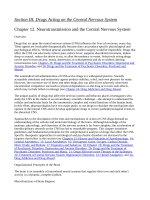
Fig. 1. Phylogenetic tree showing the lineage of snakes.
In the current phylogeny, snakes belong to the Animalia kingdom, Chordata
phylum, Vertebrata subphylum, the Reptilia class and Squamata order (Evans,
2003).
Venomous Snakes
There are many different and diverse species of animals across all phyla of the
animal kingdom that are venomous. Around 600 species are recognized to be
venomous (King et al., 2008; White, 2000), constituting more than a third of all
snake species (Harvey, 1991).
6
Groups of snakes that are mentioned below (Table 1) can be violent and inflict
dangerous, even likely lethal bites:
Family
Atractaspididae (atractaspidids)
Explanation (Tree)
Burrowing asps, mole vipers, stiletto
snakes.
Colubridae (colubrids)
Most are harmless, but others have
toxic saliva and at least five species,
including the boomslang (Dispholidus
typus), have caused human fatalities.
Elapidae (elapids)
Cobras, coral snakes, kraits, mambas,
sea snakes, sea kraits and Australian
elapids.
Viperidae (viperids)
True vipers and pit vipers, including
rattlesnakes.
Table 1 Snakes that known to cause dangerous and/or lethal bites
Cobras, vipers, and closely related species use venom to immobilize or kill their
prey. The saliva of such species have been modified through evolution to develop
venom, and is delivered through fangs (Calvete et al., 2007; Kochva and Gans,
1970). In all venomous snakes salivary glands of these species open through
ducts into grooved or hollow fangs in the upper jaw.
An examination of a fang
from any of the above families will show a non-functional anterior groove, which is
7
no more than an enamel-sealed seam. Calvete et al. (2007) labels this external
groove as such. The continual reference to grooved front fangs, and the incorrect
inference that the venom flows down this groove, probably originated by mistake
as a typo error, but has persisted through the years. Imagine a scientist presenting
a paper on the evolutionary development of snake fangs. They explained how the
ancestral state was an open anterior canal, or groove, and illustrates this with a
several drawings of an open canal, an elapid or hydrophiid fang (the more primitive
state in front-fanged snakes) and a viper's fang. As they point to the most obvious
closed seam in an elapid or hydrophiid fang they said, see how it was grooved.
The possible typo error was the transposing of it was with it's.
'Groove' is a misnomer that is when describing the fangs in front-fanged snakes.
The only fangs with a functional groove (ectoglyphous) for envenomation are those
in back-fanged snakes (opisthoglyphous). All others have an enclosed venom
canal and are effectively hollow (endoglyphous). The outer enamel layer of the
tooth is continuous across the front face of this tubular fang (Zahradnicek et al.,
2008). Subcutaneous fluid delivery is an effective method of rehydration and of
opioid administration, and can prevent the need for intravenous catheterization. It
is a simple procedure to initiate.
The fangs of 'advanced' venomous snakes like the Viperidae and Elapidae family
are hollow in order to inject venom more effectively, while the fangs of rear-fanged
8
snakes such as Dispholidus typus (Boomslang) merely have a groove on the
posterior edge to channel venom into the wound.
Snake Venoms and Their Pharmacology
Snake venoms are often prey specific, its role in self-defense is secondary.
Venom, being a salivary secretion, is a pre-digestant which initiates the breakdown
of food into soluble compounds allowing for proper digestion. We can also see
that "non-venomous" snake bites (like any animal bite) will cause tissue damage
from the salivary components alone (Calvete et al., 2007; Kochva and Gans,
1970).
Venomous snakes include five families of snakes. The Colubridae, Elapidae,
Hydrophiidae, Viperidae and Crotalidae.
As mentioned earlier, snake venoms are evolutionarily modified components within
the salivary secretions. These venoms are complex mixtures of proteins and are
stored in modified salivary glands at the back of the head. These proteins can
potentially be a mixture of neurotoxins (which attack the nervous system),
hemotoxins (which attack the circulatory system), cytotoxins and many other toxins
that affect the body in different ways (Calvete et al., 2007; Harvey, 1991; Kochva
and Gans, 1970; Lu et al., 2005; Markland, 1998). They are produced by special
glands of certain species of snakes.
9
Importance of studying Snake Venom
Snake venoms are complex mixtures of bioactive proteins and polypeptides which
produce many different pharmacological activities, including coagulopathy,
neurotoxicity and necrosis (Harvey, 1991; Tan and Hj, 1989; Pung et al., 2005;
Pung et al., 2006; Calvete et al., 2007; Lu et al., 2005; Markland, 1998). These
toxins are used in the immobilization and capture of their prey as well as for
defense against predators. Many of these proteins are non-toxic to humans, and
few are toxins.
A number of snake venom toxins have been isolated and characterized. Due to
their highly specific interactions with a particular receptor or ion channel, they are
useful in the investigation of physiological and cellular processes (Menez, 1998;
Torres et al., 2003; Hodgson and Wickramaratna, 2002; DiMattio et al., 1985;
Junqueira-de-Azevedo et al., 2006; Kini, 2002; Adams and Olivera, 1994; Kini et
al., 2001; Calvete et al., 2007). Some of the snake venom toxins have contributed
significantly in the studies of neuronal receptors and ion channels.
Until recently, most of the studies focused on toxins that are either highly abundant
or most toxic. With the advent of new technologies, snake venom proteins that are
less abundant are isolated and investigated for their activity. This has led to the
discovery of novel proteins or polypeptides that could be used as lead compounds
10
for drug discovery (Adams and Olivera, 1994; Kini and Evans, 1995; Kini, 2004;
Kini, 2005; Menez, 1998).
Ophiophagus hannah – King Cobra
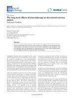
The King Cobra (Ophiophagus hannah) is the world's longest venomous snake,
with a length that can be as large as 5.6 m. This species is found throughout
South-Eastern Asia and into Pakistan and India. The skin is either olive-green,
tan, or black and it has faint, pale yellow cross bands down the length of the body.
The underbelly is cream or pale yellow, and the scales are smooth. The head of
mature snake can be quite massive and bulky in appearance.
Fig 2. King Cobra, Ophiophagus hannah. Reproduced from Wikimedia (Open
Source)
11
King Cobras, like other snakes, receive chemical information via their forked
tongues, which pick up scent particles and transfer them to a special sensory
receptor (Jacobson's Organ) located in the roof of its mouth. When the scent of a
potential meal has been detected, the snake will continue to flick its tongue to
gauge the prey's direction (Young, 1993); it will also rely on its eyesight, and
sensitivity to earth-borne vibration to track its prey. Following envenomation, the
King Cobra will begin to swallow its struggling prey while its toxins begin the
digestion of its victim. King Cobras are able to hunt at all times of day, although it
is rarely seen at night, leading most herpetologists to classify it as a diurnal
species (Goldsmith, 1990).
The King Cobra's diet is mainly composed of other snakes (ophiophagy): both nonvenomous snakes such as pythons and venomous snakes including kraits and
Indian Cobras. Like all snakes, they can expand their jaws to swallow large prey
items.
When food is scarce, King Cobras may also feed on other small
vertebrates such as lizards, birds, and rodents (Harvey, 1991; Chanhome et al.,
1998; Jackson et al., 2004). After a large meal the snake may live for many
months without another one due to its slow metabolic rate. After eating, snakes
become dormant while the process of digestion takes place.
Digestion is an
intense activity, especially after consumption of very large prey. In species that
feed only sporadically, the entire intestine enters a reduced state between meals
to conserve energy, and the digestive system is 'up-regulated' to full capacity
12
within 48 hours of prey consumption. Being ectothermic, the surrounding
temperature plays a large role in a snake's digestion. 30 degrees Celsius is the
ideal temperature for snakes to digest their food (Wang et al., 2001).
When
undisturbed, the digestive process is highly efficient, with the snake's digestive
enzymes dissolving and absorbing the prey, excreting only its hair and claws along
with waste.
The venom of the King Cobra is primarily neurotoxic, and the snake is fully
capable of killing a human with a single bite. However, most bites actually involve
non-fatal amounts of venom (Chanhome et al., 1998; Pochanugool et al., 1998).
Ohanin – A Specialised Neurotoxin found in Ophiophagus hannah
The King Cobra's venom is primarily neurotoxic and thus attacks the victim's
nervous system and quickly induces severe pain, blurred vision, vertigo,
drowsiness, and paralysis. In one to two minutes, cardiovascular collapse occurs,
and the victim falls into a coma. Death soon follows due to respiratory failure
(Chanhome et al., 1998). There are two types of antivenin made specifically to
treat
Ophiophagus
hannah
envenomation.
The
Red
Cross
in
Thailand
manufactures one, and the Central Research Institute in India manufactures the
other, however both are made in small quantities, and are not widely available.
(Tun et al., 1995; Pochanugool et al., 1998; Chanhome et al., 1998)
13
Snake venom proteins and peptides that are found in abundance or that are highly
toxic have been isolated and characterized. Most of them belong to a few
structural superfamily of proteins.
We have recently isolated and characterized a novel protein, ohanin, from King
Cobra venom. (Pung et al., 2005; Pung et al., 2006) Ohanin, a novel protein from
the king cobra crude venom that shows relatively good homology (54% similarity
and 44% identity) to PRY and partial SPRY domains, which are domains of
unknown function in a variety of proteins (Pung et al., 2005). We have named this
new family of proteins as vespryns (venom PRY-SPRY domain-containing
proteins). Additional members of vespryns family from cobra and Lachesis muta
venoms have been identified (Junqueira-de-Azevedo et al., 2006; Li et al., 2004).
Ohanin induces significant dose-dependent hypolocomotion and hyperalgesia in
mice (Pung et al., 2005). The amount of ohanin required to exert its
pharmacological action is ~6500-fold less when injected intracerebroventricularly
(i.c.v.) compared to intraperitoneal (i.p.) administration, suggesting that ohanin
acts primarily on the CNS. Ohanin is synthesized as a precursor named proohanin with the C-terminal propeptide segment which completes the SPRY domain
(Pung et al., 2006).
14
Binding Studies of Ohanin and Pro-ohanin
The lack of natural abundance of ohanin (~1 mg/g of crude venom) and absence
of pro-ohanin in the venom necessitates the use of recombinant proteins for
studies. In addition, the cDNA encoding for ohanin was cloned and sequenced.
The deduced amino acid sequence demonstrates that ohanin has a propeptide
segment at the C-terminal extension. Sequence analysis also shows that ohanin
has the complete PRY-SPRY domains (B30.2-like domain) with the presence of
the C-terminal propeptide segment. The precursor protein was named pro-ohanin
(Pung et al., 2006).
To understand the importance of maturation, we compared the hypolocomotory
effects of pro-ohanin and ohanin by administering them through i.p. and i.c.v.
routes. The specific binding sites of these proteins in mouse brain were identified
using immunofluorescent detection of their N-terminal hexahistidine tags under
both ex vivo and in vivo conditions. The results indicate that both forms of ohanin
specifically bind to hippocampus and cerebellum. Further, significantly higher
amount of mature ohanin crosses the blood-brain barrier (BBB) compared to the
precursor pro-ohanin without affecting its permeability, suggesting that maturation
of the protein is crucial for its biological function.
To date, a number of neurotoxic peptides and proteins that are found to affect
primarily the peripheral nervous system in a physiological situation have been well
15
characterized (Hodgson and Wickramaratna, 2002; Calvete et al., 2007).
However, only a few snake venom proteins and peptides are known to show
pharmacological effects on the central nervous system (CNS). In the 1980’s, a
number of experiments were carried out with radiolabelled iodine or glucose to test
the ability of various venoms and purified toxins to penetrate the BBB (DiMattio et
al., 1985; Gubensek et al., 1982; Silveira et al., 1988).
The intraperitoneal
administration of dendrotoxin from Dendroaspis augusticeps venom led to
behavioural patterns that suggested CNS penetration and phospholipase A from
Vipera ammodytes venom led to penetration of the radiolabelled compounds into
the brain. However, mechanisms of penetration and actions in the brain were not
yet fully understood. Although dendrotoxin binds to specific neural potassium
channels in the CNS, it is not clear whether their localization shown to be
necessary in the physiological function (Chiappinelli, 1991).
However, there is no direct evidence for their transport across the blood-brain
barrier (BBB) and their in vivo binding specificities. Also, it is not clear whether
their specific localization in the CNS is necessary for the physiological functions
(Chiappinelli, 1991). Nevertheless, dendrotoxins and apamin, along with a number
of toxins are used to identify and study specific ion channels/receptors in the brain
(Dolly et al., 1984; Halliwell et al., 1986). Beta-Bungarotoxin from Bungarus
multicinctus venom affects primary cultures of neurons, although these effects
were not demonstrated in vivo (Chen, 2005; Shakhman et al., 2003). Although
crude venoms were shown to affect the transport of radiolabeled glucose across
16
the BBB (DiMattio et al., 1985), neither
the mechanisms of penetration nor the
individual components responsible for such transport are known.
Currently, a number of toxins such as dendrotoxins and apamin are used to
identify and study specific ion channels/receptors in brain.
A number of presynaptic and postsynaptic neurotoxins from snake venoms that
block neuromuscular junction have been isolated and characterized (Black et al.,
1988; Bon et al., 1979; Chang et al., 1977; Chang and Lee, 1977). Because of
their specific interactions, several snake venom toxins have contributed
significantly to the studies of neuronal receptors and ion channels. Most of them
affect primarily the peripheral nervous system and only a few affect the central
nervous system (CNS).
For example, intraperitoneal (i.p.) administration of
dendrotoxin from Dendroaspis angusticeps venom led to behavioral patterns that
suggested CNS penetration (Silveira et al., 1988). Ex vivo binding studies
demonstrate that dendrotoxin binds to distinct neuronal potassium channels in the
CNS (Mehraban et al., 1984). Similarly, ex vivo binding studies indicate that
apamin from bee venom binds to specific, but a different class of neuronal
potassium channels in the CNS (Mourre et al., 1986).
There have been only a limited number of proteins reported in snake venoms
which were postulated to pass the BBB to effect their actions (Gubensek et al.,
1982; Silveira et al., 1988). It is important to study ohanin and its precursor to
17
understand the actual processes that allow the 12 kDa protein to transverse the
BBB, and to study in detail the regions of interaction of the protein with
components in the brain, as this might be useful to the understanding of the control
of locomotion and pain responses.
The identification of the receptor(s), and the identification of the specific regions of
the receptor(s) and ohanin specific to binding for the observed neurological effects
could prove to be the basis in not only understanding the receptor(s) involved in
locomotion and pain sensations within the brain, but also into lead to the
developing of future pharmacological peptides that would have potential
therapeutic uses in locomotion or pain pathologies. (Rossetto and Montecucco,
2008; Albuquerque et al., 2009).
18
MATERIALS AND METHODS
All chemicals were purchased from Sigma-Aldrich (St. Louis, MO, USA) with the
exception of the following: reagents for Edman Degradation N-terminal sequencing
(Applied Biosystems, Foster City, CA, USA), acetonitrile (Merck KGaA, Darmstadt,
Germany), Luria Bertani broth and agar (Q.BIOgene, Irvine, CA, USA), Prestained
broad range SDS-PAGE standards and Precision plus prestained dual-color
standards (Bio-Rad Laboratories, Hercules, CA, USA), RP-Jupiter C18 (10 m, 300
Å, 10 mm X 250 mm) column (Phenomenex, Torrance, CA, USA), Nickel-NTA
agarose
(Qiagen
GmbH,
Hilden,
Germany),
oligonucleotides
(1stBase,
Singapore), Platinum Taq polymerase, dNTP mix and DNA ladders (100 bp and 1
Kb Plus) (GIBCO BRL, Carlsbad, CA, USA), restriction endonucleases (New
England Biolabs Beverly, MA, USA), pGEMT-easy vector (Madison, WI, USA),
CØmplete
protease
inhibitor
cocktail
tablets
(Roche
Applied
Sciences,
Indianapolis, IN, USA), Superfrost Plus slides (Menzel-Gläser, Braunschweig,
Germany), rabbit anti-hexahistidine antibodies (Cat. #29673, Anaspec Inc, San
Jose, CA, USA), and Alexa Fluor 488-conjugated anti-rabbit antibodies (Cat.
#A11034) and Prolong Gold antifade reagent with DAPI (Cat #P36935) (Molecular
Probes Inc, Eugene, OR, USA), trypan blue 0.4% (Invitrogen). Water was purified
with a MilliQ system (Millipore, Billerica, MA, USA).
Animals.
Swiss albino male mice (20 ± 2 g) were used for the animal experiments. In order
to reduce the impact caused by environmental changes and handling during
19
behavioral studies, mice were acclimatized to the Laboratory Animal Holding
Center and laboratory surroundings for 3 days and at least 1 h prior to
experiments, respectively. Animals were kept under standard conditions with food
(low protein diet) and water available ad lib. The animals were housed 4 per cage
in a light-controlled room (12 h light/dark cycles, lights on at 07:00 h) at 23° C and
60 % relative humidity. All behavioral experiments were performed between 08:30
h to 13:00 h. Each test group consisted of at least seven mice and each mouse
was used only once. All the animal experiments were conducted according to
guidelines set by the Laboratory Animal Holding Center of the National University
of Singapore.
Isolation and purification of native ohanin.
Native ohanin was purified via a two-step chromatography technique involving gel
filtration and reverse phase-HPLC as described earlier (Pung et al., 2005).
Lyophilized crude venom was dissolved in 2 ml of MilliQ water and loaded onto a
Superdex 30 column pre-equilibrated with 50 mM of Tris-HCl (pH 7.4).
The
proteins were eluted with 50 mM of Tris-HCl (pH 7.4) at a flow rate of 1 ml/min on
a Fast Protein Liquid Chromatography system (FPLC).
Protein elution was
monitored at 280 nm.
The fraction of interest was then loaded onto an RP-Jupiter C18 semi-preparative
column equilibrated with 0.1% trifluoroacetic acid (v/v) on Vision Work station (PE
Applied Biosystems, Foster City, CA). The bound proteins were eluted using a