Applied molecular genetics
Bạn đang xem bản rút gọn của tài liệu. Xem và tải ngay bản đầy đủ của tài liệu tại đây (1.7 MB, 120 trang )
3
Advancesin BiochemicalEngineering/
Biotechnology
Managing Editor: A. Fiechter
Applied
Molecular Genetics
Guest Editor: J. Reiser
With contributions by
C. L. Cooney, R. L. Dabora, S.-O.Enfors,
V. Glumoff, H. Hellebust, H. Heslot,
M. Kiilin, K. K6hler, U. Ochsner, S.B. Primrose,
J. Reiser, L. Strandberg, A.Veide
With 19 Figures and 12 Tables
Springer-Verlag
Berlin Heidelberg NewYork London
Paris Tokyo Hong Kong Barcelona
ISBN 3-540-52794-X Springer-Verlag Berlin Heidelberg New York
ISBN 0-387-52794-X Springer-Verlag New York Berlin Heidelberg
This work is subject to copyright. All rights are reserved, whether the whole or part of
the material is concerned, specificallythe rights of translation, reprinting, re-use of illustrations, recitation, broadcasting, reproduction on microfilms or in other ways, and storage
in data banks. Duplication of this publication or parts thereof is only permitted under
the provisions of the German Copyright Law of September 9, 1965, in its current version,
and a copyright fee must always be paid.
© Springer-Verlag Berlin - Heidelberg t990
Library of Congress Catalog Coard Number 72-152360
Printed of Germany
The use of registered names, trademarks, etc. in this publication does not imply, even
in the absenCe of a specific statement, that such names are exempt from the relevant
protective laws and regulations and therefore free for general use.
Typesetting: Th. Miintzer, Bad Langensalza; Printing: Heenemann, Berlin;
Bookbinding: Liideritz & Bauer, Berlin
2152/3020-543210 -- Printed on acid-free paper
Managing Editor
Professor Dr. A. Fiechter
Institut ftir Biotechnologie, Eidgen6ssische Technische Hochschule
ETH -- H6nggerberg, CH-8093 Ziirich
Guest Editor
Dr. J. Reiser
Institut fiir Biotechnologie, Eidgen6ssische Technische Hochschule
ETH -- H6nggerberg, CH-8093 Ztirich
Editorial Board
Prof. Dr. S. Aiba
Prof. Dr. H. R. Bungay
Prof. Dr. Ch. L. Cooney
Prof. Dr. A. L. Demain
Prof. Dr. S. Fukui
Prof. Dr. K. Kieslich
Prof. Dr. A. M. Klibanov
Prof. Dr. R. M. Lafferty
Prof. Dr. S. B. Primrose
Prof. Dr. H. J. Rehm
Prof. Dr. P. g Rogers
Prof. Dr. H. Sahm
Prof. Dr. K. Schiigerl
Prof. Dr. S. Suzuki
Prof. Dr. G. T. Tsao
Dr. K. Venkat
Prof. Dr. E.-L. Winnacker
Department of Fermentation Technology, Faculty of
Engineering, Osaka University, Yamada-Kami, Suita-Shi,
Osaka 565, Japan
Rensselaer Polytechnic Institute, Dept. of Chem. and
Environment. Engineering, Troy, NY 12180-3590/USA
Massachusetts Institute of Technology,
Department of Chemical Engineering,
Cambridge, Massachusetts 02139/USA
Massachusetts Institute of Technology, Dept. of
Biology, Room 56-123
Cambridge, Massachusetts 02139/USA
Dept. of Industrial Chemistry, Faculty of
Engineering, Sakyo-Ku, Kyoto 606, Japan
Gesellschaft fiir Biotechnologie, Forschung mbH,
Mascheroder Weg 1, D-3300 Braunschweig
Massachusetts Institute of Technology, Dept. of
Chemistry, Cambridge, Massachusetts 02139/USA
Techn. Hochschule Graz, Institut ftir
Biochem. Technol., Schl6gelgasse 9, A-8010 Graz
General Manager, Molecular Biology Division,
Amersham International plc., White Lion Road
Amersham, Buckinghamshire HP7 9LL, England
Westf. Wilhelms Universit/it, Institut ffir
Mikrobiologie, Corrensstr. 3, D-4400 Mfinster
School of Biological Technology, The University
of New South Wales, P.O. Box 1,
Kensington, New South Wales, Australia 2033
Institut f/Jr Biotechnologie, Kernforschungsanlage
Jiilich, D-5170 Jtilich
Institut fiir Technische Chemic, Universitgt Hannover,
Callinstrage 3, D-3000 Hannover
Tokyo Institute of Technology,
Nagatsuta Campus, Res. Lab. of Resources Utilization,
4259, Nagatsuta, Midori-ku, Yokohama 227/Japan
Director, Lab. of Renewable Resources Eng., A. A. Potter
Eng. Center, Purdue University, West Lafayette,
IN 47907/USA
Corporate Director Science and Technology, H. J. Heinz
Company U.S. Steel Building, P.O. Box 57, Pittsburgh,
PA 15230/USA
Universitgt Miinchen, Institut f. Biochemie, Karlsstr. 23,
D-8000 M/inchen 2
Editorial
This volume of Advances in Biochemical Engineering/Biotechnology as well as the following one are dedicated to Professor
Armin Fiechter on the occasion of his 65th birthday. Contributions
were solicited from Armin's colleagues but were limited to subjects
which related to the biotechnology scope of this series.
One might judge a successful scientist by several criteria and
Attain fullfills them all. One criterion of success is one's record
of scientific achievement. Armin's work on yeast physiology,
process development and instrumentation has been very fruitful
and the necessity of adopting new technology or applying concepts
from other disciplines has never hindered progress in his laboratory.
One very important measure of achievement and the one which
is likely to be the most important in both professional and human
terms, is the training of successful students. Armin is one of the
most eminent promoters of biotechnology in Switzerland and
abroad and he was instrumental in establishing the Institute for
Biotechnology at the ETH in 1982 of which he is still director.
By pioneering the combination of fundamental and applied aspects
he has greatly influenced the advancement of biotechnology in
teaching and research in Switzerland.
At the age of 65, the pace of Armin's activities shows no sign of
slackening. At the latest count there are some 20 doctoral students
working under his supervision. He is currently the editor-in-chief
of the Journal of Biotechnology and of this series, both of which
were co-founded by him and have gained momentum under his
influence.
This special issue is dedicated to Armin Fiechter with admiration.
Zfirich, July 1990
Jakob Reiser
Armin Fiechter
Table of Contents
Controlling Bacteriophage Infections in Industrial Bioprocesses
9S. B. Primrose . . . . . . . . . . . . . . . . . . . .
Intracellular Lyric Enzyme Systems and Their Use for
Disruption of Escherichia coli
R. L. Dabora, C. L. Cooney . . . . . . . . . . . . . .
11
Impact of Genetic Engineering on Downstream Processing of
Proteins Produced in E. coli
S.-O. Enfors, H. Hellebust, K. K6hler, L. Strandberg, A. Veide
31
Genetics and Genetic Engineering of the Industrial Yeast
Yarrowia lipolytica
H. Heslot . . . . . . . . . . . . . . . . . . . . . .
43
Transfer and Expression of Heterologous Genes in Yeasts
Other Than Saccharomyces cerevisiae
J. Reiser, V. Glumoff, M. K~lin, U. Ochsner . . . . . . .
75
Author Index Volumes 1-43 . . . . . . . . . . . . . . .
103
Controlling Bacteriophage Infections
in Industrial Bioprocesses
S. B. P r i m r o s e
Life Sciences Division, A m e r s h a m I n t e r n a t i o n a l plc, W h i t e L i o n R o a d , A m e r s h a m ,
Bucks H P 7 9LL, U K
1 Introduction . . . . . . . . . . . . . . . . . . . . . . . . . . . . . . . . . . . . . . . . . . . . . . . . . . . . . . . . . . . . . . . . . .
2 The Biology of Bacteriophages . . . . . . . . . . . . . . . . . . . . . . . . . . . . . . . . . . . . . . . . . . . . . . . . . .
2.1 Physical Properties of Bacteriophages . . . . . . . . . . . . . . . . . . . . . . . . . . . . . . . . . . . . . . . . .
2.2 The Process of Infection . . . . . . . . . . . . . . . . . . . . . . . . . . . . . . . . . . . . . . . . . . . . . . . . . . . .
2.3 The Genetics of Bacteriophage Resistance . . . . . . . . . . . . . . . . . . . . . . . . . . . . . . . . . . . . .
2.4 Bacteriophage Ecology . . . . . . . . . . . . . . . . . . . . . . . . . . . . . . . . . . . . . . . . . . . . . . . . . . . . . .
3 Recognizing a Bacteriophage Infection . . . . . . . . . . . . . . . . . . . . . . . . . . . . . . . . . . . . . . . . . . . .
4 Cleaning Up a Bacteriophage Infected Plant . . . . . . . . . . . . . . . . . . . . . . . . . . . . . . . . . . . . . . .
5 Prevention of Bacteriophage Attack . . . . . . . . . . . . . . . . . . . . . . . . . . . . . . . . . . . . . . . . . . . . . .
5.1 Facility Location . . . . . . . . . . . . . . . . . . . . . . . . . . . . . . . . . . . . . . . . . . . . . . . . . . . . . . . . . . .
5.2 Plant Design . . . . . . . . . . . . . . . . . . . . . . . . . . . . . . . . . . . . . . . . . . . . . . . . . . . . . . . . . . . . . .
5.3 Strain Selection . . . . . . . . . . . . . . . . . . . . . . . . . . . . . . . . . . . . . . . . . . . . . . . . . . . . . . . . . . . .
5.4 Operating Practice . . . . . . . . . . . . . . . . . . . . . . . . . . . . . . . . . . . . . . . . . . . . . . . . . . . . . . . . .
6 References . . . . . . . . . . . . . . . . . . . . . . . . . . . . . . . . . . . . . . . . . . . . . . . . . . . . . . . . . . . . . . . . . . . .
1
2
2
3
4
5
6
6
7
7
8
8
9
9
Bacteriophage infections of microbial processes are relatively common but can be economically
devasting. If the designers and operators of bioreactors have an understanding of the biology
and ecology of bacteriophages the risks and hazards of bacteriophage infections can be minimised.
If an infection should occur it is important to recognize the fact as soon as possible. The symptoms
of a phage infection and "procedures for cleaning-up infected bioprocess plants are described.
1 Introduction
B a c t e r i o p h a g e s are viruses w h i c h infect a n d lyse bacteria and as such can, and do,
w r e a k h a v o c o n industrial bioprocesses. D e s p i t e this there is no b o d y o f scientific
or technical literature on the subject. B i o c h e m i c a l engineers and b i o p r o c e s s technologists are s e l d o m t a u g h t the basics o f p h a g e biology. C o n s e q u e n t l y , those
c h a r g e d with the o p e r a t i o n o f bioprocesses d o n o t recognize the early signs of
a phage infection, d o n o t k n o w h o w to c a r r y out a post-infection clean-up, n o r
h o w to m i n i m i z e o r p r e v e n t future attacks. A p h a g e a t t a c k can be c a t a s t r o p h i c
and result in lack o f p r o d u c t i o n for periods r a n g i n g f r o m a few days to m a n y
m o n t h s . F o r t u n a t e l y for the industry, yeast a n d fungal cultures do not suffer
Advances in Biochemical Engineering/
Biotechnology, Vol. 43
Managing Editor: A. Fiechter
9 Springer-Verlag Berlin Heidelberg 1990
2
S.B. Primrose
from viral attacks. Fungal viruses are known but their infective properties are
such that they do not cause widespread culture lysis.
There is little published information on the importance of phages in industrial
bioprocesses, with only the dairy industry freely admitting to the problem [1-4].
Other processes known to be seriously affected are the bioproduction of acetonebutanol [5, 6] and monosodium glutamate [7] and processes utilizing actinomycetes [8, 9], and Escherichia coli and its close relatives. The microbial processing
of milk products probably is the most important industrial process affected by
phages.
2 The Biology of Bacteriophages
An understanding of the biology of bacteriophages is fundamental if infection is
to be prevented or controlled. There are many specialized texts on the subject but
only a few [10-12] are appropriate for the non-specialist.
2.1 Physical Properties of Bacteriophages
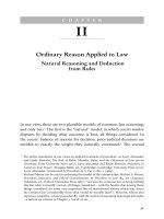
Bacteriophage particles exhibit considerable diversity in size and shape (Fig. 1).
The vast majority of phages have a head-tail morphology, an architectural principle not found in other groups of viruses and which may be a reflection on the
way bacterial viruses infect susceptible cells. Many different structural variations
have been noted, e.g. contractile versus non-contractile tails, presence or absence
of base-plates, collars, etc, but this morphological diversity is irrelevant as far
as infection of industrial processes is concerned. On analysis, practically all phage
t
j
1000 n m
Fig. 1. Relative size of a bacterium (Escherichia coli) and
an assortment of bacteriophageswhich infect it
Controlling BacteriophageInfections in Industrial Bioprocesses
3
particles consist of protein and nucleic acid only although some do have a lipid
coat. The nucleic acid can be either DNA or RNA, but not both, and in tailed
phages is located in the head.
The overall length of a phage is in the general range of 50-200 millimicrons
and the head averages 50-90 mu in width. Thus phages are small enough to pass
through most bacteriological filters which have pore sizes in excess of 0.2 microns.
The phage particle is essentially a survival mechanism designed to protect the
phage genome from the rigours of the environment to which it is exposed when it
destroys one host and before it infects another. It is clearly efficient otherwise
phages would long ago have become extinct. Most phages will survive in cell
lysates for long periods (months to years) at normal environmental temperatures
provided they do not dry out. Many phages also are resistant to drying. Although
freezing and thawing can reduce viral titres, survival is still very high. Bacteriophages are about as sensitive to heat as the majority of non-sporing bacteria.
At 100 ~ they are inactivated almost instantly. Between 65 ~ and 85 ~ inactivation rates can be determined conveniently under laboratory conditions. Below
65 ~ some phages are inactivated very rapidly but most hardly at all. The medium
in which phages are heated has a great influence on the rate of inactivation. Inactivation is most rapid in pure water; the addition of salts, especially calcium or
magnesium, and proteins reduces the rate of inactivation considerably.
Many of the classical disinfectants active against bacteria also inactivate bacteriophages and can be used in plant clean-up. Thus compounds which react
with proteins or nucleic acids should destroy phages. However, some of the newer
surface-active agents may have little effect on the viability of phages. Under
certain conditions phage particles may not exist as monodisperse units but as
aggregates. The application of disinfectants to phage aggregates may not result
in complete inactivation.
Some well-studied bacteriophages, e.g. coliphage T1, have acquired notoriety
because of the ease with which they spread through laboratory complexes. What
property or properties of a phage confer upon it the ability to be readily disseminated is not known. Apparent ease of spread may be related to drying. Alternatively
it could be related to the ability to form aggregates and the impact aggregate size
has on gravitational removal from aerosols. Unfortunately, those phages which
most easily infect a bioprocess plant are those which spread the easiest, making
their elimination more difficult. Many of them also are very heat resistant !
2.2 The Process of Infection
The process of infection begins when a bacteriophage particle undergoes a chance
collision with a host cell. If the phage possesses an adsorption site that is chemically
complementary to a specific receptor on the bacterial cell surface then irreversible
adsorption occurs. The receptors for most phages are located on a cell wall although
for some phages cellular appendages such a flagella or pili can act as adsorption
sites. Following adsorption, the viral nucleic acid enters the cell but for most
phages the mechanism whereby this occurs is not known.
4
s.B. Primrose
Once inside the cell, the phage nucleic acid undergoes replication. As the number
of phage nucleic acid molecules builds up inside the cell, viral genes are transcribed
and translated and structural proteins are synthethized. Eventually the nucleic
acid and protein assemble into complete phage particles. Coincident with phage
assembly is the synthesis of a virally-specified lysozyme. This enzyme attacks the
peptidoglycan layer of bacterial cell walls. The wall is progressively weakened
until it is ruptured by the internal osmotic pressure of the cell, and the progeny
phages are liberated into the environment along with the other contents of the
cell.
The average number of new phage particles liberated by each infected cell is
known as the burst size. The value of the burst size varies from 30-300 and the time
from infection to lysis ranges from 20-100 minutes. The exact value of each
depends on the particular host-phage system and the environmental conditions.
The particles liberated by lysis can in turn infect other ceils in the population with
a repetition of the same cycle. Consequently, even if only a single infectious phage
particle is introduced into a culture, practically the entire bacterial population
may be destroyed in a few hours.
The last cycle of infection is particularly dramatic. When as many as 1-5
of the cells in the culture are lysing there are no obvious physical or physiological
signs of infection. However, the phage particles released can infect all the remaining
cells and shortly thereafter the entire culture becomes glass-clear. In aerated
cultures excessive foaming also may occur as a result of release of host cell proteins
into the growth medium. When the entire culture finally lyses the number of
phage particles present is very high. Thus in a 10000 L culture whose density
was 10l~ cells m1-1 prior to lysis by a phage with a burst size of 100, the total
number of phage particles will be 1 0 1 9 . As will be seen later, the magnitude of
this phage count makes clean-up very difficult.
2.3 The Genetics of Bacteriophage Resistance
The rate of spontaneous mutation at any genetic locus is of the order of 10-5-10-s
per cell per generation. Thus as a culture increases in density it becomes increasingly
heterogenous. If a bacterium is susceptible to a particular phage then high cell
density bacterial cultures will contain a proportion of phage-resistant mutant cells.
These mutant cells are easily isolated and can be shown to have an inheritable
inability to adsorb the phage as a result of an alteration in the structure of their
cell walls. There is a common misconception that such mutants provide a defence
against future infections with the same phage. Nothing could be further from the
truth. Just as bacteria mutate, so do phage. The rate of mutation of phage genes
is similar to that of bacterial genes. If the phage population exceeds 108 particles,
as it usually does (see above), host-range mutants will be present which can infect
the cells resistant to wild-type phage. Further bacterial mutants resistant to the
host-range phage mutant can be isolated but new phage mutants which infect
it also can be isolated [13] (Table 1). Because one always is dealing with numbers
of bacteria and phage in excess of 10l~ it is virtually impossible to stop the alternate
Controlling Bacteriophage Infections in Industrial Bioprocesses
Table 1. Infectivity of host-range (hr) mutants
of a bacteriophage for wild-type and various
resistant mutants (R1, R2) of the best bacterium
Phage
Bacteria
Wild-type
Wild-type
+
hrl
hr2
+
+
R1
R2
+
+
+
selection of host and phage mutants. An example of this in an industrial bioprocess
has been reported [14].
Even where suitable phage-resistant mutants can be isolated following infection
this is not usually a viable option. Most of the bacterial strains used in bioprocesses
are selected for high yield of product. The requisite high yields are obtained only
under precise culture conditions and any genetic alteration to the cell can reduce
the yield. This is particularly true with phage-resistant mutants which result from
cell wall alterations, many of which can affect cell permeability.
Although bacteriophages have been isolated for almost all known bacteria
there is wide variation in the susceptibility of different strains of any one bacterial
species. For example, phages infecting E. coli are extremely easy to isolate but
a few strains of E. coli have been identified for which it has proved impossible
to isolate phages [151. A number of factors in combination, e.g. presence of multiple
host-restriction and modification systems plus abnormal wall structure, probably
contribute to the extreme phage resistance of certain strains [16].
In the case of the lactic streptococci, phage-resistance can be plasmid-encoded
[17] and resistance can result from inhibition of phage adsorption, restriction of
D N A and inhibition of phage maturation. In most instances the mechanism of
resistance has been determined for only a few phage isolates: it remains to be seen
if any of these resistance mechanisms will operate against a wide range of lactic
bacteriophages or in unrelated bacterial species.
2.4 Bacteriophage Ecology
Bacteriophages, like all viruses, are obligate intracellular parasites. In order to
multiply they need not only susceptible host cells but cells which are actively
growing. Thus the numbers of a particular phage in the environment can be
correlated with the numbers of its host cells. Not surprisingly, as the external
environment warms up in spring and summer the numbers of bacterial cells and
their associated phages increase I18]. As the external environment cools in autumn
and winter the numbers of phage decline. However, many of these phage are not
inactivated, at least not rapidly. Rather they adsorb to clay minerals [19] in soil
and water as a result of seasonal changes in pH.
6
S.B. Primrose
The physical location of a bioprocess plant can greatly influence the probability
of phage infections occurring. Thus plants where soil microorganisms such as
actinomycetes (antibiotics) or Brevibacterium (mono-sodium glutamate) are
cultured on a large scale will be more susceptible to phage infections if located
in the middle of an agricultural region. With the advent of genetic engineering,
bioprocesses utilizing E. coli increasingly are being used. Such bioprocesses
should not be conducted close to sewage disposal plants. Coliphages are present
in high numbers in sewage and the aerosols produced by activated sludge plants
can result in phage particles being dispersed many miles downwind. Even plants
in agricultural areas are not safe if the local farmers spread sewage sludge or
farmyard waste on their land.
At a more local level, plant operators should ensure that pools of the production organism do not occur below sampling ports or in waste channels. Such
locations are breeding grounds for unwanted phages.
3 Recognizing a Bacteriophage Infection
As indicated earlier, ifa phage is multiplying in a bacterial culture then the number
of infected cells can increase from a few percent to nearly 100~o in as little as
30 minutes. Even the best-trained operator is unlikely to notice any changes in
the bioprocess operating parameters when less than 5 ~o of the cells are infected.
However, during the last cycle of infection the cells metabolic rate will decrease
sharply. This is most easily detected as a sharp increase in the dissolved oxygen
tension. If mass spectrometry is being used to analyse exit gases then a rapid
decrease in CO 2 concentration also should be noted. If the pH of the growth
medium is being controlled then a decrease in the rate of alkali addition might
be noted [9].
As the cells begin to lyse the turbidity of the culture will decline. On line monitoring of culture density is unusual but if plant operators suspect a phage infection
they should monitor turbidity every few minutes (the reason will become clear
later). Because lysing cells liberate a lot of protein the amount of foam will increase
rapidly as will the consumption of anti-foam [9].
As with any contaminant, bacteriophages result in clearly observable differences
in the temporal profiles of the various culture parameters being monitored. The
difference between phages and other contaminants is the rapidity with which they
cause very large deviations from normality.
4 Cleaning Up a Bacteriophage Infected Plant
As soon as a phage infection is suspected the bioreactor should be shut down
immediately. As noted earlier, in a 10000 L vessel there can be as many as 1019
phage particles although this will depend on the cell density when lysis occurs.
Continued aeration of the culture medium will only disseminate large numbers
of phage particles into the environment of the bioprocess plant making clean-up
Controlling BacteriophageInfections in Industrial Bioprocesses
7
far more difficult. This dispersal will be facilitated if excess foaming of lysed cells
has overwhelmed the anti-foam system. Spread of phage particles will be facilitated
if the exit gas lines from a number of bioreactors are manifolded. Although it
is fundamentally bad practice to design a bioprocess plant with the air intakes and
air exits in close proximity, this author has seen numerous examples. In such
situations the entire aeration system, e.g. compressor, filters, etc, also will be
contaminated.
The first task is to develop an assay for the phage particles. This is very easy
to do and should take only one day [20]. The second task is to heat sterilize the
contents of the bioreactor. The exact time-temperature profile will depend on
the design of the bioprocess plant but clearly the hotter and the longer the treatment, the better. Before discarding the reactor contents, the newly developed
phage assay procedure should be used to ensure the absence of viable phage.
Because bacteriophages can pass through most, if not all, bacteria-proof filters,
any pipework connected to the fermenter, however remotely, needs to be extensively steamed. This includes air inlet lines both upstream and downstream of
the compressor. Much more rigour in sterilization of pipework and other plant
is needed at this stage than would be the case if a bacterial contaminant had
predominated in the culture. Where possible liquid samples or surface swabs
should be taken and assayed for phage.
The next step is to decontaminate the exterior surfaces of all pipes and plant.
The ease with which this can be done will depend on the size and design of the
plant. Where possible the entire plant should be washed down with water, which
is as hot as is practicable. Again, samples of washings should be checked with the
phage assay procedure to determine the extent of contamination and the efficacy
of its reduction, particularly if open drains exist in which pools of liquid might
collect and serve as reservoirs for infection. Nor should cooling ponds be ignored.
5 Prevention of Bacteriophage Attack
With an understanding of the biology of bacteriophages it is possible to design,
build and operate a bioprocess facility in which the probability of bacteriophage
infection will be very low. The extent to which this can be done will depend entirely
on whether a new plant and process is to be operated or whether an existing plant
and process is to continue.
5.1 Facifity Location
If a new facility is being built consideration should be given to its location. If
enteric organisms are to be grown the plant should be sited well away from sources
of coliphages. If soil organisms are to be grown then the facility should not be
too close to cultivated land. The local weather also can have an effect. The normal
British climate with its regular rainfall means that airborne phages will be
"grounded". The long dry summers in the US Midwest are conducive to the
spread of soil bacteriophages in dust.
8
S.B. Primrose
5.2 Plant Design
As with other contaminants, bacteriophages usually gain access to the culture
medium via the air handling system. Whereas bacteria and othe microbial contaminants can be excluded by careful selection and maintenance of an inlet air
filter this is not necessarily the case for bacteriophages. The size of bacteriophages
is such that they will pass through membranes whose pore size is small enough
to remove bacteria. Man 5, of the air filters in common use are not absolute filters
but rely on the tortuous path of gas molecules through a packed bed of fibres to
remove contaminating microbes. The performance of such fibrous filters depends
on air velocity and particle size [21]. At the high flow rates used in most largescale microbial cultures inertial forces are crucial to particle removal. Unfortunately bacteriophages are too small to have a significant moment of inertia and
are not removed [22].
The choice of air compressor can facilitate sterilisation. If compression is done
adiabatically the subsequent rise in temperature in the compressed air may be
sufficient to inactivate any phages present. The efficiency of this dry heat sterilization can be improved by passing the air through a lagged retention chamber.
Bartholomew et al. [7] have presented data on the exposure time/temperature
combinations necessary to reduce the probability of phage penetration to less
than 10 -15 . Alternatively the compressed air can be flash heated to at least
90-95 ~ by steam injection and then cooled to remove condensate before filtration. Although not an easy solution from a design point of view it does afford
the best protection.
The air inlet should be well-separated from the air outlet, to prevent circulation
of any contaminants [19]. Raising the air inlet as high as possible also may be
beneficial. A series of coarse filters should be fitted to remove dust particles and
other debris and, if possible, a hydrophobic filter to remove phage being transmitted in water droplets. Some commercially-available filters, e.g. Pall Emflon,
may be particularly effective at removing bacteriophages. Maintenance of these
filters must be rigorous.
The other design feature of importance relates to waste disposal. There should
be no pools of liquid containing live culture anywhere in the vicinity of the bioprocess plant. All sampling points should be fitted with tun dishes and any spillage
piped to a kill tank. There should be no open drains, no drains with horizontal
sections where culture medium containing live organisms can collect, and all
drains should flow to a kill tauk. Finally, downstream processing operations should
be designed such that there isno waste containing any live organisms.
5.3 Strain Selection
Where a microbial process has been in operation for some time there is little that
can be done to select a naturally phage-resistant strain. Where a new process is
being developed careful tought should be given to the choice of starting organism.
For example, E. eoli mutants which have lost most of the "dispensable" layers
Controlling BacteriophageInfections in Industrial Bioprocesses
9
of the cell wall are likely to be less susceptible to phage infection than wild-type
strains since most of the potential phage receptors have been eliminated.
If a genetically-engineered organism is to be cultured then thought should be
given to the deliberate inclusion in the genetic construct of one or more hostrestriction and modification (HRM) systems. HRM is a mechanism whereby
foreign nucleic acids which gain entry to a cell can be destroyed. Most HRM
systems reduce the infectivity of bacteriophage DNA by a factor of 10z to 104
and so can provide a defence against bacteriophage attack. The genes for many
different HRM systems have been cloned and can be inserted into other bacteria
almost at will.
Another advantage of genetically-engineered organisms is that they offer the
potential for strain rotation. In the dairy industry, the recurring problem of phage
infection can be minimised by constantly changing the bacterial strains used to
ones with different phage sensitivities [1, 2]. This cannot be done with most of
the other traditional bioprocesses where there is a single long lineage of improved
strains, all derived from the same parent. However, if the key genetic information
is plasmid borne, as in genetically-engineered organisms, it is possible to move
the plasmid to host strains with different phage sensitivities. Although similar
yields of product cannot be guaranteed, strain rotation in this way can minimise
the downtime following a serious phage attack.
5.4 Operating Practice
There are three simple procedures which should be introduced in all microbial
processes which are susceptible to phage attack. The first of these is to develop
an emergency control procedure which can be implemented in the event of a
phage infection. The key aspects of this procedure were detailed in Sect. 4.
Second is the development of a suitable phage assay procedure. Once this procedure is established, samples for phage assay should be withdrawn regularly
from all culture vessels when the density of cells within them is such that there is
visible turbidity. Hopefully, no phage will ever be detected. If it is, the emergency
control procedures can be implemented immediately. Environmental samples also
should be examined for the presence of bacteriophages capable of infecting the
production strain. These samples should be taken only from aqueous environments, e.g. cooling ponds, semi-permanent puddles in the grounds, waste-disposal
systems, etc. A build-up of phages in the environment should be taken as an indication that a plant clean-up is required if infection of the bioreactors is to be avoided.
Finally, plant operators should be taught to recognize the key symptoms of
a phage infection (Sect. 3) and to take immediate remedial action.
6
References
1. Daly, C (1983) Antonie van Leeuwenhoek49:297
2. Lawrence, RC, Thomas, TD (1979) The Fermentation of Milk with Lactic Acid Bacteria.
In: Microbial Technology: Current State, Future Prospects (eds. Bull, AT, Ellwood, DC,
Ratledge, C), P. 187, Cambridge, Cambridge University Press
10
S.B. Primrose
3. Klaenhammer, TR (1984) Advances in Applied Microbiology 30, 1
4. Vedamuthu, ER, Washam, C (1983) Cheese. In: Biotechnology, Volume 5, Food and Food
Production with microorganisms. (eds. Rehm, H-J, Reed, G), p 231, Basel, Verlag Chemic
5. McCoy, E, McDaniel, LE, Sylvester, JC (1944) J. Bacteriol. 47, 433
6. Ogata, S, Hongo, M (1979) Advances in Applied Microbiology 25, 241
7. Barthomomew, WH, Engstrom, DE, Goodman, NS, O'Toole, AL, Shelton, JL and Tannen,
LP (1974) Biotechnology and Bioengineering 16, 1005
8. Casida, LE Jr (1968) Industrial Microbiology. New York, John Wiley
9. Hongo, M, Oki, T, Ogata, S (1973) Phage contamination and control. In: The Microbial
Production of Amino Acids (eds. Yamada K, Kinoshita S, Tsunoda T and Aida K) p67.
New York, John Wiley
10. Douglas, J (1975) Bacteriophages. London, Chapman and Hall
11. Dimmock, NJ, Primrose SB (1987) Introduction to Modern Virology. Oxford, Blackwell
Scientific Publications
12. Smith, KM, Ritchie, DA (1980) Introduction to Virology. London, Chapman and Hall
13. Baylor, MB, Hurst, DD, Allen, SL, Bertani, ET (1967) Genetics 42, 104
14. Oki, T, Matsui, T, Ozaki, A (1967) Agric. biol. Chem. 31,861
15. Primrose, SB unpublished observations
16. Primrose, SB, Seeley, ND, Logan, KB, Nicholson, JW (1982) Applied and Environmental
Microbiology 43, 694
17. Daly, T, Fitzgerald, G (1987) Mechanisms of bacteriophage insensitivity in the lactic streptococci. In: Streptococcol Genetics (eds. Ferretti JJ and Curtiss III, R) p. 259 Washington,
American Society for Microbiology
18: Primrose, SB, Seeley, ND, Logan, KB (1981) The Recovery of Viruses from Water: Methods
and Applications. In Viruses and Waste Water Treatment (eds. Goddard, M, Butler, M)
p. 211, Oxford Pergamon Press
19. Stotzky, G, Schiffenbauer, M, Lipson, SM, Yu, BH (1981) Surface Interactions between
Viruses and Clay Minerals and Microbes: Mechanisms and Implications. In: Viruses and
Waste Water Treatment (eds. Goddard, M, Butler, M) p. 199, Oxford, Pergamon Press
20. Primrose, SB, Seeley, ND, Logan, KB (1982) Methods for the Study of Virus Ecology. In:
Experimental Microbial Ecology (eds. Burns, RG, Slater, JH) p. 66, Oxford, Blackwell
ScientificPublications
21. Humphrey, AE (1960) Advances in Applied Microbiology 2, 301
22. Sadoff, HL, Almlof, JW (1956) Industrial and Engineering Chemistry 48, 2199
Intracellular Lytic Enzyme Systems and Their Use
for Disruption of Escherichia coil
R. L. D a b o r a ~ and C. L. Cooney 2' 3
1 Introduction . . . . . . . . . . . . . . . . . . . . . . . . . . . . . . . . . . . . . . . . . . . . . . . . . . . . . . . . . . . . . . . . . .
2 Methods of Microbial Cell Disruption . . . . . . . . . . . . . . . . . . . . . . . . . . . . . . . . . . . . . . . . . . . .
3 Autolytic Enzymes . . . . . . . . . . . . . . . . . . . . . . . . . . . . . . . . . . . . . . . . . . . . . . . . . . . . . . . . . . . . .
3.1 Structure of the E. coli Cell Envelope . . . . . . . . . . . . . . . . . . . . . . . . . . . . . . . . . . . . . . . . .
3.2 Types of Autolysins . . . . . . . . . . . . . . . . . . . . . . . . . . . . . . . . . . . . . . . . . . . . . . . . . . . . . . . .
3.3 Use of Autolytic Enzymes in Cell Disruption . . . . . . . . . . . . . . . . . . . . . . . . . . . . . . . . . .
3.4 Role o f the Autolysins . . . . . . . . . . . . . . . . . . . . . . . . . . . . . . . . . . . . . . . . . . . . . . . . . . . . . .
3.5 Triggering of Autolysis in E. coli . . . . . . . . . . . . . . . . . . . . . . . . . . . . . . . . . . . . . . . . . . . . .
3.6 Characteristics of Autolysis . . . . . . . . . . . . . . . . . . . . . . . . . . . . . . . . . . . . . . . . . . . . . . . . . .
4 Colicin Lytic Enzymes . . . . . . . . . . . . . . . . . . . . . . . . . . . . . . . . . . . . . . . . . . . . . . . . . . . . . . . . . .
5 Bacteriophage Lytic Enzymes . . . . . . . . . . . . . . . . . . . . . . . . . . . . . . . . . . . . . . . . . . . . . . . . . . .
5.1 Bacteriophage L a m b d a . . . . . . . . . . . . . . . . . . . . . . . . . . . . . . . . . . . . . . . . . . . . . . . . . . . . .
5.2 Bacteriophage T4 . . . . . . . . . . . . . . . . . . . . . . . . . . . . . . . . . . . . . . . . . . . . . . . . . . . . . . . . . .
5.3 Bacteriophage phiX174 . . . . . . . . . . . . . . . . . . . . . . . . . . . . . . . . . . . . . . . . . . . . . . . . . . . . .
5.4 Bacteriophage MS2 . . . . . . . . . . . . . . . . . . . . . . . . . . . . . . . . . . . . . . . . . . . . . . . . . . . . . . . .
5.5 Bacteriophage QI3 . . . . . . . . . . . . . . . . . . . . . . . . . . . . . . . . . . . . . . . . . . . . . . . . . . . . . . . . . .
6 "Designer" Lytic Proteins . . . . . . . . . . . . . . . . . . . . . . . . . . . . . . . . . . . . . . . . . . . . . . . . . . . . . .
7 Directions and Potential for Future Research . . . . . . . . . . . . . . . . . . . . . . . . . . . . . . . . . . . . . .
8 References . . . . . . . . . . . . . . . . . . . . . . . . . . . . . . . . . . . . . . . . . . . . . . . . . . . . . . . . . . . . . . . . . . . .
12
12
13
13
13
14
14
15
15
16
19
19
21
22
25
25
26
26
28
This article focusses on lytic enzyme systems available in E. coli and their potential use for cellular
disruption. In the systems described here the genetic information for lysis would be carried within
the microbial host, either integrated or naturally occurring on chromosomal D N A , or on extrac h r o m o s o m a l elements such as plasmids. Each microbe would carry complete information for
endogenous enzymatic lysis, and lysis would occur in a controlled m a n n e r after being triggered
by an external factor such as temperature or inducer addition. The lytic systems explored in this
review include the autolytic enzymes, colicin lytic enzymes, and bacteriophage lytic enzymes
from phage phiX174, T4, lambda, MS2 and QI3. M a n y of the colicin lytic enzymes a n d all of the
bacteriophage lytic enzymes described here have been cloned, and in some instances examined
as cellular disruption methods. None of the E. coli autolytic enzymes have been cloned, but
information pertinent for use as a disruption method is described.
1 Merck and Co., Inc., P.O. Box 7, Elkton, VA 22827;
2 Department of Chemical Engineering and Biotechnology Process Engineering Center,
Massachusetts Institute of Technology, Cambridge, M A 02139;
3 To w h o m correspondence should be addressed
Advancesin BiochemicalEngineering/
Biotechnology,Vol. 43
ManagingFditor: A. Fiechter
9 Springer-VerlagBerlinHeidelberg1990
12
R.L. Dabora and C. L. Cooney
1 Introduction
Many problems of downstream processing are created by the biological system
and the methods of culturing. Many of these same problems often can be resolved
through genetic manipulation and/or biochemical control. One example is the
recovery of intracellular proteins from a bacterial cell, namely Escherichia coli.
The usual approach to the initial release of protein is the use of mechanical or
chemical methods which exposes proteins to shear and creates a polydispersed
mixture of cell debris. An alternative approach is the design of a process utilizing
the natural autolytic capabilities of the cell by placing them under external control
by an environmental factor such as temperature or chemical induction. Alternatively, lytic enzymes from bacteriophage or colicin systems c,mld be used in
a similar manner. In both cases, lysis of the bacterial cell occurs from lytic enzymes
produced within the cell, affording gentle, controlled, and efficient cellular disruption.
Although the use of lytic enzymes as an alternative method of cell disruption
has been suggested, there are few studies examining the potential of these systems
[1]. The use of these enzymes for large-scale disruption in plasmid or other cloned
systems has not been fully explored, in part because the lytic genes have not been
identified or characterized. Also, special growth and media conditions, as well
as lysis triggering may be required for efficient lysis. This review focusses on
several lytic systems available for use in E. coli. While use of these systems may
not be a replacement for existing cell disruption methods, they offer an alternative
to, or may be used in conjunction with, current practices. Analysis of lytic systems
may aid in the design or improvement of an alternative, integrative, and efficient
cellular disruption process.
2 Methods of Microbial Cell Disruption
A number of different methods of microbial cell disruption are in use today;
these are described in several reviews on the theory and practice of both small
and large-scale disruption processes [1, 2, 3, 4]. In general, these processes can
be divided into two classes: physical and chemical. For large-scale processes the
primary physical methods include liquid shear (French press, homogenization),
agitation with abrasives (such as glass beads), and solid shear. The primary largescale chemical methods include osmotic shock, detergents, alkali, and enzymatic
methods such as lysozyme addition. Liquid shear is the most commonly reported
large-scale microbial cell disruption method [3].
Constraints placed on the disruption method include the choice of microorganism, the equipment available, and the product being recovered. Since
continued emphasis is being placed on integrative recovery and purification
processes [2], the procedure choice may not be one which releases the product
in highest yield, but may be one which preferentially releases the desired product
over other proteins present, thus, effecting purification.
Intracellular Lytic EnzymeSystemsand Their Use for Disruption
13
The use of enzymes and particularly cloned lytic enzymes as alternative disruption methods has been considered only in an exploratory manner. There are
only a few references to the use of recombinant DNA for potential improvement
or replacement of current disruption unit operations [5, 6, 7]. Some of the potential enzymatic methods for cell disruption processes which could be adapted
to large-scale, but have not been fully developed, include the use of: autolytic
enzymes, colicin lyric enzymes, and bacteriophage lytic enzymes.
Much of the review of the above processes will focus on E. coli. The motivation
for studying the potential of enzymatic alternatives in this microorganism is the
following: the genetics and recombinant DNA techniques of E. coli are well
characterized and defined [8], several lytic enzyme systems have been identified
in this microorganism although the potential for disruption processes has not
been fully developed [1], it is a good model system for intracellular protein recovery, and many recombinant-derived proteins are currently produced in E.
coli [9]. Development of alternative cell disruption methods in this microorganism
may therefore be of industrial importance.
3 Autolytic Enzymes
3.1 Structure of the E. coli Cell Envelope
The cell envelope of E. eoli and of all Gram negative bacteria is made up of several
layers: an inner or cytoplasmic membrane, cell wall, periplasmic space, and an
outer membrane [10]. The cell wall is particularly important for maintaining the
shape and integrity of the cell. Degradation or destruction of the wall by an intra
or extracellular method will usually result in cell lysis [11].
The E. coti cell wall is known to consist of a peptidoglycan layer containing
chains of alternating N-acetylglucosamine and N-acetylmuramic acid' residues
in J3-1,4 linkages. These long glucan chains are crosslinked by short peptides
which are attached to the carboxyl groups of N-acetylmuramic acid [10]. The cell
wall is thin and is not highly cross-linked (only 50 % of the peptide chains are
crosslinked [12]). For this reason only limited degradation of the wall should be
necessary for cell lysis [13].
3.2 Types of Autolysins
A number of enzymes present in E. coli which can degrade the peptidoglycan
layer have been identified. These are the murein hydrolases or "autolytic enzymes".
Autolytic enzymes which have been identified in E. coli include: muramidase,
glucosaminidase, amidase, endopeptidase, transglycosylase, and carboxypeptidase [14, 15, 16].
14
R.L. Dabora and C. L. Cooney
3.3 Use of Autolytic Enzymes in Cell Disruption
The use of autolysis as a method of cell disruption is not common, particularly
in E. coli. Several microorganisms such as Bacillis, Clostridia, Staphylococcus
aureus, and yeast are more readily autolysed. Yeast autolysis in particular is
practiced on an industrial scale ; yeast autolysates are used as : a source of protein
[17], a source of amino acids [18], or a food flavoring component [19]. The primary reason for using autolysis in yeast is that the cell wall is very thick and rigid
due to the beta-glucan component of the cell wall. Yeast contain endo-[3-(1,3)glucanases as well as nonspecific exo-[3-glucanases which are able to hydrolyze
the glucan layer [17]; these enzymes can be induced by aging, increasing temperature, or adding organic compounds such as butanol, ethyl acetate or toluene
[201.
There is one example of the use of ampicillin-induced autolysis in E. coli for
the recovery of protein products [21]. Human leukocyte interferon was recovered
after autolysis of the culture by the addition of 75 mg/L ampicillin and precipitation of the product by lowering the pH to 1.9.
3.4 Role of the Autolysins
A variety of different roles for the autolytic enzymes have been proposed. One
is their r01e in growth, particularly in cell wall synthesis, where it is believed the
cell wall must be broken to allow for further cell wall biosynthesis. Evidence
from this system in Streptococcus faecalis suggests that this is clearly true for
this microorganism [22]. However, it would be expected from this hypothesis
that autolysis-deficient mutants would not be able to grow and divide normally.
In fact, autolytic mutants have been described in which growth and division
occur normally [23], making it difficult to clearly identify cell wall synthesis as
the primary role of the autolysins.
Another role suggested for the autolytic enzymes has been in cell division and
motility. In several bacterial strains, decrease or loss of autolytic activity results
in a failure of cell separation and/or loss of motility [22]. In organisms such as
B. subtilis the decrease or loss ofautolytic activity can result in long chain formation
and loss of flagella while in Staphylococcus, large clumps of cells are detected.
Autolytic mutants which do not exhibit altered growth or division have been
isolated. For this reason it has been proposed that the autolytic enzymes are
separate from the enzymes involved in cell wall synthesis and growth and that
the autolytic enzymes are responsible only for the pathological response to [3lactam antibiotics or other autolytic triggering systems [24]. There remains an
incomplete understanding of the involvement of the autolytic enzymes in various
cell processes.
Intracellular Lyric EnzymeSystemsand Their Use for Disruption
15
3.5 Triggering of Autolysis in E. coil
There are three ways to trigger autolysis in E. coli. These include: subjecting the
cells to osmotic shock, inhibiting peptidoglycan synthesis, and infection with
phage phiX174 or induction of the cloned phiX174 lysis gene.
Different methods have been used to bring about autolysis by osmotic shock;
these include: the addition of 1.0 mM EDTA, distilled water, 0.5 M sodium
acetate, or distilled water followed quickly by 0.5 M sodium acetate [25]. In all
cases there seems to be a correlation between the extent of autolysis and the
extent of peptidoglycan degradation. Within 2 h of autolysis onset, there was
a 20-60 % decrease in optical density and 40 % of the cell wall label was released
into the supernatant [25].
Several methods have been used to inhibit cell wall synthesis and hence trigger
autolysis. One method is with antibiotics. Those which have been reported to
be effective are D-cycloserine, moenomycin and J3-1actams (specifically cephaloridine, ampicillin, and penicillin G). With the use of [3-1actam antibiotics, autolysis is known to proceed after penetration of the antibiotic through the outer
membrane and binding of the antibiotic to penicillin binding proteins (for review
see Ref. [16]). A second method of inhibition of cell wall synthesis is by depriving
the cell of a cell wall component such as diaminopimelate (DAP) or glucosamine
in a strain which is auxotrophic for that component. Third, synthesis can be
inhibited by transferring a strain which is temperature sensitive in some cell
wall biosynthetic step to the nonpermissive temperature.
The third way to trigger autolysis in E. coli is by infection with the phage
phiX174 or by inducing the cloned phage lysis gene E [26], although the role
of the host cell autolytic enzymes in this lytic system has not been elucidated
completely. While a functional autolytic system appears to be required for lysis
by cloned phiX174, the autolysis triggering pathway may be different than that
induced by [3-1actam antibiotics [27]. Lysis by bacteriophage phiX174 gene E
is discussed in further detail in a later section.
3.6 Characteristics of Autolysis
The rate and extent of autolysis is dependent upon growth conditions as well
as the induction method used. The highest rates of autolysis are seen in exponential phase cultures using EDTA, cephaloridine, or moenomycin as the triggering agents [28]. The initial killing and lysis rates were comparable with all
three systems [23]. A 99 % decrease in viability was observed within 60 rain of
induction of exponentially growing cultures. Experiments with chemostats
indicate that longer generation times are associated with increasing tolerance to
autolysis induced by 13-1actam antibiotics as measured by both lysis and loss of
cell viability [29]. The rate of killing of E. coli by [3-1actam antibiotics is strictly
proportional to the rate of bacterial growth.
16
R.L. Dabora and C. L. Cooney
All three methods of inducing autolysis have common features. First, all require exponentially growing cells to bring about lysis. Second, autolysis can be
inhibited by the addition of 10 mM Mg § to the culture medium [25, 26, 28, 30].
One exception to the growth requirement for autolysis induction is in r e l A
mutants [31]. When placed under non-growth conditions (for example, by amino
acid starvation), most autolysin inducers are not capable of triggering autolysis.
However, several compounds (NocardicinA, MT141, CGP14233, and imipenem) were capable of triggering autolysis in r e l A mutants which had been
starved up to 30 min. Analysis of the cell wall products revealed that the autolysins of non-growing versus growing cultures were very similar.
One difference between the autolysin triggering methods is the extent ofpeptidoglycan degradation after autolysis induction. Autolysis by osmotic shock is
accompanied by extensive peptidoglycan degradation. It was found that 65-70 %
of the peptidoglycan was degraded in EDTA-induced cells [32], 45 % in sodium
phosphate treated cells [28], while only 20-35 % was degraded in cephaloridine
or moenomycin treated cells [33]. In cells infected with phage phiX174, very
little peptidoglycan degradation occurs [34]. The results indicate that extensive
peptidoglycan degradation is not a prerequisite for efficient cell lysis [32].
The primary problem with using the autolysins as a method for cell disruption
is the need for exponentially growing cells. Efficient cell lysis may not be achievable with high cell densities. In addition, the triggering method may require
resuspending cultures in large volumes of aqueous solutions or the undesirable
addition of antibiotics. Special strains such as DAP- mutants could be used,
but induction of autolysis would require addition of DAP for growth initially,
and the removal of the component for lysis induction. Since the autolytic enzymes
act upon the cell wall it may be possible to clone one or more enzyme and induce
lysis through induction of a specific autolysin. This requires the elucidation of
the genes and/or enzymes most useful for this purpose.
4 Colicin Lytic Enzymes
The colicins are a class of antibiotic proteins some of which are produced in E.
coli. There are different classes of colicins and they are characterized by different
modes of action [35]. Colicins El, A, Ia, Ib, and K form ion-permeable channels
in the cytoplasmic membrane causing secondary effects such as decrease in active
transport, loss of motility, reduction in ATP levels, and the arrest of protein
and nucleic acid synthesis (for reviews see Ref. [36, 37]). Colicin E3 and Cloacin
DF13 inhibit protein synthesis by ribosome inactivation while Colicin E2 causes
degradation of bacterial DNA through a nonspecific endonuclease (reviews in
Refs. [36 and 38]). Similarities which have been found between the three different
mechanisms include: the production and release of colicin upon induction with
mitomycin C or UV light, the immunity of the colicin producing cells to the colicin
they produce, and sensitivity to other colicins (see review in Ref. [39]). The rever-
Intracellular Lytic Enzyme Systemsand Their Use for Disruption
17
sibility of killing action by trypsin suggests that the primary site of action of the
colicins is the bacterial membrane [35]. E. coli mutants have been isolated which
prevent colicin production. One class of mutants lacks a recepter on the outer
membrane; the other class shows a tolerance to the lethal effects of colicins and
is characterized by changes in the outer membrane [37].
The lethal and lytic effects which occur during colicin release have been attributed to the presence of a lethality gene within the colicin operon. The kil gene
of ColE1 [40], the H gene of Cloacin DF13 [41, 42], celB gene of ColE2 [43],
hic/eelC genes of ColE3 [44, 45], and cal from ColA [46] have all been identified.
They all show some homology in their DNA sequences [44, 47, 48, 49]. The site
of action of the lethality genes seems to be the bacterial membrane; no activity
against the cell wall has been reported.
Several of the lysis genes have been cloned under controllable promotors.
The celB gene of ColE2, when placed under lac promoter control, was sufficient
for lysis upon induction [50]. The gene seemed to be very lethal even in strains
with lacIq background. The culture had to be cultivated at 30 ~ (a growth temperature of 37 ~ was lethal); temperature shift to 42 ~ resulted in lysis of the
culture as detected by halos surrounding colonies on X-gal (5-bromo-4-chloro3-indolyl-13-D-galactosidase) plates. When grown in liquid culture, turbidity decreased within one to two hours after induction. The release of intracellular
f3-galactosidase was slow; only 10-20 ~ of the total activity was released after 3 h
of induction. In contrast, Colicin E2 comprised 60-70 ~ of the total cell protein
released into the culture [43]. The authors suggest that the preferential release
of colicin and other cell proteins (such as alkaline phosphatase) over intracellular
~-galactosidase may be the result of altered membrane permeability after celB
induction [43]. Although the reason for selectivity of release is not known, it is
possible that size, hydrophobicity, compartmentalization, or charge of the protein is an important characteristic. Additional evidence for altered membrane
permeability is given by the increased sensitivity of induced cultures to exogenously added lysozyme [43]. Although lysozyme is normally excluded by the
outer membrane, mitomycin C-induced cultures showed increased sensitivity to
lysozyme action as measured by a decrease in turbidity.
Other characteristics of the culture upon induction of the celB gene include
the activation of E. coli chromosomal phospholipase, and elimination of turbidity decrease (but not colicin production or release) in the presence of 10 to
20 mM Mg 2+ [43]. Colicin release and the decrease in culture turbidity upon
celB induction therefore may not be coupled. No host cell genes seem to be
involved in the lysis mechanism as established by the lack of E. coli mutants
recovered which are resistant to celB gene product action [50].
Induction of the cloned kil gene from the ColE1 operon under lac promoter
control shows some physiological similarities to that of celB [51]. Strains containing plasmids with the cloned kil gene also could not be grown at temperatures
greater than 30 ~ Within thirty minutes of induction, culture turbidity and
viability decreased (greater than 95 ~), there was a decrease in protein synthesis,
and a decreased ability to accumulate a-methylglucoside (a measure of active
transport). In addition, the decrease in turbidity was eliminated in medium con-
18
R.L. Dabora and C. L. Cooney
taining 10 or 20 mM Mg 2+. Several classes of E. coli mutants were isolated with
increased resistance to kil function, suggesting the involvement of host cell proteins with the lytic effects of the kil gene product. These mutants also showed
altered sensitivity to various detergents and antibiotics. The mutants were normal
for phage lambda and T4 production, suggesting different lysis mechanisms
for the two systems [51].
The hic gene from Colicin E3 has been cloned and characterized under control
of its own promoter. The N-terminal region of the hic gene product is hydrophobic and may therefore associate with the membrane. A twenty-fold reduction
in viability occurs within ninety minutes after mitomycin C induction of the
hie gene. Release of cellular proteins appears to be nonspecific [44].
Other colicin lytic genes which have been identified are gene H of CloDF13
[41] and the lys gene of ColE3 [45]. The gene product of gene H (6 kDa) was
found to associate with the membrane. This protein contains a hydrophobic
region in its C-terminus. High concentrations of gene product H in mitomycininduced cells bring about cell lysis [42]. Gene H has been shown to be necessary
for the both the lytic and killing effects upon induction.
Autolytic mutants defective in EDTA or ~-lactam induced lysis were examined
for the effect of the ColA lysis gene. It was found that lysis and production of
colicin A occurred normally in these mutants suggesting alternate lysis pathway
systems [52].
One interesting practical use of the colicin lysis genes is in the construction of
an excretion vector in E. coli [53]. Using this vector the cloned penicillinase from
Bacillus sp. was excreted along with the periplasmic enzyme alkaline phosphatase.
Up to 97 ~ of the total penicillinase activity was excreted into the culture medium,
while no [3-galactosidase activity was detected in the medium [54]. Excretion was
believed to occur due to weak expression (activation) of the kil gene on the plasmid.
No cell lysis was observed. In a separate study, human growth hormone attached
to a Bacillus signal sequence along with weak expression of the kil gene caused
excretion of growth hormone through the outer membrane. The signal sequence
allows passage of the protein through the inner membrane while weak expression
of kil causes permeability of the outer membrane without lysis [55]. Using this
excretion vector 11.2 mg/L (55 ~ ) of the human growth hormone was recovered
in the culture medium after 24 h of cultivation. Forty-two percent of the total
remained in the periplasm. The excreted protein appeared to be processed correctly and showed similar activity to authentic human growth hormone.
Problems which may be encountered with use of the colicin lytic genes include
high lethalit5 and instability. Strains containing cloned genes under lac promoter
control show instability at high growth temperatures (over 30 ~ in addition
to loss of viability even in the absence of induction. At the present time, the mode
of action of many of these enzymes has not been elucidated, making control
of their action difficult.
Promising results using this system include the differential release of proteins.
In addition, the increased sensitivity to lysozyme makes use of these genes in
conjunction with other chemical or physical means of disruption a viable alternative. Additional information including the study of factors controlling differential