Intein mediated generation of n terminal cysteine proteins and their applications in live cell bioimaging and protein microarray
Bạn đang xem bản rút gọn của tài liệu. Xem và tải ngay bản đầy đủ của tài liệu tại đây (826.17 KB, 77 trang )
INTEIN-MEDIATED GENERATION OF N-TERMINAL
CYSTEINE PROTEINS AND THEIR APPLICATIONS IN
LIVE CELL BIOIMAGING AND PROTEIN MICROARRAY
YEO SU-YIN DAWN
(B. Sc. (Hons.), NUS)
A THESIS SUBMITTED
FOR THE DEGREE OF MASTER OF SCIENCE
DEPARTMENT OF BIOLOGICAL SCIENCE
NATIONAL UNIVERSITY OF SINGAPORE
2004
Acknowledgements
The past two years foraging into research in the exciting and rapidly growing field of
chemical biology has opened my eyes to the life of a scientist - the long hours behind the
bench, repeated experiments, bursts of ingenious ideas, and a sense of achievement when
finally accomplishing what we had set out to do. But none of these would have had been
possible on my own. It is the brains and hearts of the people around me who have
molded my critical thinking and provided me with moral support along this journey. I
would like to credit much of it to my supervisor, Dr Yao Shao Qin, for playing out his
role as principal investigator and mentor in a manner that I could not think of better. He
has allowed me much freedom and encouraged initiative on my part, but at the same time
always giving thoughtful and thought-provoking advice and guidance along the way. Not
forgetting my fellow lab-mates for their invaluable aid, ideas, company and fruitful
discussions. All of them have had a hand in the successful completion of my master
thesis - Souvik for providing laughs, Lay Pheng whom I can always count on, Hu Yi for
his quirky-ness, Rina for her steadfastness, Aparna, Hong Yan, Huang Xuan, Eunice,
Raja, Elaine, Resmi, Wang Gang, Zhu Qing, Grace, Mahesh, Keith and Marie. Every
single individual has marked my life both on an academic level as well as on a personal
basis. I sincerely thank them all.
i
Table of contents
Acknowledgements
i
Table of Contents
ii
Summary
iv
List of Figures
vi
Abbreviations
viii
1. Introduction
1
1.1. Inteins and protein splicing
1
1.2. Native chemical ligation
3
1.3. Bioimaging
5
1.4. Protein microarrays
8
1.4.1. Immobilization strategies
9
1.5. Objectives
12
1.5.1. Site-specific protein labeling with small molecule probes for
12
bioimaging applications
1.5.2. Site-specific immobilization strategy for protein microarray
2. Materials and Methods
15
17
2.1. Construction of expression plasmids
17
2.2. Protein expression and purification
20
2.3. Small molecule probes
21
2.4. In vitro labeling
22
2.5. In vivo labeling in bacteria
23
ii
2.6. Protein expression and in vivo labeling in mammalian cells
24
2.7. Probe-toxicity assay
25
2.8. Fluorescence microscopy
25
2.9. Protein microarray
26
2.9.1. Derivatization of thioester slides
26
2.9.2. Protein immobilization and detection
27
3. Results and Discussion
28
3.1. One-step affinity column intein cleavage and protein purification
28
3.2. Specific covalent labeling of N-terminal Cys proteins for Bioimaging
30
3.2.1. In vitro labeling
30
3.2.2. Expression and in vivo cleavage of intein-fusion to generate
33
N-terminal Cys proteins in bacteria and mammalian cells
3.2.3. In vivo labeling in bacteria
36
3.2.4. Fluorescence microscopy of bacteria cells labeled with different
39
probes
3.2.5. In vivo labeling of N-terminal Cys proteins in mammalian cells
43
3.2.6. Fluorescence microscopy of mammalian cells
47
3.2.7. Probe toxicity
50
3.3. Protein microarray
52
3.3.1. Purified proteins
52
3.3.2. Crude cell lysates
56
4. Conclusion
60
5. References
62
iii
Summary
The post-genomic era heralds a multitude of challenges for chemists and biologists
alike, with the study of protein functions at the heart of much research. The elucidation
of protein structure, localization, stability, post-translational modifications and protein
interactions will steadily unveil the role of each protein and its associated biological
function in the cell.
The push to develop new technologies has necessitated the
integration of various disciplines in science. Consequently, the role of chemistry has
never been so profound in the study of biological processes. By combining the strengths
of recombinant DNA technology, protein splicing, organic chemistry and the
chemoselective chemistry of native chemical ligation, various strategies have been
successfully developed and applied to chemoselectively label proteins, both in vitro and
in live cells, with biotin, fluorescent and other small molecule probes. The site-specific
incorporation of molecular entities with unique chemical functionalities in proteins has
many potential applications in chemical and biological studies of proteins. In this study,
we present an intein-mediated strategy to generate N-terminal cysteine containing
proteins both in vitro and in vivo, and its applications in 2 different areas related to
proteomics and chemical biology, namely protein microarray technologies for large-scale
protein analysis and live cell bioimaging.
In the first application for live cell bioimaging, a protein of interest having an Nterminal cysteine was expressed inside a live cell using intein-mediated protein splicing.
Our choice of intein meant that no external factors (e.g. proteases) were required for
splicing, with the splicing activity occurring spontaneously inside the cell and affected
primarily by the identity of amino acids at the splice junction. Incubation of the cell with
iv
a thioester-containing, cell-permeable small molecule probe allowed the probe to
efficiently penetrate through the cell membrane into the cell, where the chemoselective
native chemical ligation reaction occurred between the thioester of the small molecule
and the N-terminal cysteine of the protein, giving rise to the resulting labeled protein.
Other endogenous molecules, such as cysteine and cystamine, are present in the cell and
will also react with the probe. However, their reaction products are also small molecules
in nature, and can be easily removed, together with any excessive unreacted probe, by
extensive washing of the cells after labeling. This is a simple and elegant approach for
site-specific labeling of proteins in live cells with minimal modifications to the target
protein, apart from the introduction of a few extra amino acid residues at the N-terminus
of the target protein. We have shown that the strategy may be readily applied to both
bacterial and mammalian cells with a variety of thioester-containing small molecule
probes.
In the second application, N-terminal cysteine-containing proteins generated by the
same intein-based approach were immobilized onto thioester functionalized glass slides
to generate a protein microarray. The N-terminal cysteine residue of the protein reacts
chemoselectively with the thioester to form a native peptide bond while the presence of
other reactive amino acid side chains, including internal cysteines, is tolerated in this
reaction.
We have demonstrated that the site-specific immobilization of proteins
orientates the proteins in a uniform fashion, allowing the full retention of their biological
activities. We have also shown that the strategy is extremely versatile, applicable to the
immobilization of N-terminal cysteine proteins which are either purified prior to spotting,
or present in crude cell lysates (e.g. unpurified).
v
List of Figures
Figure
Page
1.
13
Mechanism of intein splicing at the C-terminal junction of the Ssp
DnaB intein
2.
Site-specific protein labeling strategy with small molecule probes
14
3.
Intein-mediated strategy for the site-specific protein immobilization
16
on a microarray
4.
Cloning of target gene into pTWIN vector
18
5.
Expression and purification of N-terminal Cys EGFP by in vitro intein
29
-mediated cleavage on a chitin affinity column
6.
SDS-PAGE of purified N-terminal Cys EGFP labeled with various
31
probes in vitro
7.
% completion of EGFP labeling over a 24-hour interval
32
8.
Control labeling reactions
32
9.
In vivo cleavage efficiency of intein-fused proteins in different organisms
35
10. In vivo labeling of bacterial cells
38
11. Specific in vivo labeling of N-terminal Cys-containing GST with TMR
39
12. Fluorescence microscopy of live bacteria after labeling
42
13. FRET analysis of N-terminal Cys EGFP-expressing cells labeled with TMR 43
14. Specific in vivo labeling of N-terminal Cys-containing proteins in HEK293
46
mammalian cells
15. Fluorescence microscopy of N-terminal Cys ECFP-NLS-expressing
vi
49
HEK293 cells labeled with TMR
16. Assessment of probe toxicity on HEK293 cells expressing N-terminal Cys
51
ECFP-NLS proteins
17. Immobilization and detection of purified N-terminal Cys proteins on
54
a glass slide
18. Native fluorescence of immobilized N-terminal Cys EGFP monitored before 55
and after washing for different lengths of time in PBST
19. Native fluorescence of N-terminal Cys EGFP monitored after protein
55
immobilization and storing the slide at 4°C for 15 days
20. Direct spotting of whole cell lysates containing N-terminal Cys proteins
and negative controls
vii
57
Abbreviations
Asn
Asparagine
CBD
Chitin binding domain
CF
Carboxynapthofluorescein
CM
Coumarin
Cys
Cysteine
DHFR
Dihydrofolate reductase
DIEA
Diisopropylethylamine
DMEM
Dulbecco’s modified Eagle’s medium
DMF
N,N-dimethylformamide
DMSO
Dimethyl sulfoxide
DNA
Deoxyribonucleic acid
DTT
1,4-dithiothreitol
E. coli
Escherichia coli
ECFP
Enhanced cyan fluorescent protein
ECL
Enhanced ChemiLuminescent
EDTA
Ethylenediaminetetraacetic acid
EGFP
Enhanced green fluorescent protein
EPL
Expressed protein ligation
FITC
Fluorescein isothiocynate
FL
Fluorescein
FLIP
Fluorescence loss in photobleaching
FRAP
Fluorescence recovery after photobleaching
viii
FRET
Fluorescence resonance energy transfer
GFP
Green fluorescent protein
GSH
Glutathione
GST
Glutathione-S-transferase
hAGT
Human O6-alkylguanine-DNA alkyltransferase
His
Histidine
HOBt
1-hydroxy 1H-benzotriazole
HRP
Horse-radish peroxidase
IPTG
Isopropyl-β-D-thiogalactoside
LB
Luria Bertani
Mtx
Methotrexate
NHS
N-hydroxysuccinimide
Ni-NTA
Nickel nitrilotriacetate
NLS
Nuclear localization sequence
PBS
Phosphate buffer saline
PEG
Polyethylene glycol
PCR
Polymerase chain reaction
PVDF
Poly-vinylidene fluoride
RNA
Ribonucleic acid
SDS-PAGE
Sodium dodecyl sulfate polyacrylamide gel electrophoresis
TBTU
N,N,N'-tributyl thiourea
TEV
Tobacco etch virus
TMR
Tetramethylrhodamine
ix
Tris
(Hydroxymethyl)-amoinmethane
UV
Ultraviolet
x
1. Introduction
Genetic engineering of inteins and clever manipulations of their unique protein
splicing chemistry have allowed researchers to developed powerful tools for
biotechnological applications. Putting together N-terminal cysteine proteins generated by
intein-fusion methods with a highly chemoselective chemistry known as native chemical
ligation, we can site-specifically incorporate molecular entities with unique chemical
functionalities into proteins. We have thus identified two important applications in the
field of chemical biology, namely protein microarrays and bioimaging. The following
paragraphs will cover relevant background information on the biological as well as
chemical aspects of the technologies that we have utilized in this work.
1.1. Inteins and protein splicing
Inteins and their protein splicing abilities are becoming increasing invaluable tools in
protein engineering.[1] Protein splicing is a cellular processing event that occurs posttranslationally at a polypeptide level.
The initial nonfunctional protein precursor
undergoes a series of intramolecular reactions and rearrangements, resulting in the
excision of an internal polypeptide fragment, the intein, and the concurrent ligation of the
two flanking polypeptide sequences, termed the N- and C-exteins. The product is the
ligation of the two exteins through a native peptide bond to form a functionally mature
protein. Inteins are thus analogues of the well-known self-splicing RNA introns. The
first intein was discovered 1987 and more than 100 inteins are listed to date.[2,3] Inteins
have been found in organisms from eubacteria, archaea, and eucarya, as well as in viral
and phage proteins.
They are predominantly found in enzymes involved in DNA
1
replication and repair. Inteins can be divided into four classes: 1) the maxi inteins, with
integrated endonuclease domain, 2) mini inteins, lacking the endonuclease domain, 3)
trans-splicing inteins, where the splicing junctions are not covalently linked and 4)
Alanine inteins, where alanine is the N-terminal amino acid).
Protein splicing is an intramolecular process, involving bond rearrangement rather
than bond cleavage and resynthesis and is catalyzed entirely by the amino acid residues
contained in the intein.[1] The biochemical mechanism of protein splicing includes the
formation of a (thio)ester intermediate and the final step of a N→O or N→S acyl shift to
form the final amide-linked product. This extremely complex process is autocatalytic,
requiring neither cofactors nor auxiliary enzymes.
The elucidation of the splicing
mechanism and the identification of the key amino acid residues involved in the scission
and ligation of the peptide bonds have facilitated the molecular engineering of artificial
inteins as tools for different applications in protein chemistry. These intein-mediated
recombinant approaches provide a biological alternative to traditional chemical means for
protein semi-synthesis of proteins.[4] This so called “Expressed Protein Ligation” (EPL),
or intein-mediated protein ligation,[5] has found many applications in biotechnology.
Briefly, by generating proteins containing either a C-terminal thioester or an N-terminal
Cys residue using protein expression systems with self-cleavable affinity tags based on
modified inteins, it is now possible to introduce unnatural functional groups into large
proteins using semi-synthetic approaches.
Many important proteins have been
successfully synthesized in vitro, including the 600 amino acid N-terminal segment of the
σ70 subunit of E. coli RNA polymerase.[6]
Trans-splicing inteins, in which the
functionally mature inteins are split into two smaller intein pieces, regain their activity
2
upon reconstitution of the fragments.[1] They have found a variety of applications in
vitro, including protein semi-synthesis[5, 7] and segmental isotopic labeling.[8] These split
inteins have even been used to cyclize proteins in vivo,[9-11] and to study protein-protein
interactions in living cells.[12, 13] Indeed, protein trans-splicing has many of the attributes
necessary for the semi-synthesis of unnatural proteins in vivo. Recently, Muir et al.
cleverly adopted the trans-splicing property of the Ssp DnaE intein for the semi-synthesis
of proteins in live cells,[14] where the specific incorporation of chemical probes into the
protein was successfully demonstrated possible to introduce unnatural functional groups
into large proteins using semi-synthetic approaches. In this study, we take advantage of
the self-splicing ability of genetically modified inteins coupled with a unique chemical
reaction described in the following section.
1.2. Native chemical ligation
Covalent chemical reactions compatible with physiological environments and capable
of achieving high selectivity have a myriad of applications in biotechnology, biomedical
research and chemical biology. For such reactions to work inside complex cellular
environments, they have to 1) proceed efficiently in aqueous conditions, 2) have
participating functional groups that are carefully tuned such that their reaction is highly
specific and devoid of any interference from other chemical entities present in
surrounding molecules (e.g. proteins, DNA/RNA, etc.) and 3) generate a product which is
highly stable in its physiological environments. Very few highly selective and in vivocompatible reactions for the in vivo labeling of biomolecules are known to date.[4, 5, 15-19]
One such reaction is the native chemical ligation.
3
The well-established chemistry of native chemical ligation was first described by
Dawson et al. in 1994,[15] and recently reviewed,[4] as a general synthetic route for the
semi-synthesis of native proteins. While many other ligation chemistries exist which
result in the formation of a non-native bond at the ligation site of the protein,[16] the
native chemical ligation is one of the very few non-enzymatic reactions known, which
efficiently join two unprotected peptide segments, containing appropriately installed
chemical functionalities, to generate a ligated peptide/protein product having a native
peptide bond at the reaction site.[15] This highly chemoselective reaction occurs in an
aqueous solution at physiological pH and involves a peptide fragment with an N-terminal
Cys residue and a second peptide fragment containing a C-terminal thioester group. The
essence of the native chemical ligation reaction lies in the trans-thioesterification step
between the thioester in one peptide and the sulfydryl group from the N-terminal Cys
residue in the other to generate a ligated thioester intermediate, which then undergoes
spontaneous S→N acyl rearrangement to give rise to the final ligated product containing
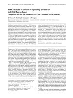
a native peptide bond at the ligation junction (Fig. 2A).
The first-step trans-
thioesterification reaction is catalyzed by a suitable thiol additive, and is reversible under
physiological conditions. The subsequent intramolecular nucleophilic attack by the αamino group of the N-terminal Cys to form the final amide bond is irreversible, and
highly favorable due to the intramolecular five-member ring formation. Consequently,
all of the freely equilibrating thioester intermediates (i.e. from the first-step reaction) will
eventually be depleted by the irreversible second-step reaction, giving rise to only a
single stable, ligated product.
A key feature of this reaction is that it is highly
chemoselective – the reaction occurs exclusively at the N-terminal Cys of the peptide,
4
even in the presence of other unprotected side-chain residues including internal cysteine
residues.[4, 15]
1.3. Bioimaging
One potential application of the above described biotechniques is in bioimaging, a
field that is still in its stages of infancy but is currently seeing rapid development.
Studying the dynamic movement and interactions of proteins inside living cells is critical
for a better understanding of cellular mechanisms and functions. Traditionally this has
been done by in vitro labeling of proteins with fluorescent and other molecular probes,
followed by monitoring them inside live cells. Recent advances in genetic engineering
have made it possible to directly generate fluorescent proteins in living cells or even in
live animals by fusion of fluorescent proteins such as GFP (green fluorescent protein) to
the protein of interest.[20-22] GFP and its variants, some of which possessing enhanced
fluorescent properties, improved pH-resistance etc., have been of late, a popular choice
for tracking protein movement and interactions.[22] However, the main drawbacks of
GFP-like proteins include their large sizes (e.g. 27 kDa for GFP), obligate
oligomerization which may affect the native biological activity of the fused protein, and
sometimes slow or incomplete maturation.
In addition, relatively few “colors” are
available amongst existing fluorescent proteins and they are not always ideal
fluorophores, as many have broad excitation/emission spectra, low quantum yields and
are susceptible to photobleaching. Lastly, the use of fluorescent proteins limits protein
labeling to only fluorescent and not any other molecular tags (e.g. biotin).
5
A good labeling strategy should ideally satisfy the following criteria: 1) a high signalto-noise ratio (i.e. high specificity for target protein); 2) uphold the integrity of the
labeled protein; 3) non-interference with the biochemical functions or cellular
localization of the labeled protein and 4) have minimal perturbation of cellular
processes.[20, 21]
Chemical methods for live cell labeling with a plethora of novel fluorescent labels,
many of which are much smaller and chemically tailored to specifically label proteins are
a promising alternative to fluorescent proteins. Numerous novel strategies for specific
labeling of proteins with small molecular probes in live cells have recently been
reported.[23-30] Each strategy is based on well-known chemistries and bio-interactions.
Cell permeability, non-toxicity, specific reactivity, good fluorescent properties, should be
carefully noted during the design and tailoring of small molecule probes. Compared to
the fluorescent proteins like GFP, small molecule probes possess a myriad of advantages
including the above as well as a wide spectral color. Their small size ensures minimal
perturbation of protein function other cellular components, which makes them attractive
over fluorescent proteins.
One of the first strategies for site-specific labeling of recombinant protein with small
organic molecules within live cells was developed by Tsien’s group.[23, 24] This method
exploits the high affinity of organoarsenicals with pairs of thiols. A tetracysteine motif,
CCXXCC (in which X is a non-cysteine amino acid) was genetically fused to the protein
and labeled with biarsenical probes. Their utility in live cell imaging was demonstrated
in visualizing the translocation of connexin in and out of gap junctions.[25] Non-covalent
interactions of small molecule ligands with streptavidin- and antibody-conjugated fusions
6
have also been used for in vivo labeling of proteins.[26, 27] Johnsson et al. described an
enzymatic approach to label proteins fused to the human O6-alkylguanine-DNA
alkyltransferase (hAGT) with small molecular substrates as probes.[28] A recent report of
small molecule probes for live cell labeling was based on the noncovalent interaction
between E. coli dihydrofolate reductase (DHFR) and methotrexate (Mtx) conjugates.[29]
Vogel et al. reported a generic method for the site-selective and reversible labeling of
membrane proteins containing a polyhistidine sequence in live cells with small organic
fluorophores conjugated to a metal ion (e.g. Ni2+) chelating nitrilotriacetate (NTA)
moiety.[30] The labeling is based on the well-known interaction between polyhistidine
sequences and the Ni2+-NTA moiety.[31]
This fast and reversible approach was
successfully applied to determine the topology of the membrane proteins in living cells
and is suitable for studying the protein-protein interactions within cellular signaling.
The above methods provide a site-specific means to label proteins in vivo, with
relatively fast reaction rates, with labeling being both covalent irreversible and
noncovalent
reversible.
Each strategy,
however,
possess their
own
inherent
disadvantages. For example, hAGT and DHFR are both full-length proteins (21 kDa and
18 kDa respectively) required for fusion to the target protein, while the biarsenical
labeling requires addition of high amounts of dithiols additive (micromolar range) to
reduce the background signal.[23-25] The main disadvantage of fluorescent NTA probes is
that direct visualization is not possible with a His6 tag and had to be indirectly inferred
from FRET (fluorescence resonance energy transfer) data due to the relatively low
affinity of the interaction (1-10µM). The affinity of the interaction improved with 10
7
histidines (~200nM), and although shown to be give a better signal-to-noise ratio for
detection, is still probably not as high as one would desire.[30]
1.4. Protein microarrays
The second application of this work is in the bludgeoning field of microarrays, in
which protein microarrays hold great promise. In the post-genomic era, the primary aim
for researchers around the world is to fully characterize and understand all proteins
encoded by the genome, or the so-called “proteome”.[32] Over the past few years, a
variety of proteomic techniques have been developed, allowing many thousands of
proteins to be studied based on either their relative abundance,[33] or their enzymatic
activities.[34] Most of these technologies, however, are based on the traditional protein
separation technique, the 2-dimensional gel electrophoresis, which requires downstream
instrumentations such as mass spectrometry in order to identify the proteins of interest
individually. They are therefore time-consuming and not easily automatable. Newer
technologies, especially those based on microarray platforms, have the potential to
rapidly profile the entire proteome, thus are capable of revealing novel protein functions
and mapping out comprehensive protein interaction networks of an organism.[34] The
very first report on microarray technologies was credited to Fodor and coworkers in
1991.[35]
This novel idea demonstrated the feasibility of simultaneously generating
thousands of µm-size spots on a small glass slide, leading to potential miniaturization and
high-throughput screenings of biological assays. In 1999, MacBeath and Schreiber,[36]
generated a high-density microarray of proteins and peptides in a 3” x 1” area using an
automatic robotic spotter.[37] Their seminal work, although conceptually simple and
8
primitive by today’s standard, has inspired the rapid development of many other types of
related technologies in the subsequent years.[38-48]
1.4.1. Immobilization strategies
The miniaturization of high-throughput screening on a single microscope-sized glass
slide has the undeniable advantage of needing only minute quantities of expensive
reagents for most biological assays. Nevertheless, the challenges when dealing with
proteins are numerous and complex. Proteins, in general being polymers of amino acids
and possessing immense chemical, physical and structural diversity, present additional
problems when immobilized in a microarray. They require intricate manipulation and
care to ensure preservation of features such as spot uniformity, stable immobilization and
preservation of desired protein activity in a microarray.[34]
The immobilization of biomolecules onto a glass surface while maintaining their
native properties is a critical area where researchers focus on generating different
chemical surfaces on a plain glass, allowing efficient protein/peptide immobilization
using appropriately chosen functional groups present on these biomolecules. For most
biological assays to be successfully carried out in a microarray, it is crucial that
immobilized proteins and peptides are oriented on the glass surface in an active state and
with a high density. Traditional surfaces used for protein/peptide immobilization in a
standard biochemical assay, including polystyrene, poly-vinylidene fluoride (PVDF),
agarose thin film and nitrocellulose membranes, could not be adopted easily in a
microarray format, primarily because these surfaces use non-covalent forces (e.g.
hydrophobic interactions) for immobilization, resulting in the generation of low-density
9
arrays of biomolecules which are randomly oriented on the surface. As a consequence,
these surfaces often give rise to relatively low signal-to-noise ratios in downstream
protein/peptide screening assays.
Glass slides, however, have the ideal surface for
microarray applications because they are inexpensive and with low intrinsic fluorescence,
at the same time also possessing a relatively homogeneous chemical surface, which,
when used with appropriate bioconjugate chemistry, are capable of immobilizing
biomolecules at very high densities. This directly translates into highly sensitive detection
of proteins/peptides in most microarray assays.
The surface of the glass slide is usually derivatized with chemicals to generate
different types of molecular layers.
Immobilization of proteins/peptides is then
subsequently carried out either by covalent linkage or non-covalent adsorption. One of
the most crucial disadvantages of non-specific adsorption is insufficient exposure of
functional domains, largely due to a variety of unpredictable orientations the immobilized
peptides/proteins can adopt upon binding to the glass surface. This often results in
binding of an unnecessary fraction of biomolecules with improper orientation, thus
impeding their binding with ligands and subsequent downstream biological assays.
Another possible drawback is that noncovalent binding by hydrophobic interaction may
cause protein denaturation and the loss of its functional activity.
The molecules
immobilized on the array may also be vulnerable to further manipulation, which may
result in the graduate depletion of proteins adsorbed noncovalently.
In order to ensure that all biomolecules are functionally active, it is imperative that
they are aligned uniformly and optimally upon immobilization onto the glass surface. A
variety of immobilization techniques have therefore been developed in the past few years
10
which allow site-specific immobilization of different molecules. These involve various
chemoselective chemistries including the oxime/thiazolidine formation,[38] the DielsAlder reaction,[39, 40] the Staudinger ligation,[41] the α-oxo semicarbone ligation.[42] Zhu
and Snyder were the first to successfully spot 6000 yeast proteins onto a single glass slide
to generate the so-called “proteome array”.[43]
Their work was made possible by
introducing site-specific immobilization of their proteins, which are all (His)6-tagged,
onto a glass slide functionalized with Ni-NTA. Our group developed two strategies for
site-specific immobilization of peptides using the high affinity biotin-avidin interaction
and native chemical ligation.[44] The use of the biotin-avidin interaction has recently been
extended by our group to the specific immobilization of proteins in a microarray format,
while preserving the proteins’ function and integrity.[45-48]
11
1.5. Objectives
1.5.1. Site-specific protein labeling with small molecule probes for bioimaging
applications
We propose a novel bioimaging strategy using intein-mediated splicing and small
molecule probes to specifically label live cells.[40, 41] We have recombinantly engineered
N-terminal Cys containing proteins at the C-terminus of the Ssp DnaB mini intein
(17kDa) using the pTWIN vector (NEB, USA).
Intein-mediated cleavage occurs
between the last amino acid of the mini intein and the first residue (i.e. cysteine) of the
target protein (Fig. 1). Incubation of the cell with a thioester-containing, cell-permeable
small molecule probe (Fig. 2C) allows the probe to efficiently penetrate through the cell
membrane into the cell, where the chemoselective native chemical ligation reaction
occurs between the thioester of the small molecule and the N-terminal Cys of the protein,
giving rise to the resulting labeled protein (Fig. 2A & B).[49, 50] The presence of other
reactive amino acid side chains, including internal cysteines, is tolerated.[15] The inteinmediated approach requires no external factors (e.g. proteases)[51] or addition of thiols,
with the splicing activity affected primarily by the identity of amino acids at the splice
junction, pH and temperature.[52, 53]
Our strategy provides an elegant and simple approach for the site-specific labeling of
proteins in live cells with minimal modifications to the target protein. We first showed
that the strategy could be applied to the specific labeling of proteins in vitro, followed by
labeling of proteins expressed inside live bacterial cells. We then further demonstrate the
generality and versatility of our approach for bioimaging by extending it to site-specific,
12
in vivo labeling of proteins expressed inside live mammalian cells, which are
considerably more complex in terms of their cellular environments.
HS
CYS
O
Intein
Protein
N
H
NH2
ASN
O
O
HS
CYS
Intein
NH
+
H2N
Protein
ASN
O
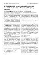
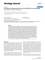
Figure 1. Mechanism of intein splicing at the C-terminal junction of the Ssp DnaB intein,
with Asn as the last amino acid of the intein and Cys as the first residue of the target
protein. Self-cleavage can be induced by a shift in temperature and pH conditions and
generates the protein of interest with an N-terminal Cys.
13
O
A
S
tag
O
O
+
H 2N
tag
H2 N
Protein
O
Protein
O
HS
O
HN
S
tag
Protein
HS
O
B
S
tag
Intein
tag
Target protein In vivo
cleavage
C
O
tag
O
O
O
N
O
N
O
O
H
N
CM
RO
O
O
O
O
O
O
O
OR
O
O
O
N
H
O
TMR
O
O
O
CF
N
H
H
HN
FL: R =
O
O
C2FL: R =
S
H
N
N
H H
O
Biotin
NO2
D
14
N
H