Vitamin k dependent γ carboxylation in chronic kidney disease
Bạn đang xem bản rút gọn của tài liệu. Xem và tải ngay bản đầy đủ của tài liệu tại đây (9.63 MB, 106 trang )
Institut für Tierwissenschaften
Abteilung Biochemie der Rheinischen Friedrich-Wilhelms-Universität Bonn
Vitamin K dependent γ-carboxylation in chronic kidney disease
Inaugural-Dissertation
zur
Erlangung des Grades
Doktor der Ernährungswissenschaften
(Dr. troph)
der
Landwirtschatlichen Fakultät
der
Rheinischen Friedrich-Wilhelms-Universität
Bonn
vorgelegt im Juli 2013
von Nadine Kaesler
aus
Aachen
Referent:
Prof. Dr. rer. nat. Brigitte Schmitz
Koreferenten:
Prof. Dr. rer. nat. Simone Diestel
PD. Dr. med. Vincent Brandenburg
Tag der mündlichen Prüfung:
19.11.2013
Erscheinungsjahr:
2014
Veröffentlichungen im Rahmen dieser Arbeit
I Originalarbeiten
Kaesler N, Schettgen T, Mutucumarana VP, Brandenburg V, Jahnen-Dechent W, Schurgers
LJ, Krüger T.: A fluorescent method to determine vitamin K-dependent gamma-glutamyl
carboxylase activity. Anal Biochem. 2012 Feb 15;421(2):411-6. doi:
10.1016/j.ab.2011.11.036. Epub 2011 Dec 2.
Kaesler N, Magdeleyns E, Herfs M, Schettgen T, Brandenburg V, Vermeer C, Floege J,
Schlieper G, Krüger T: Impaired vitamin K recycling in uremia is rescued by vitamin K
supplementation; Kidney International, in press
Kaesler N, Krüger T: Vitamin K, mehr als nur Koagulation, akzeptiert bei Ernährung und
Medizin 03/2013
Kaesler N, Immendorf S, Ouyang C, Herfs M, Magdeleyns E, Carmeliet P, Floege J, Krüger
T, Schlieper G: Gas6 Protein and its Role in Vascular Calcification; under revision, PLOSone
II Kongressbeiträge und Meetings
a) Vorträge
The Vitamin K cycle and vascular calcification
o Fellows Meeting, Abbvie Symposium on behalf of the 50th ERA.EDTA, Istanbul,
Turkey, 2013
“Gas6 protein and its role in vascular calcification”
o 45th Annual Meeting of the American Society of Nephrology, Kidney Week, San
Diego, USA, 2012
“Reduced γ-carboxylase Activity in Uremia - a Possible Mechanism of Uremic Vascular
Calcification”
o Fellows Meeting, Abbott Symposium on behalf of the 49th ERA-EDTA, Paris,
France, 2012
o 4th Conference of Fat Soluble Vitamins, Kalabaka, Greece, 2012
b) Ausgewählte Poster
“Increased level of vitamin K in ApoE-/- and LDL-/- mice”, 20th Congress of Nutrition,
Granada, Spain, 2013
“Gas6 protein and its role in vascular calcification”
o International Society of Nephrology (ISN) Nexus, Kidney and Bone, Copenhagen,
Denmark, 2012
“Reduced Activity of the gamma-Carboxylase in Uremia – Possible Mechanism of Uremic
Vascular Calcification”
o 49th ERA-EDTA, Paris, France, 2012; winner of Travel Grant
o 44th Annual Meeting of the American Society of Nephrology, Kidney Week,
Philadelphia, USA, 2011
Summary
Vascular calcification is present in atherosclerosis, ageing, chronic kidney diseases and
diabetes and is strongly associated with an increased morbidity and mortality. Calcification of
arteries occurs at the tunica intima and the tunica media. Thereby, vascular smooth muscles
cells (VSMC) transdifferentiate into an osteoblastic phenotype. In contrast to the antiquated
opinion that calcification of soft tissues is a passive process it is now known that actively
regulated processes play a major role. Modifiable calcification inhibitors were identified of
which matrix gla protein (MGP) is regarded as the most potent one being expressed in the
vascular wall. MGP gets posttranslationally gamma carboxylated at 5 glutamic acid residues,
which achieve calcium binding properties. This carboxylation step requires reduced vitamin K
as a cofactor. It is provided and recycled in the so called vitamin K cycle, which consists of
the vitamin K epoxid reductase (VKOR), DT-diaphorase and γ-glutamyl carboxylase
(GGCX). The VKOR is inhibitable by warfarin and other coumarins. High levels of
uncarboxylated MGP (ucMGP) were found in VSMC after treatment with vitamin K
antagonists like warfarin, which is frequently used for anticoagulation. Besides MGP, other
vitamin K dependent proteins are known. One is the Gas6 protein, which is also expressed by
VSMC, but its role is not yet fully understood. Gas6 binds to the Axl receptor, a receptor
tyrosine kinase which gets autophosphorylated after binding to its ligand. Gas6 offers one nterminal carboxylation site. The effects of uremia on vitamin K recycling via the vitamin K
cycle are unknown.
Aim of this thesis was to characterize the activities of three enzymes of the vitamin K cycle
and the role of vitamin K dependet Gas6 protein under uremic conditions.
First, a fluorescence method for the quantification of GGCX activity in vitro in tissue samples
was developed. This method employs a fluorescein isothiocyanate (FITC) labelled Glu
containing hexapeptide which gets carboxylated by the GGCX. The generated Gla-peptide
can be easily quantified using a reversed phase HPLC setup. For further proteomic analysis
mass spectrometry was applied.
Second, the influences of uremia and pharmacological doses of vitamin K supplementation on
the activity of the vitamin K cycle and extraosseous calcification were investigated. Uremia
was induced in rats by adenine diet, in part supplemented with vitamin K1 or K2 for 4 or 7
weeks. After 4 weeks of adenine, the activity of the vitamin-K cycle enzyme GGCX but not
DT diaphorase or VKOR was reduced. Serum levels of ucMGP increased, indicating
functional vitamin K deficiency. No histological calcification was detected at this stage but
aortic and renal calcium content increased. Seven weeks of adenine induced histological
calcification in the aorta, heart and kidneys. The addition of vitamin K restored the intrarenal
gamma-carboxylase activity and over-stimulated it in the liver and aorta. Moreover, vitamin
K treatment decreased tissue calcium content. Uremic functional vitamin K deficiency, at
least results from a reduction of the gamma-carboxylase activity which possibly contributes to
calcification.
Third, the influence of Gas6 protein on vascular calcification was investigated in murine in
vitro VSMC culture and different in vivo models using a) Warfarin diet, b) uninephrectomy or
c) electrocautery of the kidney as well d) ageing mice.
In vitro VSMC exposed to warfarin calcified and showed increased apoptosis without
differences between wildtype (WT) and Gas6-/- mice. In vivo, after electrocautery, serum
calcium increased similarly in WT and Gas6-/- mice but no significant difference in aortic
calcium content was observed between the groups. In all groups von Kossa staining revealed
only a weak positive vascular staining in WT and Gas6-/- mice. In ageing mice no significant
differences in vascular calcification could be identified between Gas6-/- and WT mice. No
differences were found in left ventricular (LV) mass, stroke volume or pulse wave velocity
(PWV) in all treatment groups. Gas6-/- mice showed no up regulation of MGP. This does not
support a role of Gas6 in the pathogenesis of vascular calcification.
Zusammenfassung
Vaskuläre Kalzifizierung tritt als eine Begleiterscheinung von Atherosklerose, Alter,
chronischen Nierenerkrankungen und Diabetes auf und geht mit einer stark erhöhten
Morbidität und Mortalität einher. Arterielle Kalzifizierungen erfolgen in der Tunica intima
und der Tunica media. Hier transdifferenzieren glatte Gefäßmuskelzellen (vascular smooth
muscle cells, VSMC) in einen osteoblastären Phänotyp. Entgegen der tradierten Auffassung,
dass die Gewebeverkalkung ein passiver Prozess ist, weiß man nun, dass es sich um einen
aktiv regulierten Prozess handelt. Es konnten regulierbare Verkalkungsinhibitoren identifiziert
werden. Ein potenter Kalzifizierungsinhibitor ist das Matrix Gla Protein (MGP), welches
insbesondere in der Gefäßwand von VSMC exprimiert wird. MGP wird an 5 Glutamatresten
posttranslational γ-carboxyliert, wodurch eine Kalziumbindung ermöglicht wird. Zur γCarboxylierung wird reduziertes Vitamin K als Cofaktor benötigt. Dieses wird im
sogenannten Vitamin K Zyklus bereitgestellt und recycelt. Die beteiligten Enzyme sind die
Vitamin K Epoxid-Reduktase (VKOR), DT-Diaphorase und γ-glutmayl-Carboxylase
(GGCX). Die VKOR wird durch Coumarine wie Warfarin inhibiert.
Erhöhte Level an uncarboxyliertem MGP (ucMGP) werden in VSMCs nach Warfarin
Exposition gefunden, welches weitverbreitet therapeutisch als Antikoagulanz Einsatz findet.
Neben MGP sind weitere Vitamin K abhängige Proteine bekannt. Hierzu zählt auch das Gas6
Protein, welches ebenfalls von VSMC exprimiert wird, aber dessen Funktion noch nicht
vollständig geklärt ist. Gas6 bindet an den Axl-Rezeptor, eine Rezeptor-Tyrosinkinase die
nach Ligandenbindung autophosphoryliert wird. Gas6 verfügt über einen n-terminale Gla
Rest. Das Ziel dieser Arbeit war die Charakterisierung der Enzymaktivitäten im Vitamin K
Zyklus und die Rolle des Vitamin K abhängigen Gas6 Proteins in der experimentellen
Urämie.
Dazu wurde zunächst eine Fluoreszenz-gestützte Methode entwickelt, zur Bestimmung der
GGCX Aktivität in Gewebeproben. Verwendet wurde ein Fluorescein Isothiocanat (FITC)
gekoppeltes Glu-haltiges Hexapeptid, welches durch die GGCX carboxyliert wird. Ein
reversed phase (rp) HPLC gestütztes Setup ermöglicht eine einfache Qunatifizierung des
generierten
Gla-Peptids.
Zur
weiterführenden
Proteom-Analyse
wurde
eine
Massenspektometrie durchgeführt.
Zweitens wurde der Einfluss einer Urämie sowie die Verabreichung pharmakologischer
Dosen Vitamin K auf die Enzyme des Vitamin K Zyklus und extraossäre Kalzifikation
untersucht. Durch Gabe von Adenin über einen Zeitraum von 4 oder 7 Wochen wurde in
Ratten eine Urämie induziert, teilweise unter Supplemtentation mit Vitamin K1 oder K2. Nach
7
4-wöchiger Adenin Behandlung war die Aktivität der GGCX reduziert, nicht jedoch der DTDiaphorase oder der VKOR. Die Serumwerte von ucMGP waren erhöht, woraus auf eine
funktionale Vitamin K Defizienz geschlossen werden kann. Histologisch konnte keine
Kalzifiaktion nachgewiesen werden, es zeigten sich jedoch erhöhte renale und aortale
Calcium Gehalte. Eine 7-wöchige Verabreichung von Adenin induzierte histologische
Kalzifikation von Aorta, Herz und Niere. Durch Zugabe von Vitamin K wurde die erniedrigte
renale GGCX Aktivität zurückgesetzt und in Leber und Aorta überstimuliert. Darüber hinaus
senkte Vitamin K den Gehalt an Calcium im Gewebe. Möglicherweise resultiert die
funktionale Vitamin K Defizienz in urämischen Patienten zum Teil aus einer erniedrigten
GGCX Aktivität mit einhergehenden Kalzifikationen.
Drittens wurde der Einfluss des Gas6 Proteins auf die Gefäßkalzifikation in murinen in vitro
VSMC Kultur und in verschiedenen in vivo Modellen untersucht: a) Warfarin Diät b)
Uninephrektomie c) Elektrokoagulation der Niere sowie d) alternde Mäuse. In vivo erhöhte
sich nach Elektrokauterisation der Serum Calcium Gehalt in WT und Gas6-/- ohne
signifikanten Unterschied zwischen den Gruppen. In allen Gruppen zeigte sich lediglich eine
schwach positive vaskuläre von Kossa Färbung in WT und Gas6-/- Mäusen. In alternden,
unbehandelten Mäusen gab es keine signifikanten Unterschiede bezüglich vaskulärer
Kalzifikation zwischen WT und Gas6-/- Mäusen. Echokardiographisch zeigten sich keine
Unterschiede
in
der
linksventrikulären
(LV)
Masse,
Schlagvolumen
oder
Pulswellengeschwindigkeit (PWV) in allen behandelten Gruppen. In Gas6-/- Mäusen lag keine
Heraufregulierung von MGP vor. Diese Daten unterstüzen keine Rolle von Gas6 in der
Pathogenese der vaskulären Kalzifikation.
8
Table of contents
page
13
I List of Tables
II List of Figures
14
III List of Formulas
17
IV Abbreviations
18
Chapter 1: INTRODUCTION
1.1 General Introduction
20
1.2 Aims
25
Chapter 2: MATERIALS AND METHODS
2.1 Chemicals
26
2.2 Instruments
29
2.3 Materials
30
2.4 Software
31
2.5 Methods for aim 1
32
2.5.1 Peptide design
32
2.5.2 Animals
32
2.5.3 Preparation of microsomes
32
2.5.4 Protein determination
33
2.5.5 GGCX activity assay
33
2.5.6 Purification of FLELFK-FITC
34
2.5.7 rp-HPLC
34
2.5.8 MS by LC/ESI-MS
34
2.5.9 14CO2 incorporation
35
2.6 Methods for aims 2-6
2.6.1 Rats
36
2.6.2 Blood Pressure
37
9
2.6.3 Biochemistry
37
2.6.4 ucMGP ELISA
38
2.6.5 Enzyme activites
38
2.6.5.1 GGCX activity assay
38
2.6.5.2 VKOR activity assay
38
2.6.5.3 DT-diaphorase activity assay
39
2.6.6 Calcium measurement
41
2.6.7 Histochemistry
42
2.6.8 Real time PCR
42
2.7 Methods for aim 7
2.7.1 Mice
44
2.7.2 VSMC culture
44
2.7.3 Protein determination
44
2.7.3 TUNEL assay
44
2.7.4 Mice: surgery and diets
45
2.7.5 Genotyping
46
2.7.6 Biochemistry
47
2.7.7 Calcium measurement
47
2.7.8 Echocardiography
47
2.7.9 Histochemistry
50
2.7.10 Real time PCR
50
2.8 Statistical analysis
51
Chapter 3: RESULTS
3.1 Results for aim 1
52
3.1.1 Peptide design
52
10
3.1.1 1 Detection of the uncarboxylated FLEFLK-FITC
52
3.1.1.2 Characterization of the carboxylated FLELFK-FITC
56
3.1.2 GGCX activity
59
3.1.3 14CO2 incorporation
61
3.2 Results for aims 2-6
3.2.1 Blood pressure
63
3.2.2 Biochemistry
63
3.2.3 Enzyme activities
67
3.2.3.1 GGCX activity
67
3.2.3.2 VKOR activity
69
3.2.3.3 DT-diaphorase activity
71
3.2.4 Calcium determination
72
3.2.5 Histochemistry
74
3.2.6 GGCX gene expression
77
3.3 Results for aim 7
3.3.1 In vitro calcification model: VSMC
78
3.3.2 In vivo calcification models
80
3.3.2.1 Genotyping
80
3.3.2.2. Biochemistry
82
3.3.2.3 Calcium measurments
84
3.3.2.4 Echocardiography
88
3.3.2.5 TUNEL assay
90
3.3.2.6 Collagen staining
90
3.3.2.7 RT-PCR for MGP
90
Chapter 4: DISCUSSION
92
11
Chapter 5: CONCLUSIONS
99
i: Reference list
100
ii: Danksagungen
106
12
I List of Tables
Table 1: Composition of diets for rats and treatment duration
Table 2: Molecular weights of MS fragments
Table 3: Biochemical results of rat serum at the end of the experiment
Table 4: VKOR activity in rat kidney and liver
Table 5: Baseline biochemical and functional characteristics of healthy wildtype and Gas6-/mice at different ages
Table 6: Baseline biochemical characteristics of healthy WT and Gas6-/- mice at different ages
Table 7: Biochemical haematology and 24h urine charactersitics of WT and Gas6-/- after
different treatments
Table 8: Functional characteristics of healthy WT and Gas6-/- mice at different ages
Table 9: Functional characteristics of WT and Gas6-/- mice after different treatments
13
II List of Figures
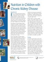
Figure 1: Chemical structure of phylloquinone
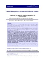
Figure 2: Chemical structure of menaquinone 4
Figure 3: Chemical structure of warfarin
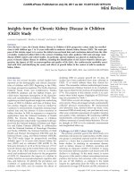
Figure 4: Schematic illustration of the γ-carboxylation
Figure 5: Chemical structure of dicoumarol
Figure 6: The vitamin K cycle
Figure 7: Structure of the FITC labelled hexapeptide FLEFLK
Figure 8: Overview of the 8 different rat treatment groups.
Figure 9: Principle of the DT diaphorase activity assay
Figure 10: Pipetting scheme of the 96-well plate for DT-diaphorase activity assay
Figure 11: Experimental design of the in vivo mouse experiments.
Figure 12: Electrocoagulation of the right kidney
Figure 13: Echocardiographic M-mode pictureof the long axis view
Figure 14: Short axis view of the diastole
Figure 15: Assessment of the pulse wave velocity in the common carotid artery
Figure 16: Rp-HPLC chromatogram of the purified reaction mixture at t=0
Figure 17: MS chromatogram of unmodified FLEFLK-FITC peptide solution
Figure 18: Linear correlation of the peak area and the uncarboxylated FLEFLK-FITC
Figure 19: Mass spectrum at 19.55 min of FLEFLK-FITC
Figure 20: HPLC chromatogram of the reaction mixture at t = 30 min
Figure 21: LC/MS chromatogram of the carboxylated peptide at t = 60 min
Figure 22: Mass spectrum of the carboxylated peptide at t = 60 min at 15.76 min
Figure 23: Linear correlation between time and GGCX activity
Figure 24: Linear correlation between the amount of microsomal protein and GGCX activity
14
Figure 25: Measurement of the activity of GGCX from rat liver and kidney without and with
NEM inhibition
Figure 26: 14CO2 incorporation of FLEFLK-FITC
Figure 27: Effect of acetonitrile on GGCX activity
Figure 28: Systolic blood pressure in CKD rats
Figure 29: Creatinine level in rat serum after 4 weeks of treatment
Figure 30: Phosphate level in rat serum after 4 weeks of treatment
Figure 31: GFR in rats at the end of the experiment
Figure 32: UcMGP measured in rat serum a) after 4 weeks; b) after 7 weeks of treatment.
Figure 33: GGCX activity (mean ± SD) in rat kidneys [a) after 4 weeks, b) after 7 weeks] and
liver [c) after 4 weeks, d) after 7 weeks]
Figure 34: GGCX activity in rat aortas after 7 weeks (mean ± SD) of treatment
Figure 35: In vitro incubation with 50 mM urea prior GGCX activity assay
Figure 36: Vitamin K1 peak at 8.7 min in rp-HPLC
Figure 37: Linear correlation of Vitamin K1 and area under the curve in rp-HPLC setup
Figure 38: DT-diaphorase activity (mean ± SD) in kidneys [a) after 4 weeks, b) after 7 weeks]
and liver [c) after 4 weeks, d) after 7 weeks]
Figure 39: Calcium content in rat aorta [a) after 4 weeks; b) after 7 weeks], heart [c) after 4
weeks; d) after 7 weeks] and kidney [e) after 4 weeks; f) after 7 weeks]
Figure 40: Quantification of von Kossa staining in rat aortic tissue (mean ± SD)
Figure 41: Von Kossa and ucMGP staining in rat aortic tissue (100 x)
Figure 42: Relative expression of GGCX in rat liver
Figure 43: a) Ca2+ deposition in VSMC culture derived from Gas6-/- and WT mice after 168
hours (h) of exposure to phosphate and calcium enriched cell culture medium. b)
TUNEL positive VSMC of Gas6-/- and WT mice after exposure to phosphate and
calcium enriched cell culture medium.
Figure 44: TUNEL and DAPI staining in VSMC from WT after 0 and 5 days of calcification
medium plus warfarin
Figure 45: Kaplan-Meier curve after electrocautery surgery in WT and Gas6-/Figure 46: DNA gel for Gas6 gene
15
Figure 47: Ca2+ content in mice aortas after warfarin diet, UniNx, EC or in healthy aging WT
(C57BL/6) and Gas6-/- mice
Figure 48: Von Kossa staining of aorta, heart and kidney after uninephrectomy in WT
compared to Gas6-/- mice
Figure 49: Collagen staining by sirius red in warfarin treated WT and Gas6-/- mice
Figure 50: MGP gene expression in WT versus Gas6-/- mice
16
III List of Formulas
Formula 1: Michaleis Menten equation
Formula 2: Glomerular Filtration Rate (GFR)
Formula 3: Difference of extinction of reduced MTT
Formula 4: Extinction of the specific DT-diaphorase activity
Formula 5: Beer Lambert Law
Formula 6: DT diaphorase activity
Formula 7: Devereux formula for LV mass
Formula 8: Ejection fraction
Formula 9: Stroke volume
Formula 10: Pulse wave velocity
17
IV Abbreviations
A
bp
BSA
B-mode
C
CAPS
CKD
DAPI
DNA
DTT
E
EC
EDTA
EDV
EF
ELISA
ESD
FAD
FITC
FLEEL
FLEFLK-FITC
G
GAPDH
GFR
GGCX
Gla
Glu
HEPES
K1
KM
LVID
LVM
LVPW
LVSW
MGP
MK4
M-mode
MS
MTT
MW
NADPH
NEM
Adenine
base pairs
Bovine Serum Albumin
Brightness mode
Cytosine
N-cyclohexyl-3-aminopropanesulfonic acid
Chronic Kidney Disease
4’,6-Diamidino-2-Phenylindole
Desoxyribonucleic Acid
Dithiotreitol
Extinction
Electrocautery
Ethylendiamintetraacetate
(left ventricular) end diastolic volume
Ejection Fraction
Enzyme Linked Immunosorbent Assay
(left ventricular) end systolic volume
Flavin Adenine Dinucleotide
Fluorescein Isothiocyanate
Phe-Leu-Glu-Glu-Leu
Phe-Leu-Glu-Phe-Leu-Lys-Fluorescein Isothiocyanate
Guanine
Glycerinaldehyde-3-phosphat dehydrogenase
Glomerular Filtration Rate
Gamma Glutamyl Carboxylase
Gamma-carboxy glutamic acid
Glutamic acid
2-(4-(2-Hydroxyethyl)-1-piperazinyl)-ethansulfon acid
Vitamin K1 epoxide
catalytic production of product
Michaelis constant
Left Ventricular Inner Diameter
Left Ventricular Mass
Left Ventricular Posterior Wall
Left Ventricular Septal Wall
Matrix Gla Protein
Menaquinone 4
Motion mode
Mass Spectrometry
3-(4,5-Dimethylthiazol-2-yl)-2,5-Diphenyltetrazolium Bromide
Molecular Weight
Nicotinamide Adenine Dinucleotide Phosphate
N-Ethylmaleimide
18
Ocn
PBS
PCR
PIVKA
PWV
RNA
rp-HPLC
S
SD
SV
T
TFA
TUNEL
ucMGP
UniNx
VACC
Osteocalcin
Phosphate Buffered Saline
Polymerase Chain Reaction
Prothrombin/ Protein Induced by Vitamin K Absence
Pulse Wave Velocity
Ribonucleic Acid
reversed phase High Performance Liquid Chromatography
Substrate
Standard Deviation
Stroke Volume
Thymine
Trifluoroacetic acid
Terminal deoxynucleotidyl transferase dUTP nick end labeling
uncarboxylated Matrix Gla Protein
Uninephrectomy
Pulse wave Velocity over the Arteria Carotis Communis
VKOR
Vmax
VSMC
WT
Vitamin K Epoxid Reductase
Maximum Velocity
Vascular Smooth Muscle Cells
Wildtype
19
Chapter 1___________________________________________________________________
1.1 General Introduction
Fat soluble vitamin K exists in 4 major different forms, all based on the 2-methyl 1,4naphtochinon (K3). The phylloquinone (K1; Figure 1), a 3 phytyl substituent, is located in
membranes of chloroplasts. It is the major component of vitamin K uptake in human nutrition.
Figure 1: Chemical structure of phylloquinone (Vitamin K1)
High contents of vitamin K1 can be found for example in herbs (cress: 600 µg/100 g; chive:
570 µg/100 g ), green leafy vegetables (corn salad: 200; chard 441 µg/100 g) or broccoli (129
µg/100 g) (1). Menaquinones (K2) contain an unsaturated isoprenoid side chain at the C3
position (MK4 - MK10, Figure 2).
Figure 2: Chemical structure of menaquinone 4 (MK4)
They are formed by bacterial fermentation, for example by bacillus subtilis in natto (2). Natto,
made from fermented soy beans, is the richest known source of vitamin K2. Menadione (K3)
and menadione esther (K4) are synthetic compounds and play only a role in animal nutrition.
Vitamin K1 is mainly found in the liver to serve as a cofactor for γ-carboxylation of the blood
coagulation factors II, VII, IX, X, protein C and S (3). Vitamin K2 is mainly distributed in
extrahepatic tissues (4) and contributes to γ-carboxylation of vitamin K dependent proteins
like MGP, Osteocalcin (Ocn) or Gas6. The function of all vitamin K dependent proteins is
mediated by binding of calcium to the γ-carboxylated form (5). Increased amounts of
uncarboxylated prothrombin (PIVKA), Ocn and MGP were detected in dialysis patients
compared to healthy controls (6;7). This indicates a functional vitamin K deficiency in this
20
Chapter 1___________________________________________________________________
population. The origin of the functional vitamin K deficiency in CKD is only partially
understood. Reduced vitamin K intake has been described in dialysis patients (7) but we
reasoned that this cannot fully explain the marked functional vitamin K deficiency.
Low levels of carboxylated MGP predict mortality in such patients (6;8). MGP knockout mice
develop spontaneous calcification of arteries (9). The mechanism by which MGP inhibits
vascular calcification may involve BMP-2 antagonism and a direct calcium-complexing effect
(10). MGP is expressed predominantly by vascular smooth muscle cells (VSMC) in the
arterial media and chondrocytes. It contains five glutamic acid residues that can be γcarboxylated (Glu → Gla) by the vitamin K-dependent γ-carboxylase (GGCX). MGP potently
inhibits precipitation of hydroxyapatite crystals in uremia. Abnormalities in mineral
metabolism and vascular calcification are highly present in chronic kidney diseases (CKD)
(11). Increased ucMGP is associated with increased coronary artery calcification (12).
Calcification can occur at the intimal (atherosclerosis) or at medial layer (arteriosclerosis) of
an arterial vessel wall (13). In CKD, defined as a decreased kidney function with a glomerular
filtration rate < 60 mL/min per 1.73 m2 (14), the arterial tunica media gets predominantly
calcified (13). It can be visualized by computed tomography (13;15). An increased coronary
artery calcification score is highly related to mortality in haemodialysis patients, independent
of the traditional risk factors (16). Possible unique contributors to the development of vascular
calcification in CKD are an increased calcium-phosphate product, parathyroid hormone and
as well as reduced levels of inhibitors of vascular calcification like fetuin-A and insufficient
activity of MGP (17).
In contrast to MGP, the role of Gas6 in vascular calcification is not well established. VitaminK dependent carboxylation of Gas6 is essential for its binding to the Axl receptor (18).
Tyrosine phosphorylation of Axl induces cell proliferation (19). Gas6 is known to protect
endothelial cells and VSMC against apoptosis (20;21), the latter is known to be associated
with vascular calcifications. Another potential link between Gas6 and vascular calcification is
demonstrated by in vitro data showing that phosphate- induced calcification of VSMC is
associated with a downregulation of Gas6 expression (21). In addition, antiapoptotic effects
and protection of calcification of VSMC by statins were mediated through Gas6 mRNA
stabilization (21). So far no in vivo data are available on the role of Gas6 in vascular
calcification. To clarify this, I assessed Gas6 knockout (Gas6-/-) mice and Gas6-/- derived
VSMC in in vitro and in vivo vascular calcification models.
21
Chapter 1___________________________________________________________________
Existing animal models contribute to a better understanding of the pathogenesis in vascular
calcification processes. Experimental uremia can be created by reducing the kidney mass
(uninephrectomy, 5/6 nephrectomy, electrocautery), which mimics the progressive nephron
loss occurring in patients with chronic renal failure (22;23). Noninvasively, dietary adenine
causes an overload of the converting adenine phosphoribosyltransferase and leads to
deposition of 2,8 dihydroxyadenosine crystals in the tubulo-interstitium of the kidney (22;23).
Another approach to induce vascular calcification is oral administration of coumarins like
warfarin (Figure 3) or phenprocoumon. This was shown both in rats (24) and humans (25)
(26). Coumarins directly inhibit the activity of the vitamin K oxidoreductase (VKOR; EC
1.1.4.1)(27). This leads to an insufficient activation of blood coagulation factors, a desirable
effect for patients with artificial heart valves or after thromboembolism. But the drawback is a
lesser γ-carboxylation of extrahepatic proteins like the vessel derived calcification inhibitor
MGP. Replacing coumarins with alternative thrombin inhibitors is under actual debate
(28;29). In turn, a high intake of vitamin K2 (MK4) was capable of regressing warfarininduced medial calcification in Wistar rats (30).
Figure 3: Chemical structure of warfarin
The GGCX (EC code 4.1.1.90) is an intrinsic membrane protein located in the endoplasmic
reticulum and requires vitamin K as a cofactor (31). It utilizes both reduced vitamin K (KH2)
and vitamin K with KH2 being the more potent one (32) (Figure 4). The amino terminus is
located on the cytoplasmic side and its carboxyl terminus on the lumen (33). The enzyme
carboxylates specific protein bound glutamate residues at the gamma position resulting in an
extra negative charge and thus potent calcium binding site (34).
22
Chapter 1___________________________________________________________________
Vit KH2
Vit K>O
GGCX
Figure 4: The gamma-carboxylation step: A peptide gets γ-carboxylated by the GGCX, which
requires the reduced form of vitamin K (Vit KH2) as a cofactor. KH2 is epoxidized to vitamin
K epoxide (Vit K>O).
Multiple proteins require γ-carboxylation to achieve full bioactivity (35). The pro-sequence,
which is the enzyme binding site, is a homologous region of several vitamin K dependent
proteins (3). Some known proteins containing this pro-sequence and thus being targets for the
GGCX are prothrombin, protein S and extrahepatic osteocalcin, Gas6 and MGP (5). Besides
the availability of vitamin K and KH2, the GGCX activity is also dependent on the
concentration of substrate and NaHCO3 (36). The enzymatic reaction produces the unusual
aminoacid Gla and vitamin K epoxide (K>O) as products, whereby K>O is recycled to K and
KH2 by the warfarin sensitive VKOR (37;38). The latter product is also generated by a
warfarin insensitive antidotal enzyme (39) the DT-diaphorase (EC 1.6.99.2 also called
NADPH-quinone oxidoreductase). The DT diaphorase using NAD(P)H as an electron
acceptor uses vitamin K as a substrate and is independent to the dithiotreitol pathway, which
antagonises the effects of warfarin (40;41). The DT-diaphorase is predominantly active in the
liver and offers an alternative pathway to provide vitamin KH2. Dicoumarol (Figure 5)
inhibits the purified DT-diaphorase by binding to the oxidized form of the enzyme (42).
Figure 5: Chemical structure of dicoumarol
23
Chapter 1___________________________________________________________________
These three enzymes form the so called vitamin K cycle (Figure 6) (43;44).
DTdiaphorase/
VKOR
Vitamin
Vitamin
KH2K
hydroquinone
protein
Vitamin K
GGCX
Warfarin
GGCX
carboxylated
carboxylated
protein
protein
VKOR
VitVitamin
K>O K
epoxide
protein
Figure 6: The vitamin K cycle (modified from Stafford, 2005 (43)). The required cofactor
vitamin KH2 for the γ-carboxylation by the GGCX is stepwise recycled by 2 additional
enzymes: the VKOR, which is directly inhibitable by warfarin and the DT-diaphorase.
In 1975, Emson and Suttie developed a method for measuring the GGCX activity by
incorporation of radioactive H14CO3- in the synthetic peptide Phe-Leu-Glu-Glu-Leu (short:
FLEEL) which is based on the sequence of prothrombin (31). Ulrich and colleagues tested 16
peptide sequences as enzyme substrates and found FLEEL to be the most active one (45). The
peptides were hydrolyzed and purified by anion exchange HPLC. Quantification was
achieved using a setup based upon a liquid scintillation for detection (46). Protocols describe
the addition of propeptide in approaches with purified enzyme (47) or in tissues without
propeptide (31;32;48). In vitro, GGCX is inhibited by N-Ethylmaleimide (NEM) (49) and 5Mercapto-1-Methyl-Thiotetrazole (5-MMT), which is used as a part of antibiotics
(moxalactam) (50).
24
Chapter 1___________________________________________________________________
1.2 Aims
1. Establishment of an appropriate method for safe and reproducible detection of γglutamyl-carboxylase activity in different tissues
2. Development of a model for uremia and vascular calcification in rats
3. Investigation of the vitamin K cycle under the conditions of uremia in rats
4. Analysis of the influence of oral vitamin K supplementation on vascular calcification
5. Investigation of the influence of oral vitamin K supplementation on vitamin K
dependent enzyme activities
6. Examination of the influence of uremic toxins on the activity of the γ -glutamyl
carboxylase activity
7. To clarify the role of Gas6 protein in vascular calcification processes
25