OCT 3 4 and SOX 2 are key factors for SDIA neurogenesis of mouse embryonic stem cells
Bạn đang xem bản rút gọn của tài liệu. Xem và tải ngay bản đầy đủ của tài liệu tại đây (6.44 MB, 88 trang )
OCT-3/4 AND SOX-2 ARE KEY FACTORS FOR SDIA
NEUROGENESIS OF MOUSE EMBRYONIC STEM
CELLS
STEPHEN WEIHUNG CHEN
(BSc.), Brown University
A THESIS SUBMITTED
FOR THE DEGREE OF MASTER OF SCIENCE
DEPARTMENT OF BIOCHEMISTRY
NATIONAL UNIVERSITY OF SINGAPORE
Acknowledgements
This work is supported by funding provided by The Bioprocessing Technology
Institute (BTI) and the Agency for Science Technology and Research (A*STAR).
Thanks to Prof. Wang Nai-Dy, Prof. Miranda Yap, Prof. Tan Bor Leung, Prof. James
Chen, and Dr. Paul Robson for their guidance, in addition to Peng Zhong Ni, Yoong
Lifoong, John Gan Wuoqiang, Theodosia Tan, and Tan Yew Chung for their technical
and personal support.
The author would also like to express gratitude for the
wonderful discussions and advice from Dr. Valerie Ng and Dr. Jason Kreisberg, the
latter providing invaluable feedback on submitted manuscripts and thesis revisions.
Thanks to MSc. supervisor, Dr. Andre Choo, for his suggestions and insight as well as
Dr. Steve Oh for his patience and providing motivation to persevere and succeed.
In particular, thank you to Prof. Too Heng-Phon for providing countless days of
humor and inspiration, and providing invaluable thoughts on science and life, and all
things in between.
2
Table of Contents
Abstract
Introduction
Results
1. Culturing of Mouse Embryonic Stem Cells (mESC)
a. Culturing Conditions
b. Expression of Oct-3/4, Alkaline Phosphatase and SSEA-1
2. Neurogenesis Using PA6 Stromal-Derived Inducing Activity (SDIA)
a. PA6 Co-Culture Method
b. Heparin Neural-Inducing Factor (Hep-NIF) SDIA Feeder-Free
Method
3. Knockdown of Oct-3/4 and Sox-2 Transcription Factors in Mouse
Embryonic Stem Cells
a. Establishment of Efficient Transfection Method
b. Oct-3/4, Sox-2 Knockdown and Differentiation Into Trophectoderm
4. Generation of Inducible Tet-Repressor (Tet-R) Short Hairpin RNA
(shRNA) mESC
a. Stable Transfection of Tet-R Protein into mESC
i.
pLenti6/TR Lentiviral Propagation and Packaging
ii.
pcDNA6/TR Plasmid
b. Generation of Tetracycline Inducible Short, Hairpin RNA (shRNA)
mESC Lines
i.
Construction and Design of shRNA Vectors
3
ii.
Stable Transfection and Selection into mESC
5. Differentiation of mESC Following Oct-3/4 and Sox-2 Knockdown
a. SDIA Differentiation Following Oct-3/4 and Sox-2 Knockdown
i.
Establishment of Precise Transcript Quantification Using
Cloned Standards
ii.
Quantification of Oct-3/4, Sox-2, Canonical Transactivation
Targets Following Knockdown
b. Screening for Alternative Cellular Fates
i.
Transcript Quantification of Lineage-Specific Differentiation
Markers
ii.
Appearance of Glial-Like Cells during SDIA Differentiation
Discussion and Future Work
Materials and Methods
References
Appendix 1 Media Formulations
Appendix 2 Related Articles by Author
4
Abstract
Utilizing a stromal-derived inducing activity (SDIA) model of neurogenesis, we
investigated the effects of the targeted knockdown of Oct-3/4 and Sox-2 by short
interfering RNAs (siRNAs) in mouse embryonic stem cells (mESC). Quantitative
real-time PCR showed a 40-90% knockdown of specific transcripts with cognate Oct3/4 or Sox-2 siRNA transfection compared to FAM-labelled negative control (FAM)
siRNAs or mock transfection, and was confirmed at the protein level by Western blot
analysis. Using PA6 SDIA co-cultures, neurogenesis was significantly diminished in
Oct-3/4 or Sox-2 targeted mESC upon differentiation. We observed that 45±12%,
65±13% and 90±8% (Mean +/- SD) of the colonies were stained with neuron-specific
β-tubulin III in Oct-3/4, Sox-2, and FAM siRNA transfected mESC respectively.
Similar results were observed when differentiating mESC with neural-inducing
factors (Hep-NIF) collected from the surface of PA6 cells using heparin. In addition,
differentiation of mESC using Hep-NIF but not Oct-3/4 and Sox-2 knockdown led to
the pronounced appearance of discrete, dark granular glial acidic fibrilary protein
(GFAP)-positive cells which also expressed the glial cell marker Vimentin. Taken
together, these results extend the role of Oct-3/4 in SDIA, implicate a similar role for
Sox-2, and support emerging observations for the role of these factors and SDIA in
gliogenesis.
5
List of Tables
Table 1 Selection of Real-Time PCR primer sets
Table 2 siRNA designs
Table 3 Primers sequences used for Real-Time PCR detection
Table 4 shRNA cassette designs
List of Figures
Figure 1 Culturing conditions of mESC cells (AB2.2)
Figure 2 Pluripotent mESC markers, Oct-3/4, Alkaline Phosphatase (AP), and StageSpecific Embryonic Antigen-1 (SSEA-1) are readily detectable in E14 cells
Figure 3 LIF withdrawal caused mESC (E14) differentiation
Figure 4 PA6 Stromal-Derived Inducing Activity (SDIA) co-culture
Figure 5 Heparin Neural-Inducing Factor (Hep-NIF) feeder-free SDIA
Figure 6 Western blot detection of TuJ1 in Hep-NIF differentiated mESC
Figure 7 Detection of FAM oligodT after two hours
Figure 8 Detection of FAM oligodT after twelve hours
Figure 9 Establishment of efficient mESC transfection method
Figure 10 Transient siRNA knockdown of Oct-3/4
Figure 11 Knockdown of Oct-3/4 and Sox-2 using siRNA
6
Figure 12 Trophectodermal differentiation following Oct-3/4 or Sox-2 knockdown
Figure 13 Tetracycline repressor (Tet-R) Lentiviral plasmid propagation
Figure 14 Tetracycline repressor (Tet-R) Lentiviral packaging and selection in ECOPak cells
Figure 15 Antibiotic selection of mESC – kill curve analysis
Figure 16 Generation of Tet-R mESC lines, establishment of transient shRNA vector
knockdown
Figure 17 Morphology of stably transfected mESC
Figure 18 Characterization of pluripotent markers (Oct-3/4, Sox-2, Nanog, SSEA-1)
in stably transfected mESC.
Figure 19 Inducible shRNA knockdown of Oct-3/4
Figure 20 Cloning of Oct-Sox transactivation targets and lineage-specific markers
Figure 21 Specific and efficient qRT-PCR detection of Oct-Sox transactivation
targets, lineage-specific markers
Figure 22 Characterization of canonical transactivation targets following Oct-3/4
knockdown
Figure 23 Characterization of canonical transactivation targets following Sox-2
knockdown
Figure 24 Attenuation of neurogenesis following Oct-3/4, Sox-2 siRNA knockdown
in PA6 SDIA co-cultures
7
Figure 25 Colony counts of PA6 SDIA co-cultures following Oct-3/4, Sox-2 siRNA
knockdown.
Figure 26 Attenuation of neurogenesis following Oct-3/4, Sox-2 siRNA knockdown
in PA6 SDIA co-cultures and Heparin feeder-free SDIA
Figure 27 Detection of lineage-specific markers
Figure 28 GFAP and TuJ1 expressions in SDIA cultures
Figure 29 GFAP and Vimentin expressions in SDIA cultures
List of Illustrations
Illustration 1 – Invitrogen BlockIT tetracycline inducible short, hairpin RNA system
8
Introduction
Mouse embryonic stem cells (mESC) are uniquely capable of differentiating into all
somatic cell types in the body, a property known as pluripotency. In addition, mESC
are known to grow indefinitely in culture without reaching senescence.
The
pluripotent capability of mESC has been particularly useful in the generation of
transgenic or knockout mice, and have allowed the establishment of in vitro
developmental and cellular differentiation models such as those for neurogenesis.
The propensity of mESC to preferentially differentiate into neurons provides evidence
for a “default model” of neurogenesis (Munoz-Sanjuan and Brivanlou, 2002). This
model proposes that embryonic stem (ES) cells for various species do not require
external factors such as fibroblast growth factors (FGFs) or wingless homologues
(WNTs) to instruct ES cells to become neuronal subtypes, but rather that ES cells
preferentially differentiate into neuronal subtypes by default, and are prevented from
doing so by the presence of neurogenesis-inhibiting bone morphogenic proteins
(BMPs). In Xenopus explant experiments, an autologously transplanted Spemann
Organizer (a potent source of various BMP inhibitors) has been shown to be able to
elicit neurogenesis in the recipient region. The default model further proposes that the
apparent capability of FGFs and WNTs to differentiate ES cells into neuronal
subtypes is largely attributable to BMP inhibition, thereby releasing ES cells to
differentiate into their preferential neuronal subtypes and not due to any direct cellular
fate specification (Tropepe et al., 2001). The latter function has been described as an
“instructive model” and is largely supported by chicken models in which explants
similar to Xenopus are incapable of eliciting a similar “default” response.
A
9
combination of FGFs and WNTs are requisite for neurogenesis, suggesting therefore
that these factors to have a deterministic rather than an accessory role in neuronal
subtype differentiation.
While exogenous factors clearly have a role in ES cellular fate specification, our
understanding of a default or instructive model would be greatly aided by a better
understanding of the transcription factors regulating ES cell pluripotency,
immortality, and neuronal differentiation.
Of great interest are pluripotent
transcription factors such as Oct-3/4 and Sox-2 and their possible roles in transiting
the boundary from an ES cell to a neuronal, or other, cellular derivatives. The PitOct-Unc (POU) transcription factor Oct-3/4, and Sox-2, a high mobility group (HMG)
protein, are thought to be important factors in maintaining ES cell identity. This
hypothesis is however controversial as some contradictory reports suggest the
necessity of a sustained expression of Oct-3/4
(also known as Pou5F1) for
neurogenesis (Shimozaki et al., 2003) and that constitutive Sox-2 expression enhances
neuronal differentiation (Sasai, 2001; Zhao et al., 2004).
The apparent pleiotropy of these transcription factors in maintaining a pluripotent
mESC identity may result from a diversity of binding to multiple transactivation
targets. While various mESC transcription factors, including Stat-3, FoxD3, Oct-3/4,
Sox-2 and Nanog have all been demonstrated to be involved in maintaining a
pluripotent phenotype (Cavaleri and Scholer, 2003), the apparent inability of any
single given factor to maintain mESC pluripotency suggests the possibility of shared
transactivation targets.
10
It is worth noting that transactivation target studies that have utilized Oct-3/4 and
Sox-2 as probes are few and some are performed in non stem cell types which may
not resemble biological responses in mESC.
Canonical targets of Oct-Sox
transactivation include Utf-1, Fgf-4, Opn, Fbx-15 (Botquin et al., 1998; Okuda et al.,
1998; Tokuzawa et al., 2003; Yuan et al., 1995). However, the overexpression of any
of these transactivation targets was neither permissive nor deterministic for a
pluripotent mESC phenotype. Thus, it is unclear how the multitude of cellular fates
potentially arising from the differentiation of mESC could be specified by these
targets.
Recent reports suggesting a significantly larger number (~2000) of putative binding
targets between Oct-3/4, Sox-2, and Nanog than previously suspected could provide a
significant clue to the biochemical dynamics underlying the choice of cell fate on
mESC differentiation (Boiani and Scholer, 2005; Boyer et al., 2005; Loh et al., 2006).
It is worthy to note that the transactivation or silencing of these binding targets, as a
result of the combinatorial interactions of Oct-Sox-Nanog binding, has yet to be
definitively shown (Remenyi et al., 2004).
This thesis explores the roles of Oct-3/4 and Sox-2 during cellular fate specification
of mESC in a neurogenesis model using the stromal cell line, PA6, as well as small,
interfering (siRNA) and short hairpin RNAs (shRNA) to inhibit the expressions of
Oct-3/4 and Sox-2 in differentiating mESC. We also measured the expression levels
of some canonical Oct-Sox targets with the intention of determining their potential
contributions to cellular fates.
11
Results
1.
Culturing of Mouse Embryonic Stem Cells (mESC)
a. Culturing Conditions
Initial culturing methods for mESC required a layer of co-cultured fibroblasts to
provide a biochemical and physical support for undifferentiated proliferation; such
cells (“feeders”) were inactivated using either mitomycin C (which crosslinks
thymidine) or radiation to prevent proliferation, overgrowth, and competition for cell
culture materials. We successfully cultured mESC on mitomycin C-treated STO
feeders in this condition (Figure 1).
An alternate culturing method, requiring
supplementation of exogenous leukemia inhibitory factor (LIF), was permissive of
undifferentiated mESC proliferation in the absence of feeders. In this condition,
mESC retained a smooth, bright, round colony morphology (Figure 1B) and could be
established following routine passaging of mESC onto gelatin-coated tissue culture
surfaces. Previously co-cultured, but unproliferative mitomycin C feeder layers were
eventually removed through 2-3 subsequent passages. mESC grown in this condition
could be spontaneously differentiated into embryoid bodies (EB) in suspension
following plating onto bacterial Petri dishes (Figure 1C); EB generation is a common
first step in establishing a heterogenous population of differentiated cells that can
subsequently be selected or channeled for derivation into the cell type of interest
(Roussa and Krieglstein, 2004).
12
A
B
10X
10X
C
25X
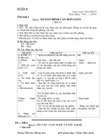
Figure 1. Culturing conditions of mESC (AB2.2). (A) mESC grown on STONeomycin-LIF feeders exhibited a bright, round, compact cluster morphology. (B)
Supplementation of exogenous Leukemia Inhibitory Factor (LIF) was sufficient to
maintain mESC morphology. Unproliferative feeders were not present after 2-3
passages. (C) mESC were placed onto bacterial petri dishes and grew in suspension as
embryoid bodies (EB), which were sequentially selected for or directed into
differentiated cell types of interest. Similar results were obtained using E14 cells.
b. Expression of Oct-3/4, Alkaline Phosphatase, and SSEA-1
Early identification and characterization of pluripotent mESC was aided by the
establishment of biochemical markers including, Oct-3/4, alkaline phosphatase (AP),
and stage-specific embryonic antigen-1 (SSEA-1), which were detectable in our
mESC cultures (Figure 2A-C, respectively). Expression of the Oct-3/4 is known to be
13
required for a pluripotent phenotype. Removal of support conditions (fetal bovine
serum or LIF) was found to result in a decrease in Oct-3/4 expression and the
inhibition of expression resulted in differentiation (see below). Interestingly, forced
expression of Oct-3/4 in concert with removal of support factors is not sufficient to
maintain a pluripotent phenotype (Chambers, 2004), suggesting Oct-3/4 alone is
insufficient to maintain stem cell identity. Other factors which are LIF dependent, but
Oct-3/4 independent, may be required for the pluripotent phenotype.
Upon LIF
removal for 1 week, AP expression was found to decrease, and the decrease in Oct3/4 expression (Figures 3A and 3B, respectively) was observed after 2-3 weeks later.
14
A Oct-3/4 Expression
B
Alk. Phos.
C
SSEA-1
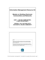
Figure 2. Pluripotent mESC markers, Oct-3/4, Alkaline Phosphatase (AP), and
Stage-Specific Embryonic Antigen-1 (SSEA-1) are readily detectable in E14 cells.
(A) mESC showed high levels of intracellular Oct-3/4 expression (dotted lines) as
compared to isotype control (solid line). (B) Immunofluorescence of AP marker for
pluripotent mESC showed the predicted expression in the cultures, as well as (C)
SSEA-1, a surface antigen specific for pluripotent mESC. Both E14 and AB2.2
showed similar profiles.
15
A
+LIF
AP 1 week
-LIF
B
Oct-3/4 Expression
2-week
58.1%
3-week
35.7%
Figure 3. LIF withdrawal caused mESC (E14) differentiation. (A) Alkaline
Phosphatase expression, viewed using phase contrast microscopy, decreased
following 1 week of LIF withdrawal from mESC cultures. (B) LIF withdrawal also
decreased Oct-3/4 expression within 2-3 weeks as shown. Dashed lines are pluripotent
mESC, solid line is mESC following LIF withdrawal, and dotted line is isotype
control. Numbers indicated are percentage of Oct-3/4 positive cells among LIF
withdrawal mESC for 2 and 3 week time points as indicated.
16
2. Neurogenesis Using PA6 Stromal-Derived Inducing Activity
a. PA6 Co-Culture Method
Mouse embryonic stem cells are able to differentiate into a wide variety of cell types,
including neuroectodermal derivatives, using a variety of methods: retinoic acid
treatment, gene trap lineage promoter-selection, or co-culture with stromal cell types
including PA6 (Kawasaki et al., 2000; Okabe et al., 1996). A skull marrow cell line,
PA6 was originally identified as enabling highly efficient (>90% colonies)
differentiation of mESC into tyrosine hydroxylase (TH)-positive neuron-like cells.
We utilized PA6 co-cultures to differentiate mESC into neuron-like cells staining
positive for Neurofilament Light Chain (NF-L), TH, and neuron-specific β-tubulin III
(Tuj1) markers (Figure 4). Neuron-like cells expressing TH are of particular interest
given their apparent destruction in Parkinson’s Disease (PD) pathology (Bjorklund et
al., 2003). The capability of PA6 to differentiate pluripotent mESC into TH+ neuron
like cells, termed stromal-derived inducing activity (SDIA), is based on the
production of unknown biological factors. Initial reports described a factor present on
the surface of PA6, as medium conditioned by PA6 is incapable of differentiating
mESC. However, it was reported that a permeable physical barrier (membrane filter)
between PA6 and mESC cells was still permissive for the differentiation of mESC to
TH+/ TuJ1+ cell types suggesting that a tethered labile factor might account for these
observations.
17
NF-L
25X
TH
TuJ1
25X
50X
2o Ab
50X
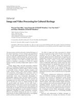
Figure 4. PA6 Stromal-Derived Inducing Activity (SDIA) co-culture. Mouse
embryonic stem cells seeded onto PA6; the latter were visible as flat, dark patches of
cells beyond the bright, round cluster morphology of differentiating neurospheres.
Extended processes were visible under bright field and immunostained for
Neurofilament Light Chain (NF-L), a neuron-specific marker. Neurospheres also
stained positive for the dopaminergic marker tyrosine hydroxylase (TH) as well as
neuron-specific TuJ1. Secondary antibody (2o Ab) negative control is shown.
18
b. Heparin Neural-Inducing Factor (Hep-NIF) SDIA Feeder-Free Method
More recently, the potency of SDIA has been extended through the use of a heparinPBS solution incubated with PA6 to isolate/collect differentiating factors from the cell
surface.
These previously incubated solutions (termed neural-inducing factors,
“NIFs”) can subsequently be added to serum-free mESC cultures for neuroectodermal
differentiation, albeit with a reduced efficiency when compared to direct plating of
mESC onto PA6 cells. Co-cultures of PA6 and heparin feeder-free (Hep-NIF) SDIA
generated 80-90% and 30-50% TH+/ TuJ1+ colony differentiation, respectively
(Yamazoe et al., 2005).
Utilizing heparin-PBS solutions of varying concentrations,
we established Hep-NIF SDIA differentiation in our mESC cultures. Consistent with
previous reports (Yamazoe et al., 2005), treatment of NIFs resulted in the formation
of neuron-like cell clusters (neurospheres) with extended processes. Only a small
number of neuron-like cell clusters was observed with PBS incubated with PA6 and
not observed at all with PBS alone (Figure 5A). These processes stained positive for
neuron-specific TuJ1, with large extensions observed between neurospheres and the
individual cells within (Figure 5B).
It was previously reported that higher
concentrations of heparin were inhibitory for efficient TuJ1+ neurosphere formation.
We observed similar results where 100 ug/mL heparin SDIA solutions resulted in
extension of processes visible under bright field, but patches of large, flat cells similar
to those generated in PBS controls were visible (Figure 5A). Expression of TuJ1 was
also significantly enhanced in mESC differentiated using Hep-NIF as compared to
PBS-NIF controls (Figure 6).
19
A
100 µg/ml Hep-NIF
10 µg/ml Hep-NIF
10X
PBS-NIF
B
PBS
TuJ1
10X
50X
Figure 5. Heparin Neural-Inducing Factor (Hep-NIF) feeder free SDIA. (A)
Heparin-SDIA differentiation using 100 or 10 µg/mL Heparin-PBS solution
previously incubated with PA6 (Hep-NIF), PBS with and without PA6 incubation
(PBS-NIF and PBS, respectively). PBS solution alone showed few processes and
increasing amounts with PBS-NIF, 100 µg / mL Hep-NIF and 10 µg / mL Hep-NIF,
respectively (similar to the original report of Yamazoe et al., 2005). (B) Usage of 10
µg / mL Hep-NIF solution added to serum-free differentiation media to differentiate
mESC resulted in brightly stained TuJ1+ colonies with processes extending between
and within differentiating neurospheres. High magnification within differentiating
neurospheres revealed polar cell bodies characteristic of neuron-like cells. All results
shown were for E14 cells.
20
E1
PB 4
SN
AB IF
PB 2 . 2
SE1 NIF
He 4
pNI
F
AB
He 2.2
pNI
F
TuJ1
Actin
Figure 6. Western blot detection of TuJ1 in Hep-NIF differentiated mESC.
Significantly higher expression of TuJ1 was detected using Western blot in mESC
differentiated using Hep-NIF compared to PBS-NIF control. This was consistently
observed in both E14 and AB2.2 cell lines.
21
3.
Knockdown of Oct-3/4 and Sox-2 Transcription Factors in Mouse
Embryonic Stem Cells
a. Establishment of Efficient Transfection Method
In order to determine potential roles for Oct-3/4 and Sox-2 in neurogenesis, we
established a robust RNAi knockdown method using siRNAs. Several factors can
contribute to the failure of an RNAi knockdown, including most commonly,
inappropriate siRNA design and poor transfection efficiency (Elbashir et al., 2002).
As an example of the latter, even if 60% of cells are properly transfected, and an 80%
knockdown occurs within that population, this would only equate to a theoretical 48%
decrease in total transcript expression. This may or may not be reproducibly detected
due to the one cycle (two-fold) differential limit of qRT-PCR detection.
We developed a simple means to optimize transfection conditions by utilizing FAM
oligodT to determine the efficiency of uptake in mESC colonies following the
transfection of several commercial reagents. These included liposomal (Invitrogen
Oligofectamine™/Lipofectamine 2000™, Bio-Rad Transfectin™, Roche Fugene 6™,
and New England Biolabs Transpass™), cationic polymer (Fermentas Exgen™), and
CaPO4 reagents, all of which were utilized according to the manufacturers’ protocols.
mESC when seeded overnight and transfected at approximately 40% confluency
failed to be transfected by Transpass™, Oligofectamine™ and Fugene 6™ when
observed at 2 and 12 hours post-transfection, while Exgen™ transfection was only
observable after 12 hours (Figure 8).
Liposomal delivery methods, including
Lipofectamine 2000™ and Transfectin™, as well as CaPO4 were able to transfect a
22
small number of colonies (<10%) when observed at 2 hours and increased marginally
at 12 hours post-transfection (Figure 7 and 8, respectively). It was observed that
CaPO4 and Exgen™ formed large deposits of FAM oligodT on the tissue culture
surface surrounding of mESC colonies (Figure 8).
Furthermore, Transfectin™
appeared to transfect slightly better than Lipofectamine™ 2000 two hours after
transfection (Figure 7). In all cases of FAM oligodT transfection, it was observed that
the transfected cells were often relegated to the periphery of mESC colonies. This is
presumably due to low accessibility of transfection complexes to membrane surfaces
within the core of mESC colonies.
23
2 hours
CaPO4
10X
FAM
Lipo2K
Transfectin
Control
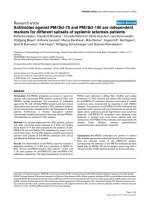
Figure 7. Detection of FAM oligodT after two hours. mESC (E14 cells) were
seeded onto 96-well plates, cultured overnight, transfected with various commercial
reagents and subsequently visualized by fluorescent microscopy at 2 hours. Except for
Bio-Rad Transfectin™, CaPO4 and Lipo2K did not show significant transfection of
FAM oligodT.
24
12 hours
CaPO4
Lipo2K
Transfectin
Exgen
Figure 8. Detection of FAM oligodT after twelve hours. Significantly more mESC
(E14) were transfected with FAM oligodT at 12 hours post-transfection for all
transfection reagents as indicated, including Exgen and CaPO4.
25