Overcoming mass transfer barriers in sandwich configuration for primary hepatocytes culture
Bạn đang xem bản rút gọn của tài liệu. Xem và tải ngay bản đầy đủ của tài liệu tại đây (3.7 MB, 105 trang )
OVERCOMING MASS TRANSFER BARRIERS IN
SANDWICH CONFIGURATION FOR PRIMARY
HEPATOCYTES CULTURE
HAN RONGBIN
NATIONAL UNIVERSITY OF SINGAPORE
2007
OVERCOMING MASS TRANSFER BARRIERS IN
SANDWICH CONFIGURATION FOR PRIMARY
HEPATOCYTES CULTURE
HAN RONGBIN
(M. Sci., TJU, China)
A THESIS SUBMITTED FOR THE DEGREE OF
MASTER OF SCIENCE
GRADUATE PROGRAM IN BIOENGINEERING
NATIONAL UNIVERSITY OF SINGAPORE
2007
ACKNOWLEDGEMENT
This research began two years ago when I settled in A/P Hanry Yu’s lab, when I
started my first lab rotation. I could never be what I am today, had there been
insufficient support and guidance from my supervisors. In the study at NUS, Prof. Yu
went extra mile to help me foster the ability to think creatively, analyze critically and
work independently. I am very grateful to him for showing me the way of research as
well as the consistent help and advice he has been providing me as close as a relative
and a good friend.
I am especially obliged to my collaborators Susanne Ng and Du Yanan who gave
countless support and help in the progress of the project. Without them I could never
explore out the way in this research field. I still want to extend my gratitude to Siew
Min, Shufang, Wen Feng, Zhang Jin, Xiaoshan, Jeff and Alex who gave me the
feeling of being at home at work.
Needless to say, that I need to thank all of my colleagues in Prof. Yu’s lab, who
provided me a lot of constructive ideas and advices during my research and
discussions of my thesis, especially Khong Yuet Mei, Toh Yi Chin, Dr Leo Hwa Liang,
Dr. Chia Ser Mien. I also want to thank Dr Sun Wanxin for his technical support on
microscopy and Chang Shi for his technical help in cell isolation.
I feel a deep sense of gratitude for my father and mother who formed part of my
vision and taught me the things that really matter in life. The encouragement of them
i
still provides a persistent inspiration for my journey in this life.
Finally I want to extend my appreciation to all of the friends who have been caring for
me and helping me during the past two years.
ii
TABLE OF CONTENTS
ACKNOWLEDGEMENT............................................................................................i
TABLE OF CONTENTS........................................................................................... iii
SUMMARY .................................................................................................................vi
LIST OF FIGURES AND TABLES ....................................................................... viii
LIST OF SYMBOLS ..................................................................................................xi
Chapter 1 Introduction................................................................................................1
1.1 Liver tissue engineering ................................................................................................... 1
1.1.1 Overview of tissue engineering .....................................................................1
1.1.2 Applications of liver tissue engineering.........................................................2
1.1.3 Liver physiology and general requirements of engineered in vitro models ..8
1.1.4 In vitro models for liver tissue engineering ................................................. 11
1.2 Primary hepatocytes in sandwich culture ................................................................... 13
1.2.1 Potential applications of sandwich culture in liver engineering ..................13
1.2.2 Polarity genesis of hepatocytes in sandwich culture....................................14
1.2.3 Functional maintenance of hepatocytes in sandwich culture.......................16
1.2.4 Inherent mass transfer barriers in sandwich configurations ........................18
1.3 Roles of flow environment in facilitation of mass transfer efficacy ..................... 19
1.3.1 Bioreactors in tissue engineering applications.............................................19
1.3.2 Increasing mass transfer efficacy by flow environment in bioreactors .......20
1.3.3 Current practice of bioreactors in liver engineering ....................................22
1.4 Synthetic ECMs in liver tissue engineering ............................................................... 23
1.4.1 Galactose-carrying synthetic ECMs ............................................................24
1.4.2 RGD motif-containing synthetic ECMs.......................................................26
iii
1.5 Project outline ................................................................................................................... 26
Chapter 2 Materials and Methods............................................................................29
2.1 Hepatocytes isolation and culture................................................................................. 29
2.2 Fabricating PET film conjugated with galactose (PET-f-Gal)................................ 29
2.3 Fabricating PET track-etched membrane conjugated with galacotose (PETm-Gal) or RGD (PET-m-RGD) ................................................................................. 30
2.4 Characterization of PET-RGD and PET-Gal substrate............................................. 31
2.5 Collagen coating and sandwich culture configuration........................................32
2.6 The bioreactor design and perfusion system .............................................................. 33
2.7 FITC-BSA transport behavior under different flow rates and diffusivity ............ 35
2.8 FITC-dextran diffusivity measurements ..................................................................... 36
2.9 Biliary excretion of fluorescein .................................................................................... 36
2.10 Immunofluorescence microscopy .............................................................................. 37
2.11 Scanning electron microscopy .................................................................................... 37
2.12 Hepatocytes functional assays .................................................................................... 38
2.13 Statistical analysis ......................................................................................................... 38
Chapter 3 Enhancing Mass Transfer Efficacy in Conventional Sandwich
Configurations by Manipulating Flow Environment ...........................39
3.1 Limited mass transfer efficacy in conventional sandwich configuration ............. 39
3.2 Effect of mass transfer efficacy on hepatocytes’ functions ..................................... 40
3.3 Regulation of mass transfer efficacy by varying perfusion flow rates ................. 42
3.4 Regulation of mass transfer efficacy by a separate drainage .................................. 46
3.5 Maintainance of hepatocytes’ fucntions and optimal mass transfer efficacy in
perfusion culture with separate draiange ................................................................ 48
Chapter 4 Engineering Novel Synthetic Sandwich Configurations with High
iv
Mass Transfer Efficacy ............................................................................53
4.1 Galactose-conjugated PET film as bottom support of sandwich configuration.. 53
4.1.1 Fabrication and characterization of PET film with Gal-ligand....................53
4.1.2 Dynamic process of self-assembly of hepatocytes on Gal-PET film...........55
4.1.3 3D monolayer on Gal-PET film...................................................................58
4.2 Overlaying of 3D monolayer with functionlized PET membrane ......................... 59
4.2.1 Permeability of selected PET membrane .....................................................59
4.2.2 Fabrication and characterization of bioactive PET membrane ....................61
4.3 Effect of various overlaying of different bioactive PET membrane ...................62
4.4 Hepatocytes sandwiched between Gal-PET membrane at the bottom and
RGD-PET membrane at the top .............................................................................. 68
4.4.1 Cell-cell interaction......................................................................................68
4.4.2 Polarity genesis ............................................................................................69
4.4.3 Functional maintenance ...............................................................................69
Chapter 5 Conclusion and Future Work .................................................................75
REFERENCES...........................................................................................................77
v
SUMMARY
This thesis explored two novel ways to encounter the inherent mass transfer barriers
of conventional sandwich configuration for primary hepatocytes culture combining
principles and technologies from tissue engineering, chemistry and bioreactor
engineering.
Sandwiching hepatocytes between two layers of extra-cellular matrix support creates
an intra-sandwich environment which differs from the extra-sandwich environment
defined by culture medium. When the intra-sandwich environment was characterized,
an albumin accumulation intra-sandwich environment in a conventional static
hepatocytes sandwich culture was identified. This indicated that the mass transfer in
the conventional sandwich configuration is limited. Further studies explored the effect
of the mass transfer limitation to hepatocytes’ functions in sandwich culture. Albumin
accumulation in the intra-sandwich environment resulted in reduced hepatocytes
functions in static culture.
To increase the mass transfer efficacy (indicated by effectively removal of albumin
out of intra-sandwich environment), hepatocytes were cultured in a perfusion
sandwich configuration by flowing culture medium at different flow rates above the
upper extra-cellular matrix support on porous membrane in a flat plate sandwich
perfusion culture bioreactor. It was found that albumin removal from the intrasandwich environment cannot be effectively achieved by varying the perfusion rates
without adversely affecting the hepatocytes functions. Based on the observation, we
have designed a novel bioreactor with a separate drainage channel directly connected
to the intra-sandwich environment, facilitating the removal of the metabolites and
vi
supply of nutrients directly. The mass transfer efficacy can be effectively regulated by
varying the drainage rates via the drainage channel without changing the perfusion
rates, as indicated by the phenomena that intra-sandwich albumin level was
effectively regulated by direct control of the drainage rates. Using the separate
drainage system, an optimal level of the drainage rates and mass transfer efficacy can
be maintained, which improved hepatocytes functions over the no-drainage controls.
Apart from the using of flow environment to improve mass transfer efficacy, we also
focused on the conventional sandwich configuration itself and tried to improve the
mass transfer efficacy by replacing the natural ECMs such as collagen, the main cause
of mass transfer limitation, with the synthetic polymers with controllable physical and
chemical properties. After trying with various functional polymers, an ideal synthetic
sandwich configuration was identified by overlaying a novel 3D monolayer developed
on galactosylated PET film with RGD conjugated polyethylene terephthalate (RGDPET) membranes, which also possessed better mass transfer properties over ECM
such as collagen. We proved that this configuration had the similar polarity genesis
process as conventional sandwich configurations: reorganization of F-actin in cell-cell
contact regions after 12h of sandwich culture; localization of bile canaliculi
transporter (MRP2) into bile channel after 24h of sandwich culture; regaining of
active bile secretion ability during the first several days of sandwich culture.
Moreover, enhanced cell-cell interaction and improved hepatocytes functions over 14
days of culture were observed in the synthetic sandwich configuration, most likely
due to the high mass transfer efficacy of this system.
vii
LIST OF FIGURES AND TABLES
Fig 1.1 Cellular architecture of the liver.
Fig 2.1 Schematic representation of perfusion circuit and separate drainage model for
perfusion sandwich culture.
Fig 3.1 Dynamic albumin accumulation in intra-sandwich environment in static
hepatocytes sandwich culture.
Fig 3.2 Effect of different sandwich culture configurations to the intra-sandwich
albumin environment and on the urea production at different culture days.
Fig 3.3 The simulation of metabolites transport process across the top collagen coated
membrane at different flow rates by a donor-receptor environment model using FITCBSA at different flow rates in a flat-bed perfusion sandwich bioreactor
Fig 3.4 Effect of different flow rates in flat-plate bioreactor for sandwich culture to
the hepatocytes functions.
Fig 3.5 The albumin level in intra-sandwich environment under flow rate of
0.25ml/min with the simulation based on the permeability coefficients.
Fig 3.6 Effect of different drainage rates to the albumin level in intra-sandwich
environment and to the urea production after four day of culture.
Fig 3.7 Hepatocytes functions in culture period of two weeks under perfusion culture
with optimized drainage rate.
Fig 3.8 Excretory function of hepatocytes indicated by FDA staining in the optimized
drainage culture condition compared with control group which do not incorporate
drainage.
Fig 3.9 The excretory function quantified by the ratio of the area of fluorescein in
intra-cellular sacs to the total area covered by cells.
viii
Fig 4.1 XPS wide scanning spectrums of PET, PET-g-AAc, PET-gal which showed
the successful grafting of acrylic acids and following immobilization of Gal ligands
onto the PET film.
Fig 4.2 Dynamic morphogenesis of hepatocytes’ self assembly on Gal-PET film using
the confocal transmission imaging.
Fig 4.3 Dynamic morphogenesis of hepatocytes’ self assembly on PET film using
SEM at different stages.
Fig 4.4 Liver specific functions and EROD activity under different culture conditions,
including 2D monolayer on collagen and 3D monolayer and spheroid.
Fig 4.5 SEM pictures of hepatocytes in various culture conditions, including 2D
monolayer, 3D monolayer and mature spheroid.
Fig 4.6 XPS C1s core-level spectra of the pristine PET track-etched membrane; the
oxidized PET membrane; the RGD conjugated PET membrane and the galactosylated
PET membrane.
Fig 4.7 Effect of overlay of 3D monolayer with Non-modified PET membrane, GalPET membrane and RGD-PET membrane on F-actin compared with no overlay group.
Fig 4.8 Effect of overlay of 3D monolayer with different functionalized PET
membrane on hepatocytes’ functions.
Fig 4.9 Morphology of hepatocytes under the sandwich configurations with Nonmodified PET membrane, RGD-PET membrane and Gal-PET membrane overlay
after one week of culture compared with no overlay group.
Fig 4.10 SEM pictures of hepatocytes cultured in the synthetic sandwich
configuration with Gal at the bottom and RGD at the top and in the conventional
collagen sandwich.
Fig 4.11 Excretory function of hepatocytes in the synthetic sandwich configuration
ix
compared with the excretory function in conventional collagen sandwich after
different culture period after overlay.
Fig 4.12 Functional maintenance of hepatocytes in the synthetic sandwich
configuration compared hepatocytes’ functions in conventional collagen sandwich.
Table 4.1 Diffusivity of dextran of various molecular weights across the modified
PET membrane and collagen layer.
x
LIST OF SYMBOLS
ECM
Extra-cellular matrix
PET
Polyethylene terephthalate
RGD
Arg-Gly-Asp
MRP2
Multidrug resistance protein 2
BSA
Bovine serum albumin
FITC
Fluorescein 5'-isothiocyanate
PBS
Phosphate buffered saline
FDA
Fluorescein diacetate
XPS
X-ray Photoelectron Spectroscopy
xi
Chapter 1 Introduction
1.1 Liver tissue engineering
1.1.1 Overview of tissue engineering
The field of tissue engineering, by integrating principles of engineering and life sciences,
exploits living cells in a variety of ways to restore, maintain, or enhance tissues and
organs [1]. Generally, the application of tissue engineering can be divided as therapeutic
application, in which the tissue is either grown in a patient or outside the patient [2,3] and
diagnostic applications, in which the tissue and culture models are engineered in vitro and
used for testing drug metabolism, uptake, toxicity and, pathogenicity, etc [4-6].
In both applications, cultured cells need to be coaxed to grow on bioactive degradable
matrix under properly engineered environment that provide the physical and chemical
cues to induce the regeneration functions needed, such as guiding cells’ differentiation
ability and assembly process into three-dimensional (3D) tissues [7]. Current progress in
tissue engineering is mainly limited in this step; those challenges include finding reliable
sources of compatible cells [8-10], engineering of proper cell culture matrix
(Biomaterials) [11-14], and the creating of novel bioreactors [15-18], which mimic the
environment of the body and that are amenable to scale-up. With fast development of
these areas recently, it is possible that laboratory-grown tissue replacements and cell
models will become a common medical therapy during the early decades of the 21st
century.
However, we need to be aware of the problems such like whether tissue
1
engineers can preserve the product so that it has a long shelf-life? Is it possible to permit
the fine control of tissue architecture for the engineered tissues to become clinically
useful without tissue rejection? All of these questions are pending solving.
1.1.2 Applications of liver tissue engineering
Liver, the largest organ in the body, serves vital roles in the body’s metabolization and
detoxification function, while liver diseases present a large portion of healthcare problem
worldwide with high incidents of cirrhosis, liver cancer and liver failure [19,20].
Although dramatic advances in surgical techniques and immuno-suppression have
permitted the use of liver transplantation in the management of liver disease, the patients
need cannot be met due to persistent donor shortage. To meet the needs, liver tissue
engineers made their efforts in both therapeutic and diagnosis approaches, namely,
extracorporeal bio-artificial liver devices and tissue-engineered constructs as therapeutic
approaches and hepatic drug testing for diagnosis uses:
1): Bio-artificial Liver Assistant Devices. The generated interest of bio-artificial liver
device (BALD) is to develop a system in which patient plasma is circulated extracorporeally through a bioreactor that houses metabolically active liver cells (hepatocytes)
sandwiched between artificial plates or capillaries to support a failing liver in the same
way that dialysis supports the failing kidney [21]. It requires keeping a large amount of
functional cells inside the engineered devices to fulfill the liver functions outside of
human body [22-25]. Those devices include hollow fiber devices, flat plate systems,
perfusion beds, and suspension reactors, which have shown encouraging results but have
been difficult to implement in the clinical setting. The most common bio-artificial liver
device design incorporates hepatocytes in hollow fiber cartridges. Hollow fiber
2
membranes provide a scaffold for cell attachment and immuno-isolation, and are well
characterized in a clinical setting, but may not provide adequate nutrient transport or the
proper environmental cues for long-term hepatocytes stabilization. Flat plate or
monolayer bioreactors have been showed to be able to offer better control of hepatocytes
microenvironment, but not ideal for scale up [26,27]. There are also many other designs,
which use perfusion environment or scaffolds to promote three-dimensional architecture
and minimize transport barriers. However, it may be difficult to provide uniform
perfusion of the packing matrix; and cells can be exposed to damaging shear forces
[27,28]. Encapsulated suspended cells or spheroid aggregates have been incorporated in
perfusion systems that would be simple to scale up, but are limited in their ability to
stabilize cells [27,29].
Although many devices include a combination of convective and diffusion transport flow
environment, mass transfer limitations of key nutrients to and from the cellular
compartment still exist due to diffusion resistance [30]. Barriers to diffusive transport, in
those cases, include membranes, collagen gels, and nonviable cells. Apart from culture
system consideration, one of the main challenges in BLAD design is to provide a proper
microenvironment for primary hepatocytes to maintain liver-specific function, which is
absent in many current device designs [31]. One of the essential requirements for BLAD
is to recapture the in vivo liver structure in vitro. In attempts to improve the hepatocytes
microenvironment, investigators have used micro-carriers; gel entrapment, both intraluminal and in the extra-capillary space; multi-compartment interwoven fibers; and multicoaxial configurations [27]. However, much more efforts are needed in the design and
3
optimization of culture models that are able to stabilize hepatocytes with cell–cell
interactions, cell–matrix interactions, and chemical cues.
2): Tissue engineering constructs. Although the approach remains largely experimental
and must overcome a number of significant hurdles before it will become a viable clinical
modality, tissue engineering of implantable cellular constructs become more and more
attractive as an emerging strategy for liver disease. Similar to cell transplantation,
hepatocytes are transplanted to perform liver functions; however, due to anchorage
dependent property of hepatocytes, it needs to be immobilized on scaffolds, encapsulated
in aggregates, or cultured ex vivo to form liver “organoids” and surgically transplanted
[32]. Most of proposed constructs need to utilize scaffolds of various chemical
compositions, both synthetic and biological compositions including biodegradable
polyesters, polysaccharides etc [33-35] and hyaluronic acid, collagen etc [36-38]
respectively. It has been reported that scaffold architecture and chemistry play essential
roles in hepatocytes survival, morphogenesis, and function. Many studies showed an
advantage of three dimensional scaffold architectures over the two-dimensional; and
functionality of implantable cellular constructs may be improved by incorporating cell
culture strategies that promote three-dimensional conformations and maintain
hepatocytes polarity [39]. Some proposed constructs use the encapsulation schemes; and
hepatocytes have been encapsulated in fibers, alginate and alginate–polylysine
composites to promote cell aggregation and liver-specific function as well as provide
immuno-isolation [40-42]. Encapsulation strategies for many different cell types,
including highly metabolic hepatocytes, face a classic dilemma between restricting
transport of immuno-modulators while maximizing transport of nutrients and desired cell
4
products. Also, spherical hepatocytes aggregates, heterospheroids of hepatocytes and
nonparenchymal cells, and cocultures formed on in vitro templates have been proposed as
tissue organoids for implantation [43-45]. While still in laboratory trying, hepatocytes
have been implanted in many sites including the peritoneal cavity and mesentery, as well
as the spleen, liver, pancreas, and subcutaneous tissues [46, 47].
Despite significant progress made in vitro, tissue engineering liver construct faces many
challenges, mainly limited by cell sourcing, immune rejection, and long-term viability
maintenance with additional issues such like transport limitations, the instability of the
hepatocytes phenotype when isolated from the hepatic microenvironment and the ability
for tissue structures to reorganize over time. Accordingly, fundamental research in tissue
engineering has been in the metabolic requirements of hepatocytes during seeding and in
early stages of implantation, design of biomaterials to improve angiogenesis, effects of
hepatocytes microenvironment on phenotypic stability (by manipulating soluble signals,
cell–substrate interactions, and cell–cell interactions), and morphogenesis of hepatocytes
structures in pure cultures[48,49]. Most importantly, none of the current proposed
constructs incorporates in their designs excretory function corresponding to the biliary
system, although studies indicate that morphogenesis can be achieved in vitro. In the
future, advances in developmental biology will likely complement “brute force”
strategies to replicate the exquisite micro-architecture of the liver and its myriad
functions. For example, soluble (fibroblast growth factor) and unidentified insoluble
factors have been identified in differentiation of the endoderm along the hepatic lineage
as well as in branching morphogenesis of the primitive kidney [50].
5
3): Hepatic-drug testing: The liver is the most important organ concerning the
biotransformation of xenobiotics. It plays a major role in the conversion of lipophilic into
hydrophilic compounds which can be readily excreted. The metabolism of chemicals
usually involves two enzymatic steps commonly referred to as phase I and phase II [51,
52]. Phase I metabolism is ensured mostly by cytochrome P450 (CYP) monooxygenases
such as EROD (ethoxyresorufin-O-deethylase, CYP 1A2) and ECOD (ethoxycoumarinO-deethylase, CYP 2B6). The oxidized metabolite is further conjugated in phase II by
UGTs (UDP-glucuronosyltransferases), STs (sulfotransferases), and GSTs (glutathioneS-transferases). The different enzymes necessary for the biotransformation are easy to
induce by a high or long substrate supply or an inducing agent. Therefore, the metabolism
and consequently the influence of drugs can be essentially affected.
Because of the important roles of liver, in vitro liver preparations are increasingly used
for the study of hepatotoxicity of chemicals. In recent years, various in vitro models were
developed with their actual advantages and limitations defined. The sandwich
configuration, liver slices, and 2D hepatocytes culture system, appear to be the most
common in vitro systems used, as liver-specific functions and responsiveness to inducers
are retained either for a few days or several weeks depending on culture conditions [53].
Maintenance of phase I and phase II xenobiotic metabolizing enzyme activities have been
proved in those systems; and those systems allows various chemical investigations to be
performed, including determination of kinetic parameters, metabolic profile, interspecies
comparison, inhibition and induction effects, and drug-drug interactions [54,55]. In vitro
liver cell models also have various applications in toxicology: screening of cytotoxic and
genotoxic compounds, evaluation of chemoprotective agents, and determination of
6
characteristic liver lesions and associated biochemical mechanisms induced by toxic
compounds. Extrapolation of the results to the in vivo situation remains a matter of debate.
Recently, hepatic transport processes have been recognized as important determinants of
drug disposition. Therefore, it is not surprising that characterization of the hepatic
transport and biliary excretion properties of potential drug candidates is an important part
of the drug development process [56]. Such information also is useful in understanding
alterations in the hepatobiliary disposition of compounds due to drug interactions or
disease states. Basolateral transport systems are responsible for translocating molecules
across the sinusoidal membrane, whereas active canalicular transport systems are
responsible for the biliary excretion of drugs and metabolites [57]. Several transport
proteins involved in basolateral transport have been identified including the Na+taurocholate co-transporting polypeptide, organic anion transporting polypeptides,
multidrug resistance–associated proteins and organic anion and cation transporters.
Canalicular transport is mediated predominantly via P-glycoprotein, MRP2, the bile salt
export pump and the breast cancer resistance protein. The development of in vitro
techniques to examine hepatic drug transport processes in human liver will provide
important insights regarding hepatobiliary drug disposition in humans. Elucidating the
mechanisms involved in hepatic drug transport, defining patient-specific factors that
affect transporter function, and characterizing how xenobiotic interactions may alter these
processes, are fundamental to our knowledge of how the liver disposes of endogenous
and exogenous compounds and are prerequisites to exploiting these processes to achieve
desirable clinical outcomes.
7
1.1.3 Liver physiology and general requirement of engineered in vitro
models
As stated in the above section, these therapies share a general requirement for adequate
cell culture environment and stability of liver-specific functions. The success of cellular
therapies ultimately depends on the stability of the hepatocytes phenotype and its
regulation by micro-environmental cues.
Primary
hepatocytes
are
anchorage
dependent and notoriously difficult to
maintain in vitro. Freshly isolated cells
rapidly lose adult liver morphology and
differentiated functions when cultured in
suspension. For years, investigators have
developed culture models based on
features
of
liver
architecture
to
recapitulate the complex hepatocytes
microenvironment.
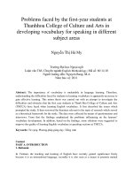
The in vivo microenvironment may
Figure 1 Celluar architecture of the liver [19]. Liver
epithelial cells called hepatocytes are arranged in cords between
the capillaries (sinusoids) of the liver. Oxygenated blood enters the
liver from the heart via the hepatic artery and from the gut via the
hepatic portal vein, mixes in the sinusoids, and drains via the
hepatic central vein back to the heart. Sinusoidal cells including
endothelial cells, Kupffer cells, and stellate cells line the sinusoids,
thus separating hepatocytes from blood. Picture use with author’s
permission.
provide a point of reference in engineering culture environments for hepatocytes in vitro.
Hepatocytes in vivo, are exposed to a variety of microenvironmental cues which are in
contact with different polarized domains of the plasma membrane associated with distinct
functions [61] (Figure 1): the sinusoidal (basal) region specialized for the exchange of
metabolites is in contact with loose ECM and sinusoidal plasma flow in the space of
Disse; the intercellular (lateral) domain whose tight junctions constitute the canaliculo-
8
sinusoidal barrier are the sites in which hepatocytes form tight cell-cell adhesions with
each other; the canalicular (apical) surface of hepatocytes highly specialized for the
secretion of bile acid and detoxification products faces a lumen which delivers bile to the
bile ductules. All of these factors, together with their interactions with non-parenchymal
cells and the exposure to acinar gradients of nutrients and xenobiotics, may work
cooperatively in vivo to supply a microenvironment which allows hepatocytes to maintain
their polarized morphology and functions, but ceases to operate when hepatocytes are
separated from their native environment.
Based on the understanding of basic liver micro-environment, the successful in vitro
models need to recapitulate the features of complex hepatocytes microenvironment [31]
to achieve: 1): Stabilization and maintenance of various liver specific functions. 2): Reestablishment of liver functional structures such like polarized structures with active bile
excretion ability. To reach these aims, there are a few features that must be incorporated
or considered in the development of in vitro culture models.
1): Cell-matrix interactions, The matrix used for liver engineering includes natural ECMs
(such like collagen, MatrigelTM, Biomatrix, laminin, fibronectin) and synthetic ECMs
[such like poly-lactic-co-glycolic acid (PLGA) and micro-carriers].The major function of
the matrix is to induce three-dimensional states in cells, essential for achieving ideal
cellular phenotype and functions. It has been shown that alterations in both the
composition and topology of the ECM have been shown to affect hepatocytes function
[62-64]. For examples, collagen enhanced hepatocytes differentiation over fibronectin,
while MatrigelTM, containing primarily laminin, collagen type IV, heparan sulfate
proteoglycan and entactin , maintained higher levels of mRNAs encoding albumin and
9
several P450 enzymes compared with gelled collagen [65]. Noteworthy, the sandwich
configuration, which mimics the matrix configuration in the Space of Disse by entrapping
cells between two layers of collagen gel, enhanced and maintained albumin secretion for
up to 6 weeks in culture, better than cells in a single layer of collagen gel [66, 67].
2): Cell-cell interactions. Cell-cell interactions, including homotypics interaction between
same cell type and hetertypic interactions between two different cell types, are crucial to
the function of several organ systems. By restoration of homotypic cell–cell interactions,
the hepatocytes spheroids and aggregates formed on non-adherent substrates have been
reported to promote the formation of bile canaliculi, gap junctions, tight junctions, and
help in stabilizing the primary hepatocytes phenotype [68]. A common feature for
hetertypic cell interaction is the interaction of parenchymal cells with nonparenchymal
neighbors resulting in the modulation of migration, cell growth and differentiation. Coculture of parenchymal cells with nonparenchymal have been shown, to varying degrees,
to induce phenotypic stability of hepatocytes for up to months in culture. These
heterotypic interactions are thought to present a highly conserved signal that greatly
augments liver-specific functions.
3): Soluble factors, such as hormones and chemical supplements [71,72]. Normally, the
soluble signals have a rapid turnover to activate transduction processes that induce a
specific physiologic process such like growth or expression of tissue specific genes. The
effect of a soluble factor is entirely dependent, both qualitatively and quantitatively, on
the matrix chemistry associated with the cell. Most of those soluble factors can be used to
help stabilize hepatocytes morphology and regulate functions.
10
4): Flow environment. A positive effect of flow environment has been proposed in in
vitro hepatocyte culture. Hepatocytes have been cultured in suspension, perfused
scaffolds and flat plate bioreactors [73]. These have not only increased hepatocytes
viability by efficient oxygenation and mass transfer of nutrients and waste products, but
have also been reported to enhance cell function and tissue morphogenesis.
1.1.4 In vitro culture models for liver tissue engineering
Based on the general requirement of in vitro models in liver tissue engineering, for years,
investigators have developed culture models based on features of liver architecture to
recapitulate the complex hepatocytes microenvironment, ranging from simple monolayer
culture to spheroids culture, to sandwich culture and co-culture system and more
sophisticated 3-D cultures [74-76].
Hepatocytes cultured as a 2D monolayer attached tightly to either plastic or ECM
proteins such as collagen I and laminin, showed deteriorating spreading morphology with
relatively low liver-specific function and nearly no native in vivo liver-like polarized
structure;
Improved spheroid culture configuration is developed based on the observation that selfassembled spherical aggregates of isolated primary hepatocytes have been obtained on
numerous moderately-adhesive substrata comprised of natural matrices such as
proteoglycan fraction from liver reticulin fibers, agarose, rigid extracellular matrix at low
concentration like Matrigel, laminin, fibronectin or collagen type I,
synthetic matrices such as positively charged
and artificially
or galactosylated substrata [77-80].
Hepatocytes spheroids with naturally formed 3D architecture showed associated cell-cell/
11
cell-matrix connectivity and ideal liver-specific functions, membrane polarities and liver
ultra-structures. However, the usefulness of 3D hepatocytes spheroids in applications is
limited due to the poor mass transport of nutrients, oxygen, xenobiotics and metabolites
into and from the core of these large cellular aggregates [81]. Cell loss is also a critical
issue in forming and maintaining these spheroids in applications due to the poor adhesion
of spheroids on the substratum [82].
Many groups have shown that hepatocytes can survive for long periods and maintain
specific functions when they are cocultured with other cell types, such as
nonparenchymal liver cells (NPCs) [69]. It was previously reported that formation of
multicellular spheroids consisting of hepatocytes and NPC in a hierarchical co-culture, in
which both cell-types were separated by a collagen layer, was very effective for the
maintenance of liver functions, such as albumin secretion, urea synthesis and induction of
tyrosine aminotransferase [83]. However, due to the system complexity and the lacking
of valid mechanisms regarding cell-cell interaction and various soluble factors involved,
these approaches, still have a long way to reach the practical uses. Also, the native liverlike structure, such as polarized structure, is hard to form due to the uncontrollable
seeding methods.
Sandwich culture, has been recognized as one of the most promising models currently
available to impact both in the studies of liver physiology/toxicology and developments
of technologies related to cell transplantation and hepatocytes bioreactors [84]. Primary
hepatocytes culture in sandwich configuration, formed by overlay of second layer of
ECMs support on monolayer cells cultured on single surface, captured the essential
characteristics of liver disse, induced the re-establishment of polarity structure with the
12