Kendig and chernicks disorders of the respiratory tract in children, 8e 2012
Bạn đang xem bản rút gọn của tài liệu. Xem và tải ngay bản đầy đủ của tài liệu tại đây (49.44 MB, 1,156 trang )
KENDIG AND CHERNICK'S
Disorders OF THE
Respiratory Tract
IN Children
EIGHTH EDITION
KENDIG AND CHERNICK'S
Disorders OF THE
Respiratory Tract
IN Children
EIGHTH EDITION
Robert W. Wilmott, MD, FRCP
IMMUNO Professor and Chair
Department of Pediatrics
St. Louis University
Pediatrician in Chief
Cardinal Glennon Children's Hospital
St. Louis, Missouri
Thomas F. Boat, MD
Christian R. Holmes Professor
Vice President for Health Affairs
Dean of the College of Medicine
University of Cincinnati
Cincinnati, Ohio
Andrew Bush, MD, FRCP, FRCPCH
Professor of Paediatric Respirology
Imperial College
Consultant Paediatric Chest Physician
Royal Brompton Hospital
London, United Kingdom
Victor Chernick, MD, FRCPC
Professor Emeritus
Department of Pediatrics and Child Health
University of Manitoba
Winnipeg, Manitoba, Canada.
Robin R. Deterding, MD
Professor of Pediatrics
Department of Pediatrics
Director, Breathing Institute
Children's Hospital Colorado
University of Colorado
Aurora, Colorado
Felix Ratjen, MD, PhD, FRCPC
Head
Division of Respiratory Medicine
Sellers Chair of Cystic Fibrosis
Professor
University of Toronto
Hospital for Sick Children
Toronto, Ontario, Canada
1600 John F. Kennedy Blvd.
Ste 1800
Philadelphia, PA 19103-2899
KENDIG AND CHERNICK'S DISORDERS
OF THE RESPIRATORY TRACT IN CHILDREN
ISBN: 978-1-4377-1984-0
Copyright © 2012, 2006, 1998, 1990, 1983, 1977, 1972, 1967 by Saunders, an imprint of Elsevier Inc.
All rights reserved. No part of this publication may be reproduced or transmitted in any form or by any
means, electronic or mechanical, including photocopying, recording, or any information storage and
retrieval system, without permission in writing from the publisher. Details on how to seek permission,
further information about the Publisher's permissions policies and our arrangements with organizations
such as the Copyright Clearance Center and the Copyright Licensing Agency, can be found at our
website: www.elsevier.com/permissions.
This book and the individual contributions contained in it are protected under copyright by the
Publisher (other than as may be noted herein).
Notices
Knowledge and best practice in this field are constantly changing. As new research and experience
broaden our understanding, changes in research methods, professional practices, or medical
treatment may become necessary.
Practitioners and researchers must always rely on their own experience and knowledge in
evaluating and using any information, methods, compounds, or experiments described herein. In
using such information or methods they should be mindful of their own safety and the safety of
others, including parties for whom they have a professional responsibility.
With respect to any drug or pharmaceutical products identified, readers are advised to check the
most current information provided (i) on procedures featured or (ii) by the manufacturer of each
product to be administered, to verify the recommended dose or formula, the method and duration
of administration, and contraindications. It is the responsibility of practitioners, relying on their
own experience and knowledge of their patients, to make diagnoses, to determine dosages and the
best treatment for each individual patient, and to take all appropriate safety precautions.
To the fullest extent of the law, neither the Publisher nor the authors, contributors, or editors,
assume any liability for any injury and/or damage to persons or property as a matter of products
liability, negligence or otherwise, or from any use or operation of any methods, products,
instructions, or ideas contained in the material herein.
Library of Congress Cataloging-in-Publication Data
Kendig and Chernick's disorders of the respiratory tract in children. – 8th ed. / [edited by] Robert W.
Wilmott … [et al.].
p. ; cm.
Disorders of the respiratory tract in children
Rev. ed. of: Kendig's disorders of the respiratory tract in children. 7th ed. c2006.
Includes bibliographical references and index.
ISBN 978-1-4377-1984-0 (hardcover : alk. paper)
I. Kendig, Edwin L., 1911- II. Wilmott, R. W. (Robert W.) III. Kendig's disorders of the respiratory
tract in children. IV. Title: Disorders of the respiratory tract in children.
[DNLM: 1. Respiratory Tract Diseases. 2. Child. 3. Infant. WS 280]
618.92'2–dc232012000458
Content Strategist: Stefanie Jewell-Thomas
Content Development Specialist: Lisa Barnes
Publishing Services Manager: Catherine Jackson
Senior Project Manager: Carol O'Connell
Design Direction: Steve Stave
Printed in China
Last digit is the print number: 9 8 7 6 5 4 3 2 1
PREFACE
In editing this, the eighth edition of Kendig and Chernick's
Disorders of the Respiratory Tract in Children, we are
struck by how much has changed since the last edition.
There have been remarkable new understandings of the
basic mechanisms of lung disease in the last 7 years. We
have recognized this by creating two new sections, each
of which has a section editor: the section on Interstitial
Lung Disease in Children edited by Robin Deterding and
the Aerodigestive Section edited by Thomas Boat. Every
chapter has been extensively updated and revised since
the last edition, and there is an increased emphasis on the
molecular mechanisms of disease and genetics. To save
space we have limited the number of references in the
paper version of the book, but the full reference lists are
available in the online version.
There are now six editors who have enjoyed the collaboration on identification of authors, review of outlines, working with the individual chapter authors, and
editing their work. With this edition we are joined by
Robin Deterding of the University of Colorado and Felix
Ratjen from the University of Toronto. Our plan is to add
two new editors with each edition to establish a rotation
that will allow some of us older ones to rotate off in the
future. However, as you might have noticed, nobody has
rotated off so far! However, we are delighted to recognize
Dr. Victor Chernick's many years of contribution to the
book with the change in its name.
There are 18 new chapters in this edition and 47 new
authors have joined the team. Thirty-two authors have
rotated off and we thank them all for their contributions.
We particularly want to recognize Dr. Mary Ellen Wohl,
who contributed several chapters to multiple editions of
the book and who passed away in 2009.
Our goal in editing this book is to publish a comprehensive textbook of pediatric respiratory diseases for a wide
audience: the established pediatric pulmonologist and
intensivist, fellows in pediatric pulmonology or intensive
care, pediatric practitioners, and residents. We also see this
book as an important resource for pediatric radiologists,
allergists, thoracic and cardiac surgeons, and others in the
allied health specialties. We have covered both common
and rare childhood diseases of the lungs and the basic science that relates to these conditions to allow for an understanding of pulmonary disease processes and their effect
on pulmonary function. Edwin Kendig founded this book,
which some say has become the bible of pediatric pulmonology, and we have strived to continue this tradition and
this degree of authority and completeness.
The staff at Elsevier, especially Lisa Barnes and Judy
Fletcher, have provided outstanding support for our
work, and we are grateful for their organization, sound
advice, attention to detail, and patience.
Finally, we must thank our families and partners for
their patience during the writing of this book, which has
been time consuming, and only their tolerance has made
the work possible.
Robert W. Wilmott
Thomas F. Boat
Andrew Bush
Victor Chernick
Robin R. Deterding
Felix Ratjen
CONTRIBUTORS
Robin Michael Abel, BSc, MBBS, PhD, FRCS (Eng
Paeds)
Consultant Paediatric and Neonatal Surgeon
Hammersmith Hospital, London, United Kingdom
Steven H. Abman, MD
Professor
Department of Pediatrics
University of Colorado School of Medicine
Director
Pediatric Heart Lung Center
Co-Director
Pulmonary Hypertension Program
Children's Hospital Colorado
Aurora, Colorado
Mutasim Abu-Hasan, MD
Associate Professor of Clinical Pediatrics
Pediatric Pulmonology and Allergy Division/
Pediatrics
University of Florida
Gainesville, Florida
Najma N. Ahmed, MD, MSc, FRCP(C)
Assistant Professor
Department of Pediatrics
McGill University
Pediatric Gastroenterology
Department of Pediatrics
Montreal Children's Hospital
McGill University Health Center
Montreal, Quebec, Canada
Samina Ali, MDCM, FRCP(C), FAAP
Associate Professor
Pediatrics and Emergency Medicine
University of Alberta
Edmonton, Canada
Adrianne Alpern, MS
Graduate Researcher
Department of Psychology
University of Miami
Miami, Florida
Eric F.W.F. Alton, FMedSci
Professor of Gene Therapy and Respiratory Medicine
National Heart and Lung Institute
Imperial College London
Honorary Consultant Physician
Royal Brompton Hospital
London, United Kingdom
Daniel R. Ambruso, MD
Professor
Department of Pediatrics
University of Colorado School of Medicine
Anschutz Medical Campus
Pediatric Hematologist
Center for Cancer and Blood Disorders
Children's Hospital Colorado
Aurora, Colorado
Medical Director
Research and Education
Bonfils Blood Center
Denver, Colorado
M. Innes Asher, BSc, MBChB, FRACP
Paediatrics
Child and Youth Health
The University of Auckland
Auckland, New Zealand
Ian M. Balfour-Lynn, BSc, MD, MBBS, FRCP,
FRCPCH, FRCS (Ed), DHMSA
Consultant in Paediatric Respiratory Medicine
Department of Paediatrics
Royal Brompton Hospital
London, United Kingdom
Peter J. Barnes, FRS, FMedSci
Professor
Imperial College London
London, United Kingdom
Robyn J. Barst, MD
Professor of Pediatrics
Department of Pediatric Cardiology
Columbia University College of Physicians
and Surgeons
Attending Pediatrician
Department of Pediatric Cardiology
Morgan Stanley Children's Hospital of New York
Presbyterian Medical Center
Director
Pulmonary Hypertension Center
New York Presbyterian Medical Center
New York, New York
Leslie L. Barton, MD
Professor Emerita
Pediatrics
University of Arizona College of Medicine
Tucson, Arizona
Contributors
Deepika Bhatla, MD
Chih-Mei Chen, MD
R. Paul Boesch, DO, MS
Lyn S. Chitty, PhD, MRCOG
Assistant Professor of Pediatrics
Saint Louis University
Bob Costas Cancer Center
Cardinal Glennon Children's Medical Center
St. Louis, Missouri
Asisstant Professor of Pediatrics
College of Medicine
University of Cincinnati
Asisstant Professor of Pediatrics
Division of Pulmonary Medicine and Aerodigestive
and Sleep Center
Cincinnati Children's Hospital Medical Center
Cincinnati, Ohio
Institute of Epidemiology
Helmholtz Zentrum München
German Research Centre for Environmental Health
Institute of Epidemiology
Neuherberg, Germany
Clinical Meolecular Genetics Unit
Institute of Child Health
Fetal Medicine Unit
University College Hospitals London
NHS Foundation Trust
London, England
Allan L. Coates, MDCM, B Eng (Elect)
Assistant Professor of Surgery and Pediatrics
Department of Surgery
Stanford University School of Medicine
Stanford, California
Senior Scientist Emeritus
Research Institute
Division of Respiratory Medicine
Department of Pediatrics
The Hospital for Sick Children
Toronto, Ontario, Canada
Andrew Bush, MD, FRCP, FRCPCH
Misty Colvin, MD
Matias Bruzoni, MD
Professor of Paediatric Respirology
Paediatric Respiratory Medicine
Imperial College and Royal Brompton
Hospital
London, United Kingdom
Michael R. Bye, MD
Professor of Clinical Pediatrics
Pediatrics
Columbia University College of Physicians and Surgeons
Attending Physician
Pediatric Pulmonary Medicine
Morgan Stanley Children's Hospital of NY
Presbyterian
New York, New York
Robert G. Castile, MD, MS
Professor of Pediatrics
Center for Perinatal Research
Nationwide Children's Hospital
Columbus, Ohio
Anne B. Chang, MBBS, FRACP, MPHTM, PhD
Professor
Child Health Division
Menzies School of Health Research
Darwin, Australia
Professor of Respiratory Medicine
Queensland Children's Medical Research Institute
Royal Children's Hospital
Brisbane, Australia
Michelle Chatwin, BSc, PhD
Clinical and Academic Department of Sleep
and Breathing
Royal Brompton Hospital
London, United Kingdom
Medical Director
Pediatric and Adult Urgent Care
Northwest Medical Center
Tucson, Arizona
Dan M. Cooper, MD
Professor
Departments of Pediatrics and
Bioengineering
GCRC Satellite Director
University of California, Irvine
Professor
Department of Pediatrics
UCI Medical Center
Professor
Department of Pediatrics
Children's Hospital of Orange County
Orange, California
Professor
Department of Pediatrics
Miller's Children's Hospital
Long Beach, California
Jonathan Corren, MD
Associate Clinical Professor
University of California, Los Angeles
Los Angeles, California
Robin T. Cotton, MD, FACS, FRCS(C)
Director
Pediatric Otolaryngology-Head and
Neck Surgery
Cincinnati Children's Hospital
Professor, Otolaryngology
University of Cincinnati College of Medicine
Cincinnati, Ohio
vii
viii
Contributors
James E. Crowe, Jr., MD
Professor of Pediatrics
Microbiology and Immunology
Vanderbilt University Medical Center
Director
Vanderbilt Vaccine Center
Nashville, Tennesee
Garry R. Cutting, MD
Professor
Institute of Genetic Medicine
Johns Hopkins School of Medicine
Baltimore, Maryland
Jane C. Davies, MB, ChB, MRCP, MRCPCH, MD
Reader in Paediatric Respiratory Medicine
and Gene Therapy
Imperial College London
Honorary Consultant in Paediatric Respiratory
Medicine
Royal Brompton Hospital
London, United Kingdom
Gwyneth Davies, MBChB
Clinical Research Fellow
Department of Gene Therapy
National Heart and Lung Institute
Imperial College
London, United Kingdom
Stephanie D. Davis, MD
Associate Professor of Pediatrics
Pediatrics, University of North Carolina at
Chapel Hill
Chapel Hill, North Carolina
Alessandro de Alarcon, MD
Assistant Professor
Department of Pediatrics
University of Cinncinatti
Director, Center for Pediatric Voice Disorders
Cincinnati Children's Hospital
Cincinnati, Ohio
Marietta M. de Guzman, MD
Assistant Professor
Department of Pediatrics
Section of Rheumatology
Baylor College of Medicine
Pediatric Rheumatologist
Texas Children's Hospital
Houston, Texas
Michael R. DeBaun, MD
Professor of Pediatrics and Medicine
J.C. Peterson Chair in Pediatric Pulmonology
Director
Vanderbilt-Meharry Center for Excellence in Sickle
Cell Disease
Vanderbilt University School of Medicine
Nashville, Tennessee
Sharon D. Dell, BEng, MD, FRCPC
Clinician Investigator
Division of Respiratory Medicine
Senior Associate Scientist
Child Health Evaluative Sciences
The Hospital for Sick Children
Assistant Professor
Department of Pediatrics
Faculty of Medicine
Unviersity of Toronto
Toronto, Canada
Robin R. Deterding, MD
Professor of Pediatrics
Department of Pediatrics
Director, Breathing Institute
Children's Hospital Colorado
University of Colorado
Aurora, Colorado
Gail H. Deutsch, MD
Associate Director
Seattle Children's Research Hospital Research
Foundation
Seattle, Washington
Michelle Duggan, MB, MD, FFARCSI
Consultant Anaesthetist
Mayo General Hospital
Castlebar, Ireland
Peter R. Durie, MD, FRCP(C)
Professor
Department of Pediatrics
University of Toronto
Senior Scientist
Research Institute
Gastroenterologist
Department of Pediatrics
The Hospital for Sick Children
Ontario, Canada
Eamon Ellwood, DipTch, DipInfo Tech
Department of Pediatrics
Child and Youth Health
The University of Auckland
Auckland, New Zealand
Leland L. Fan, MD
Professor of Pediatrics
Pediatrics
Children's Hospital Colorado
University of Colorado
Aurora, Colorado
Marie Farmer, MD
Professeure Adjoint
Pédiatrie FMSS
Université de Sherbrooke
Neurologue Pediatre
Pédiatre
CHUS
Sherbrooke, Quebec, Canada
Contributors
Albert Faro, MD
Associate Professor
Department of Pediatrics
Washington University
Physician Leader 7 East
St. Louis Children's Hospital
St. Louis, Missouri
Thomas W. Ferkol, MD
Professor
Pediatrics, and Cell Biology and Physiology
Washington University
St. Louis, Missouri
David E. Geller, MD
Associate Professor
Pediatrics
University of Central Florida
Director, Aerosol Laboratory and Cystic
Fibrosis Center
Pediatric Pulmonology
Nemours Children's Clinic
Orlando, Florida
W. Paul Glezen, MD
Professor
Molecular Virology and Microbiology,
and Pediatrics
Baylor College of Medicine
Houston, Texas
David Gozal, MD
Herbert T. Abelson Professor and Chair
Pediatrics
University of Chicago
Physician in Chief
Comer Children's Hospital
Chicago, Illinois
Anne Greenough, MD(CANTAB), MBBS, DCH,
FRCP, FRCPCH
Professor
Division of Asthma, Allergy and Lung Biology
MRC-Asthma UK Centre in Allergic Mechanisms
of Asthma
London, United Kingdom
Jonny Harcourt, FRCS
Consultant ENT Surgeon
Department of Paediatric ENT
Chelsea and Westminster Hospital
Consultant ENT Surgeon
ENT Department
Royal Brompton Hospital
London, United Kingdom
Ulrich Heininger, MD
Professor and Doctor
Division of Pediatric Infectious Diseases
University Children's Hospital
Basel, Switzerland
Marianna M. Henry, MD, MPH
Associate Professor of Pediatrics
Department of Pediatrics
University of North Carolina
Chapel Hill, North Carolina
Peter W. Heymann, MD
Head
Division of Pediatric Allergy
University of Virginia
Charlottesville, Virginia
Alan H. Jobe, MD, PhD
Professor of Pediatrics
University of cincinnati
Cincinnati, Ohio
Richard B. Johnston, Jr., MD
Associate Dean for Research Development
University of Colorado School of Medicine
Professor of Pediatrics
University of Colorado School of Medicine
and National Jewish Health
Aurora, Colorado
Sebastian L. Johnston, MBBS, PhD,
FRCP, FSB
James S. Hagood, MD
Professor and Chief
Department of Pediatrics
Division of Respiratory Medicine
University of Californi, San Diego
La Jolla, California
Professor of Respiratory Medicine
National Heart and Lung Institute
Imperial College London
Consultant Physician in Respiratory Medicine
and Allergy
Imperial College Healthcare NHS Trust
Asthma UK Clinical Professor and Director
MRC and Asthma UK Centre in Allergic
Mechanisms of Asthma
London, United Kingdom
Jürg Hammer, MD
Michael Kabesch, MD
Head
Division of Intensive Care and Pulmonology
Professor
University Children's Hospital Basel
Basel, Switzerland
Professor
Paediatric Pneumology
Allergy and Neonatology
Hannover Medical School
Hannover, Germany
ix
x
Contributors
Meyer Kattan, MD
Professor of Pediatrics
Columbia University College of Physicians
and Surgeons
Director, Pediatric Pulmonary Division
New York Presbyterian-Morgan Stanley Children's
Hospital
New York, New York
Brian P. Kavanagh, MD, FRCPC
Professor of Anesthesia, Physiology and Medicine
Department of Anesthesia
University of Toronto
Staff Physician
Critical Care Medicine
The Hospital for Sick Children
Toronto, Ontario, Canada
Lisa N. Kelchner, PhD, CCC-SLP, BRS-S
Clinical Research Speech Pathologist
Center for Pediatric Voice Disorders
Cincinnati Children's Hospital Medical Center
Cincinnati, Ohio
James S. Kemp, MD
Professor of Pediatrics
Department of Pediatrics
Washington University School of Medicine
Director of Sleep Laboratory
St. Louis Children's Hospital
St. Louis, Missouri
Terry Paul Klassen, MD, MSc, FRCPC
Director
Alberta Research Center for Health Evidence
Department of Pediatrics
University of Alberta
Edmonton, Canada
Alan P. Knutsen, MD
Director, Pediatric Allergy and Immunology
Saint Louis University
Professor
Pediatrics
Saint Louis University
St. Louis, Missouri
Alik Kornecki, MD
Associate Professor
Pediatrics
University of Western Ontario
Consultant
Pediatric Critical Care
Children's Hospital
London Health Sciences Centre
London, Canada
Thomas M. Krummel, MD
Department of Pediatric and Adolescent Medicine
Princess Margaret Hospital
Perth, Australia
Emile Holman Professor and Chair
Surgery
Stanford University School of Medicine
Susan B. Ford Surgeon-in-Chief
Lucile Packard Children's Hospital
Co-Director
Biodesign Innovation Program
Stanford University
Palo Alto, California
Carolyn M. Kercsmar, MD, MS
Geoffrey Kurland, MD
Andrew Kennedy, MD
Director, Asthma Center; Pulmonary Medicine
Cincinnati Childrens Hospital Medical Center
Professor
Pediatrics
University of Cincinnati
Cincinnati, Ohio
Leila Kheirandish-Gozal, MD
Director of Clinical Sleep Research
Section of Pediatric Sleep Medicine
Associate Professor
Pediatrics
University of Chicago
Chicago, Illinois
Cara I. Kimberg, MD
Clinical Psychologist
St. Jude's Children's Research Hospital
Memphis, Tennessee
Paul S. Kingma, MD, PhD
Neonatal Director
Fetal Care Center of Cincinnati
Assistant Professor
University of Cincinnati Department of Pediatrics
Cincinnati Children’s Hospital
Cincinnati, Ohio
Professor
Pediatrics
Children's Hospital of Pittsburgh
Pittsburgh, Pennsylvania
Claire Langston, MD
Professor, Department of Pathology
and Pediatrics
Baylor College of Medicine
Pathologist
Department of Pathology
Texas Children's Hospital
Houston, Texas
Ada Lee, MD
Attending
Department of Pediatrics
Pediatric Pulmonary Medicine
The Joseph M. Sanzari Children's Hospital
Hackensack University Medical Center
Hackensack, New Jersey
Margaret W. Leigh, MD
Professor and Vice-Chair
Pediatrics
University of North Carolina
Chapel Hill, North Carolina
Contributors
Daniel J. Lesser, MD
Clinical Assistant Professor of Pediatrics
University of California, San Diego
Pediatric Respiratory Medicine
Rady Children's Hospital
San Diego, California
Sooky Lum, PhD
Portex Unit
Respiratory Physiology and Medicine
UCL, Institute of Child Health
London, United Kingdom
Anna M. Mandalakas, MD, MS
Associate Professor, Pediatrics
Retrovirology and Global Health
Baylor College of Medicine
Texas Children's Hospital
Director of Research, Global Tuberculosis and
Mycobacteriology Program
Center for Global Health
Houston, Texas
Paulo J.C. Marostica, MD
Pediatric Emergency Section
Pediatric and Puericulture Department
Medical School of Universidade Federal do Rio Grande
do Sul
Rio Grande do Sul, Brazil
Robert B. Mellins, MD
Professor Emeritus and Special Lecturer
Columbia University
Morgan Stanley Children's Hospital of
New York
New York, New York
Peter H. Michelson, MD, MS
Associate Professor of Pediatrics
Department of Allergy, Immunology and Pulmonary
Medicine
Washington University School of Medicine
St. Louis, Missouri
Claire Kane Miller, PhD
Program Director
Aerodigestive and Sleep Center
Cincinnati Children's Hospital
Field Service Assistant Professor
Department of Otolaryngology-Head and Neck
Surgery
University of Cincinnati, College of Medicine
Clinical Speech Pathologist
Division of Speech Pathology
Cincinnati Children's Hospital
Adjunct Assistant Professor
Communication Sciences and Disorders
University of Cincinnati
Cincinnati, Ohio
Anthony D. Milner, MD, FRCP, DCH
Professor of Neonatology
Department of Pediatrics
United Medical and Dental School of Guy's
and St. Thomas's Hospital
London, United Kingdom
Ayesha Mirza, MD
Assistant Professor
Infectious Diseases and Immunology
Pediatrics
University of Florida
Jacksonville, Florida
Miriam F. Moffatt, PhD
Professor of Respiratory Genetics
National Heart and Lung Institute
Imperial College
London, United Kingdom
Mark Montgomery, MD, FRCP(C)
Clinical Associate Professor
Department of Pediatrics
University of Calgary
Calgary, Canada
Gavin C. Morrisson, MRCP
Associate Professor
Pediatrics
University of Western Ontario
Consultant
Pediatric Critical Care
Children's Hospital
London Health Sciences Centre
London, Canada
Gary A. Mueller, MD
Department of Pediatrics
Wright State University School of Medicine
Children's Medical Center
Dayton, Ohio
Vadivelam Murthy, MD
Division of Asthma, Allergy, and Lung Biology,
MRC and Asthma
United Kingdom Centre in Allergic Mechanisms of Asthma
King's College London
London, United Kingdom
Joseph J. Nania, MD
Consultant in Pediatric Infectious Diseases
Phoenix Children's Hospital, Scottsdale Healthcare
and Banner Health Network
Phoenix, Arizona
Manjith Narayanan, MD, DNB(Paediatrics),
MRCPCH, PhD
Clinical Research Fellow
Child Health Division
Depatment of Infection, Immunity, and Inflammation
University of Leicester
Specialist Registrar
Department of Paediatrics
Leicester Royal Infirmary
Leicester, United Kingdom
xi
xii
Contributors
Dan Nemet, MD, MHA
Professor of Pediatrics
Director, Child Health and Sports Center
Vice Chair of Pediatrics, Meir Medical Center
Sackler School of Medicine Tel Aviv University, Israel
Tel Aviv, Israel
Christopher Newth, MD, FRCPC, FRACP
Professor of Pediatrics
Anesthesiology and Critical Care Medicine
Children's Hospital Los Angeles
University of Southern California
Los Angeles, California
Andrew G. Nicholson, FRCPath, DM
Consultant Histopathologist specialising in thoracic
pathology
Histopathology
Royal Brompton and Harefield NHS Foundation
Trust
Professor of Respiratory Pathology
National Heart and Lung Division
Imperial College
London, United Kingdom
Terry L. Noah, MD
Professor
Pediatric Pulmonology
University of North Carolina
Chapel Hill, North Carolina
Lawrence M. Nogee, MD
Professor of Pediatrics
Pediatrics
Johsn Hopkins University School of Medicine
Baltimore, Maryland
Blakeslee Noyes, MD
Professor of Pediatrics
Department of Pediatrics
Saint Louis University School of Medicine
St. Louis, Missouri
Andrew Numa, MB, BS
Director
Intensive Care Unit
Sydney Children's Hospital
Senior Lecturer
Faculty of Medicine
University of New South Wales
Sydney, Australia
Hugh O'Brodovich, MD, FRCP(C)
Arline and Pete Harman Professor and Chairman
Department of Pediatrics
Stanford University
Stanford, California
Adalyn Jay Physician-in-Chief
Lucile Packard Children's Hospital
Palo Alto, California
Matthias Ochs, MD
Professor and Chair
Institute of Functional and Applied Anatomy
Hannover Medical School
Hannover, Germany
Øystein E. Olsen, PhD
Consultant Radiologist
Department of Radiology
Great Ormond Street Hospital for Children
NHS Trust
London, United Kingdom
Catherine M. Owens, BSC, MBBS, MRCP,
FRCR
Reader, Imaging Department
Consultant in Diagnostic Imaging
Cardiothoracic Imaging
University College London
London, United Kingdom
Howard B. Panitch, MD
Professor of Pediatrics
University of Pennsylvania School of Medicine
Director of Clinical Programs
Division of Pulmonary Medicine
The Children's Hospital of Philadelphia
Philadelphia, Pennsylvania
Nikolaos G. Papadopoulos, MD, PhD
Associate Professor
Allergy Department, Second Pediatric Clinic
University of Athens
Greece
Hans Pasterkamp, MD, FRCPC
Professor
Pediatrics and Child Health
University of Manitoba
Adjunct Professor
School of Medical Rehabilitation University
of Manitoba
Winnipeg, Canada
Donald Payne, MD, FRACP, FRCPCH
Associate Professor
Paediatric and Adolescent Medicine
Princess Margaret Hospital
Associate Professor
School of Paediatrics and Child Health
University of Western Australia
Perth, Australia
Scott Pentiuk, MD, MeD
Assistant Professor of Pediatrics
Division of Gastroenterology, Hepatology,
and Nutrition
Cincinnati Children's Hospital Medical Center
Cincinnati, Ohio
Contributors
Thomas A.E. Platts-Mills, MD, PhD
Professor of Medicine
Division Chief
Asthma and Allergic Diseases Center
University of Virginia
Charlottesville, Virginia
Timothy A. Plerhoples, MD
Resident in Surgery
Department of Surgery
Stanford University School of Medicine
Stanford, California
Amy C. Plint, MD, MSc
Pediatrics
University of Ottawa
Emeregncy Medicine
Ottawa, Canada
Jean-Paul Praud, MD, PhD
Professor
Pediatrics
Universitè de Sherbrooke
Sherbrooke, Canada
Phil E. Putnam, MD
Professor
Department of Pediatrics
University of Cincinnati
Director, Endoscopy Services
Cincinnati Children's Hospital Medical Center
Cincinnati, Ohio
Alexandra L. Quittner, PhD
Professor
Psychology
University of Miami
Coral Gables, Florida
Shlomit Radom-Aizik, PhD
Director of Research
Pediatric Exercise Research Center
University of California, Irvine
Irvine School of Medicine
Irvine, California
Mobeen H. Rathore, MD, CPE, FAAP, FIDSA, FACPE
Professor and Associate Chairman
Pediatrics
University of Florida
Chief
Pediatric Infectious Diseases and Immunology
Wolfson Children's Hospital
Chief
General Academic Pediatric
University of Florida
Medical Director
Children's Medical Services
Department of Health
Jacksonville, Florida
Gregory J. Redding, MD
Professor
Pediatrics
University of Washington School of Medicine
Chief
Pulmonary and Sleep Medicine
Seattle Children's Hospital
Seattle, Washington
Erika Berman Rosenzweig, MD
Associate Professor of Clinical Pediatrics (in Medicine)
Pediatric Cardiology
Columbia University
College of Physicians and Surgeons
New York, New York
Marc Rothenberg, MD, PhD
Director
Allergy and Immunology
Cincinnati Children's Hospital Medical Center
Professor of Pediatrics
Allergy and Immunology
University of Cincinnati
Cincinnati, Ohio
Michael J. Rutter, MD
Associate Professor of Clinical Otolaryngology-Affiliated
Department of Otolaryngology
University of Cincinnati College of Medicine
Associate Professor
Pediatric Otolaryngology
Department of Otolaryngology
Cincinnati Children's Hospital Medical Center
Cincinnati, Ohio
Rayfel Schneider, MBBCh, FRCPC
Staff Rheumatologist
Paediatrics
The Hospital for Sick Children
Associate Professor
Paediatrics
University of Toronto
Toronto, Canada
L. Barry Seltz, MD
Assistant Professor of Pediatrics
Department of Pediatrics
Section of Hospital Medicine
University of Colorado School of Medicine
The Children's Hospital
Aurora, Colorado
Hye-Won Shin, PhD
Project Scientist
Department of Pediatrics
Institute for Clinical and Translational Sciences
University of California, Irvine
Irvine, California
Michael Silverman, MD
Emeritus Professor of Child Health
Institute for Lung Health
University of Leicester
Leicester, United Kingdom
xiii
xiv
Contributors
Chrysanthi L. Skevaki, MD, PhD
Robert C. Strunk, MD
Raymond G. Slavin, MD, MS
Jennifer M.S. Sucre, MD
Research Associate
Second Department of Pediatrics
University of Athens
Athens, Greece
Professor of Internal Medicine
Saint Louis University School of Medicine
St. Louis, Missouri
Jonathan Spahr, MD
Assistant Professor of Pediatrics
Department of Pediatric Pulmonology
Children's Hospital of Pittsburgh
Pittsburgh, Pennsylvania
James M. Stark, MD, PhD
Associate Professor
Pediatrics
Wright State University
Associate Professor
Pediatrics
Dayton Children's Medical Center
Dayton, Ohio
Jeffrey R. Starke, MD
Professor of Pediatrics
Baylor College of Medicine
Infection Control Officer
Texas Children's Hospital
Chief of Pediatrics
Ben Taub General Hospital
Houston, Texas
Renato T. Stein, MD, MPH, PhD
Head
Pediatric Respirology
Department of Pediatrics
Pontificia Universidade Católica do RGS
Porto Alegre, Brazil
Janet Stocks, PhD, BSc, SRN
Professor
Portex Respiratory Unit
UCL, Institute of Child Health
London, United Kingdom
Dennis C. Stokes, MD, MPH
St. Jude Children's Research Hospital Professor
of Pediatrics (Pediatric Pulmonology)
Department of Pediatrics
University of Tennessee Health Science Center
Chief, Program in Pediatric Pulmonary Medicine
Department of Pediatrics
Le Bonheur Children's Hospital
Chief, Program in Pediatric Pulmonary Medicine
St. Jude Children's Research Hospital
Memphis, Tennessee
Strominger Professor of Pediatrics
Department of Pediatrics
Washington University School of Medicine
St. Louis, Missouri
Resident
Department of Pediatrics
St. Louis Children's Hospital
Washington University
St. Louis, Missouri
Stuart Sweet, MD, PhD
Associate Professor
Pediatric Allergy, Immunology and Pulmonary Medicine
Washington University
St. Louis, Missouri
James Temprano, MD, MHA
Assistant Professor
Director, Allergy and Immunology
Training Program
Department of Internal Medicine
Section of Allergy and Immunology
Saint Louis University
St. Louis, Missouri
Bradley T. Thach, MD
Department of Pediatrics
Washington University School of Medicine
Division of Newborn Medicine
St. Louis Children's Hospiital
St. Louis, Missouri
Bruce C. Trapnell, MD, MS
Professor
Internal Medicine University of Cincinnati
Adult Co-Director
Cincinnati Cystic Fibrosis Therapeutics Development
Network Center
Pulmonary Medicine
Cincinnati Children's Hospital Medical Center
Director, Translational Pulmonary
Medicine Research
Pulmonary Medicine
Cincinnati Children's Research Foundation
Cincinnati, Ohio
Athanassios Tsakris, MD, PhD, FRCPath
Professor
Department of Microbiology
Medical School, University of Athens
Athens, Greece
Jacob Twiss, BHB, MBChB, PhD, DipPaed, FRACP
Paediatric Respiratory and Sleep Medicine
Starship Children's Health
Auckland, New Zealand
Contributors
Timothy Vece, MD
Robert E. Wood, MD, PhD
Ruth Wakeman, BSc (Hons) Physiotherapy, MSc
Jamie L. Wooldridge, MD
Pediatrics
Baylor College of Medicine
Houston, Texas
Advanced Pediatric Practice in Acute Care
Respiratory Practitioner/Physiotherapist
Department of Paediatrics
Royal Brompton and Harefield NHS
Foundation Trust
London, UK
Colin Wallis, MD, MRCP, FRCPCH, FCP, DCH
Reader
Respiratory Unit
Institue of Child Health, University of London
Doctor
Respiratory Unit
Great Ormond Street Hospital
London, United Kingdom
Miles Weinberger, MD
Professor
Pediatric Allergy and Pulmonary Division
University of Iowa
Iowa City, Iowa
Daniel J. Weiner, MD
Children's Hospital of Pittsburgh of University
of Pittsburgh Medical Center
Pittsburgh, Pennsylvania
Susan E. Wert, PhD
Associate Professor of Pediatrics
Division of Pulmonary Biology
Section of Neonatology, Perinatal, and Pulmonary
Biology
Cincinnati Children's Hospital Medical Center
Cincinnati, Ohio
Jeffrey A. Whitsett, MD
Professor of Pediatrics
Pulmonary Biology
Cincinnati Children's Hospital Medical Center and the
University of Cincinnati College of Medicine
Cincinnati, Ohio
J. Paul Willging, MD
Professor
Otolaryngology-Head and Neck Surgery
University of Cincinnati College of Medicine
Cincinnati Children's Hopsital Medical Center
Cincinnati, Ohio
Saffron A. Willis-Owen, PhD
Molecular Genetics
National Heart and Lung Institute
London, United Kingdom
Cincinnati Children's Hospital Medical Center
Division of Pulmonary Medicine
Cincinnati, Ohio
Associate Professor of Pediatric Pulmonology
Saint Louis University School of Medicine
Cardinal Glennon Children's Medical Center
St. Louis, Missouri
Peter F. Wright, MD
Professor of Pediatrics Pathology, Immunology,
and Microbiology
Department of Pediatrics
Division of Pediatric infectious Diseases
Vanderbilt University School of Medicine
Vanderbilt Children's Hospital
Nashville, Tennessee
Sarah Wright, Grad Dip Phys
Physiotherapist
University of New Castle
New Castle, Australia
Carolyn Young, HDCR
Cardiorespiratory Unit
University College of London
Institute of Child Health
London, United Kingdom
Lisa R. Young, MD
Associate Professor of Pediatrics and Medicine
Department of Pediatrics and Department of Medicine
Vanderbilt University School of Medicine
Associate Professor; Director, Rare Lung Diseases
Program
Division of Allergy, Immunology, and Pulmonary
Medicine
Department of Pediatrics
Monroe Carell Jr. Children's Hospital at Vanderbilt
Associate Professor
Division of Allergy, Pulmonary, and Critical Care
Medicine
Department of Medicine
Vanderbilt University Medical Center
Nashville, Tennessee
Heather J. Zar, MD, PhD
Chair of Department of Paediatrics and Child Health
Director of Paediatric Pulmonology Division
Red Cross War Memorial Childrens Hospital
University of Cape Town
Cape Town, South Africa
Pamela L. Zeitlin, MD, PhD
Professor
Pediatrics
Johns Hopkins School of Medicine
Baltimore, Maryland
xv
I
General Basic Considerations
1
MOLECULAR DETERMINANTS
OF LUNG MORPHOGENESIS
Jeffrey A. Whitsett, MD, and Susan E. Wert, PhD
OVERVIEW
The adult human lung consists of a gas exchange area
of approximately 100 m2 that provides oxygen delivery
and carbon dioxide excretion required for cellular metabolism. In evolutionary terms, the lung represents a relatively late phylogenetic solution for the need to provide
efficient gas exchange for terrestrial survival of organisms of increasing size, an observation that may account
for the similarity of lung structure in vertebrates.reviewed in 1,2
The respiratory system consists of mechanical bellows
and conducting tubes that bring inhaled gases to a large
gas exchange surface that is highly vascularized. Alveolar
epithelial cells come into close apposition to pulmonary
capillaries, providing efficient transport of gases from the
alveolar space to the pulmonary circulation. The delivery
of external gases to pulmonary tissue necessitates a complex organ system that (1) keeps the airway free of pathogens and debris, (2) maintains humidification of alveolar
gases and precise hydration of the epithelial cell surface,
(3) reduces collapsing forces inherent at air-liquid interfaces within the air spaces of the lung, and (4) supplies
and regulates pulmonary blood flow to exchange oxygen
and carbon dioxide efficiently. This chapter will provide
a framework for understanding the molecular mechanisms that lead to the formation of the mammalian lung,
focusing attention to processes contributing to cell proliferation and differentiation involved in organogenesis
and postnatal respiratory adaptation. Where possible, the
pathogenesis of congenital or postnatal lung disease will
be considered in the context of the molecular determinants of pulmonary morphogenesis and function.
ORGANOGENESIS OF THE LUNG
Body Plan
Events critical to organogenesis of the lung begin with
formation of anteroposterior and dorsoventral axes in
the early embryo. The body plan is determined by genes
that control cellular proliferation and differentiation
and depends on complex interactions among many cell
types. The fundamental principles determining embryonic organization have been elucidated in simpler organisms (e.g., Drosophila melanogaster and Caenorhabditis
elegans) and applied to increasingly complex organisms
(e.g., mouse and human) as the genes determining axial
segmentation, organ formation, cellular proliferation,
and differentiation have been identified. Segmentation
and organ formation in the embryo are profoundly
influenced by sets of master control genes that include
various classes of transcription factors. Critical to formation of the axial body plan are the homeotic, or
HOX, genes.reviewed in 3–8 HOX genes are arrayed in clearly
defined spatial patterns within clusters on several chromosomes. HOX gene expression in the developing
embryo is determined in part by the position of the
individual genes within these gene clusters, which are
aligned along the chromosome in the same order as they
are expressed along the anteroposterior axis. Complex
organisms have more individual HOX genes within each
locus and have more HOX gene loci than simpler organisms. HOX genes encode nuclear proteins that bind to
DNA via a conserved homeodomain motif that modulates the transcription of specific sets of target genes. The
temporal and spatial expression of these nuclear transcription factors, in turn, controls the expression of other
HOX genes and their transcriptional targets during morphogenesis and cytodifferentiation.reviewed in 9–14 Expression
of HOX genes influences many downstream genes, such
as transcription factors, growth factors, signaling peptides, and cell adhesion molecules,13 that are critical to
the formation of the primitive endoderm from which the
respiratory epithelium is derived.15
Endoderm
The primitive endoderm develops very early in the process of embryogenesis (i.e., during gastrulation and prior
to formation of the intraembryonic mesoderm, ectoderm,
1
Section I
2
General Basic Considerations
and notochord—3 weeks postconception in the human).16
Specification of the definitive endoderm and the primitive foregut requires the activity of a number of nuclear
transcription factors that regulate gene expression in the
embryo, including (1) forkhead box A2, or FOXA2 (also
known as hepatocyte nuclear factor 3-beta, or HNF3β), (2) GATA-binding protein 6, or GATA6, (3) sexdetermining region Y (SRY)-related HMG-box (SOX)
17, or SOX17, (4) SOX2, and (5) β-catenin.17–24 Genetic
ablation of these transcription factors disrupts formation
of the primitive foregut endoderm and its developmental derivatives, including the trachea and the lung.22,24–29
Some of these transcription factors are also expressed in
the respiratory epithelium later in development when they
play important roles in the regulation of cell differentiation and organ function.reviewed in 30–34
Lung Morphogenesis
Lung morphogenesis is initiated during the embryonic
period of fetal development (3 to 4 weeks of gestation
in the human) with the formation of a small saccular
outgrowth of the ventral wall of the foregut endoderm,
a process that is induced by expression of the signaling peptide, fibroblast growth factor 10 (FGF10), in the
adjacent splanchnic mesoderm (Figure 1-1).16 This region
of the ventral foregut endoderm is delineated by epithelial cells expressing thyroid transcription factor 1, or
TTF1 (also known as NKX2.1, T/EBP, or TITF1), which
is the earliest known marker of the prospective respiratory epithelium.35 Thereafter, lung development can be
subdivided into five distinct periods of morphogenesis
based on the morphologic characteristics of the tissue
(Table 1-1; Figure 1-2). While the timing of this process
is highly species-specific, the anatomic events underlying
lung morphogenesis are shared by all mammalian species. Details of human lung development are described
in the following sections, as well as in several published
reviews.reviewed in 36–42
The Embryonic Period (3 to 6 Weeks Postconception)
Relatively undifferentiated epithelial cells of the primitive foregut endoderm form tubules that invade the
splanchnic mesoderm and undergo branching morphogenesis. This process requires highly controlled cell
proliferation and migration of the epithelium to direct
dichotomous branching of the respiratory tubules,
which forms the main stem, lobar, and segmental bronchi of the primitive lung (see Table 1-1; Figure 1-2).
Proximally, the trachea and esophagus also separate
into two distinct structures at this time. The respiratory epithelium remains relatively undifferentiated and
is lined by columnar epithelium. Experimental removal
of mesenchymal tissue from the embryonic endoderm
at this time arrests branching morphogenesis, demonstrating the critical role of mesenchyme in formation
of the respiratory tract.reviewed in 43 Interactions between
epithelial and mesenchymal cells are mediated by a
variety of signaling peptides and their associated receptors (signaling pathways), which regulate gene transcription in differentiating lung cells.30–34,42,43 These
epithelial-mesenchymal interactions involve both autocrine and paracrine signaling pathways that are critical
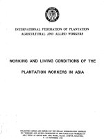
LUNG BUD FORMATION
Lung buds from ventral foregut
Early transcription factors
Endodermal
Mesodermal
Dorsal
SOX2
FIGURE 1-1. Lung bud formation. A,
Lung development is initiated during the
embryonic stage of gestation as a small,
saccular outgrowth of the ventral foregut endoderm. B, Endodermal transcription factors critical for specification of
the primitive respiratory tract include
GATA6, FOXA1, and FOXA2, which are
also expressed throughout the foregut
endoderm. At this time, SOX2 expression is limited to the dorsal aspect (future
esophagus) of the foregut endoderm,
while TTF1 expression is limited to the
ventral aspect (future trachea and lung) of
the lung bud. Mesodermal transcription
factors responsive to signaling peptides
(e.g., SHH) released from the endoderm
and critical for lung development include
GlI1/2/3 and FOXF1. C, Expression of the
signaling peptide, fibroblast growth factor 10 (FGF10), in the adjacent splanchnic mesoderm, induces outgrowth of the
lung bud. FGF10 is secreted by mesenchymal cells and binds to its receptor, FGFR2,
located on the endodermal cell surface,
inducing formation of the lung bud.
Ventral
Lung bud
Dorsal
GATA6
FOXA1, A2
FOXF1
GLI1, 2, 3
TTF1
Ventral
A
B
FGF10 Signaling induces outgrowth of the lung bud
Esophagus
Mesenchyme
Pleura
FGFR2
FGF10
Lung bud
Lung bud
C
Molecular Determinants of Lung Morphogenesis
PERIOD
AGE (WEEKS)
STRUCTURAL EVENTS
Embryonic
3 to 6
Lung buds; trachea, main
stem, lobar, and segmental
bronchi; trachea and
esophagus separate
Pseudoglandular
6 to 16
Subsegmental bronchi,
terminal bronchioles, and
acinar tubules; mucous
glands, cartilage, and
smooth muscle
Canalicular
16 to 26
Respiratory bronchioles,
acinus formation, and
vascularization; type I and
II cell differentiation
Saccular
26 to 36
Dilation and subdivision of
alveolar saccules, increase of
gas-exchange surface area,
and surfactant synthesis
Alveolar
36 to
maturity
Further growth and
alveolarization of lung;
increase of gas-exchange area
and maturation of alveolar
capillary network; increased
surfactant synthesis
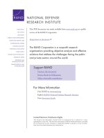
MAJOR STAGES OF LUNG DEVELOPMENT
Lung bud
Epithelium
Bronchial tubules
Epithelium
Acinar tubules
Secretory
Ciliated
Terminal saccules
Ciliated
Clara
Mesenchyme
Embryonic
3–6 wk p.c.
Mesenchyme
Vessel
Pseudoglandular
6–16 wk p.c.
Canalicular
16–26 wk p.c.
Alveoli
Type I
Vessel
Saccular
26–36 wk p.c.
Type I
Type II/
LBs
Capillary
Alveolar
36 wk p.c. to
adolescence
FIGURE 1-2. Major stages of lung development. The bronchi, bronchioles, and acinar tubules are formed by the process of branching morphogenesis
during the pseudoglandular stage of lung development (6 to 16 weeks p.c.). Formation of the capillary bed and dilation/expansion of the acinar structures is
initiated during the canalicular stage of lung development (16 to 26 weeks p.c.). Growth and subdivision of the terminal saccules and alveoli continue until
early adolescence by septation of the distal respiratory structures to form additional alveoli. Cytodifferentiation of mature bronchial epithelial cells (secretory and ciliated cells) is initiated in the proximal conducting airways during the canalicular stage of lung development, while cytodifferentiation in the distal
airways (ciliated and Clara cells) and alveoli (Type I and Type II cells) takes place later during the saccular (26 to 36 weeks p.c.) and alveolar (36 weeks p.c.
to adolescence) stages of lung development. The alveolar stage of lung development extends into the postnatal period, during which millions of additional
alveoli are formed and maturation of the microvasculature, or air-blood barrier, takes place, greatly increasing the surface area available for gas exchange.
Chapter 1
for lung morphogenesis (Figure 1-3). Paracrine signaling pathways that are important for initial formation
of the lung bud and the expansion and branching of
the primitive respiratory tubules include: (1) fibroblast
growth factor (FGF10/FGFR2), (2) sonic hedgehog
(SHH/PTCH1), (3) transforming growth factor-beta
(TGFβ/TGFβR2), (4) bone morphogenetic protein B
(BMP4/BMPR1b), (5) retinoic acid (RA/RARα, β, γ),
(6) WNT (WNT2/2b, 7b, 5a and R-spondin with their
receptors Frizzled and LRP5/6), and (7) the β-catenin
signaling pathways.30–34,42–45 Nuclear transcription factors active in the primitive respiratory epithelium during this period include: TTF1, FOXA2, GATA6, and
SOX2. Likewise, nuclear transcription factors active
in the mesenchyme at this time include: (1) the HOX
family of transcription factors (HOXA5, B3, B4); (2)
the SMAD family of transcription factors (SMAD2, 3,
4) that are downstream transducers of the TGFβ/BMP
signaling pathway; (3) the LEF/TCF family of transcription factors, downstream transducers of β-catenin;
(4) the GLI-KRUPPEL family of transcription factors
(GLI1, 2, 3), downstream transducers of SHH signaling;
(5) the hedgehog-interacting protein, HHIP1, that binds
SHH; and (6) FOXF1, another SHH target.30–34,40,43,44,47
Disruption of many of these transcription factors and
signaling pathways in experimental animals causes
impaired morphogenesis, resulting in laryngotracheal
TABLE 1-1 MORPHOGENETIC PERIODS OF HUMAN
LUNG DEVELOPMENT
3
Section I
4
General Basic Considerations
RECIPROCAL SIGNALING IN LUNG MORPHOGENESIS
Epithelium
Paracrine signaling pathways
FGFR2
EPITHELIUM
SHH
FZ/-catenin
FGFR2
FGFR2
FGF9
BMP4
BMPR1a/b
TGF2
VEGF
PDGF
MESENCHYME
PTCH1/GLI 1, 2, 3
WNT
FGF10
FGF7
FGFR1
BMPR1b
BMP4/5
TGFR2
VEGFR
PDGFR
SHH
FGF10
HHIP1
PTCH1
GLI1, 2, 3
Mesenchyme
FGF10
FIGURE 1-3. Reciprocal signaling in lung morphogenesis. Paracrine and autocrine interactions between the respiratory epithelium and the adjacent mes-
enchyme are mediated by signaling peptides and their respective receptors, influencing cellular behaviors (e.g., proliferation, migration, apoptosis, extracellular matrix deposition) that are critical to lung formation. For example, FGF10 is secreted by mesenchymal cells and binds to its receptor, FGFR10, on
the surface of epithelial cells (paracrine signaling). SHH is secreted by epithelial cells and binds to its receptor, PTCH1, on mesenchymal cells (paracrine
signaling), while HHIP1 is upregulated by SHH in mesenchymal cells, secreted, and binds back to receptors on cells in the mesenchyme (autocrine signaling). Binding of SHH to mesenchymal cells activates the transcription factors, GLI1, GLI2, and GLI3, which, in turn, inhibit FGF10 expression (negative
feedback loop). In contrast, the binding of HHIP1 to mesenchymal cells attenuates or limits the ability of SHH to inhibit FGF10 signaling. Together, these
complex, interacting, signaling pathways control branching morphogenesis of the lung, differentially influencing bronchial tubule elongation, arrest, and
subdivision into new tubules.
malformations, tracheoesophageal fistulae, esophageal
and tracheal stenosis, esophageal atresia, defects in pulmonary lobe formation, pulmonary hypoplasia, and/or
pulmonary agenesis.30–34,40,43–45
Although formation of the larger, more proximal, conducting airways, including segmental and subsegmental bronchi, is completed by the 6th week postconception
(p.c.), both epithelial and mesenchymal cells of the embryonic lung remain relatively undifferentiated. At this stage,
trachea and bronchial tubules lack underlying cartilage,
smooth muscle, or nerves, and the pulmonary and bronchial vessels are not well developed. Vascular connections
with the right and left atria are established at the end of this
period (6 to 7 weeks p.c.), creating the primitive pulmonary
vascular bed.39 Human developmental anomalies occurring during this period of morphogenesis include laryngeal,
tracheal, and esophageal atresia, tracheoesophageal fistulae, tracheal and bronchial stenosis, tracheal and bronchial
malacia, ectopic lobes, bronchogenic cysts, and pulmonary
agenesis.40,46 Some of these congenital anomalies are associated with documented mutations in the genes involved in
early lung development, such as GLI3 (tracheoesophageal
fistula found in Pallister-Hall syndrome), FGFR2 (various
laryngeal, esophageal, tracheal, and pulmonary anomalies
found in Pfeiffer, Apert, or Crouzon syndromes), and SOX2
(esophageal atresia and tracheoesophageal fistula found in
anophthalmia-esophageal-genital, or AEG, syndrome).40,46
Pseudoglandular Period (6 to 16 Weeks' Postconception)
The pseudoglandular stage is so named because of the
distinct glandular appearance of the lung from 6 to 16
weeks of gestation. During this period, the lung consists
primarily of epithelial tubules surrounded by a relatively
thick mesenchyme. Branching of the airways continues,
and formation of the terminal bronchioles and primitive
acinar structures is completed by the end of this period
(see Table 1-1; Figure 1-2). During the pseudoglandular
period, epithelial cell differentiation is increasingly apparent and deposition of cellular glycogen and expression
of a number of genes expressed selectively in the distal
respiratory epithelium is initiated. The surfactant proteins (SP), SP-B and SP-C, are first detected at 12 to 14
weeks of gestation.48,49 Tracheobronchial glands begin to
form in the proximal conducting airways; and the airway epithelium is increasingly complex, with basal,
mucous, ciliated, and nonciliated secretory cells being
detected.36,38 Neuroepithelial cells, often forming clusters
of cells, termed neuroepithelial bodies and expressing a
variety of neuropeptides and transmitters (e.g., bombesin, calcitonin-related peptide, serotonin, and others), are
increasingly apparent along the bronchial and bronchiolar epithelium.50 Smooth muscle and cartilage are now
observed adjacent to the conducting airways.51 The pulmonary vascular system develops in close relationship to
the bronchial and bronchiolar tubules between the 9th
and 12th weeks of gestation. Bronchial arteries arise
from the aorta and form along the epithelial tubules, and
smooth muscle actin and myosin can be detected in the
vascular structures.39
During this period, FGF10, BMP4, TGFβ, β-catenin,
and the WNT signaling pathway continue to be important for branching morphogenesis, along with several
other signaling peptides and growth factors, including:
(1) members of the FGF family (FGF1, FGF2, FGF7,
Molecular Determinants of Lung Morphogenesis
Canalicular Period (16 to 26 Weeks Postconception)
The canalicular period is characterized by formation of
acinar structures in the distal tubules, luminal expansion of the tubules, thinning of the mesenchyme, and
formation of the capillary bed, which comes into close
apposition to the dilating acinar tubules (see Table 1-1;
Figure 1-2). By the end of this period, the terminal
bronchioles have divided to form two or more respiratory bronchioles, and each of these have divided into
multiple acinar tubules, forming the primitive alveolar ducts and pulmonary acini. Epithelial cell differentiation becomes increasingly complex and is especially
apparent in the distal regions of the lung parenchyma.
Bronchiolar cells express differentiated features, such as
cilia, and secretory cells synthesize Clara cell secretory
protein, or CCSP (also known as CC10 or segretoglobin 1A1, SCGB1A1).49,52–54 Cells lining the distal tubules
assume cuboidal shapes and express increasing amounts
of surfactant phospholipids55 and the associated surfactant proteins, SP-A, SP-B, and SP-C.48,49,56–60 Lamellar
bodies, composed of surfactant phospholipids and proteins, are seen in association with rich glycogen stores
in the cuboidal pre–type II cells lining the distal acinar
tubules.61–64 Some cells of the acinar tubules become
squamous, acquiring features of typical type I alveolar
epithelial cells. Thinning of the pulmonary mesenchyme
continues; and the basal lamina of the epithelium and
mesenchyme fuse. Capillaries surround the distal acinar tubules, which together will ultimately form the gas
exchange region of the lung. By the end of the canalicular period in the human infant (26 to 28 weeks p.c.),
gas exchange can be supported after birth, especially
when surfactant is provided by administration of exogenous surfactants. Surfactant synthesis and mesenchymal thinning can be accelerated by glucocorticoids at this
time,60,65–67 which are administered to mothers to prevent
respiratory distress syndrome (RDS) after premature
birth.68,69 Abnormalities of lung development occurring
during the canalicular period include acinar dysplasia,
alveolar capillary dysplasia, and pulmonary hypoplasia,
the latter caused by (1) diaphragmatic hernia, (2) compression due to thoracic or abdominal masses, (3) prolonged rupture of membranes causing oligohydramnios,
or (5) renal agenesis, in which amniotic fluid production
is impaired. While postnatal gas exchange can be supported late in the canalicular stage, infants born during
this period generally suffer severe complications related
to decreased pulmonary surfactant, which causes RDS
and bronchopulmonary dysplasia, the latter a complication secondary to the therapy for RDS.70,71
Saccular (26 to 36 Weeks' Postconception) and Alveolar
Periods (36 Weeks' Postconception through Adolescence)
These periods of lung development are characterized
by increased thinning of the respiratory epithelium and
pulmonary mesenchyme, further growth of lung acini,
and development of the distal capillary network (see
Table 1-1; Figure 1-2). In the periphery of the acinus,
maturation of type II epithelial cells occurs in association with increasing numbers of lamellar bodies, as well
as increased synthesis of surfactant phospholipids,55,61 the
surfactant proteins, SP-A, SP-B, SP-C, and SP-D,48,49,56–60,72
and the ATP-binding cassette transporter, ABCA3, a
phospholipid transporter important for lamellar body
biogenesis.73 The acinar regions of the lung increase in
surface area, and proliferation of type II cells continues.
Type I cells, derived from differentiation of type II epithelial cells, line an ever-increasing proportion of the surface
area of the distal lung. Capillaries become closely associated with the squamous type I cells, decreasing the diffusion distance for oxygen and carbon dioxide between the
alveolar space and pulmonary capillaries. Basal laminae
of the epithelium and stroma fuse; the stroma contains
increasing amounts of extracellular matrix, including elastin and collagen; and the abundance of smooth muscle in
the pulmonary vasculature increases prior to birth.37 In
the human lung, the alveolar period begins near the time
of birth and continues through the first decade of life,
during which the lung grows primarily by septation and
proliferation of the alveoli,74 and by elongation and luminal enlargement of the conducting airways. Pulmonary
arteries enlarge and elongate in close relationship to
the increased growth of the lung.37 Pulmonary vascular
resistance decreases, and considerable remodeling of the
pulmonary vasculature and capillary bed continues during the postnatal period.37 Lung growth remains active
until early adolescence, when the entire complement of
approximately 300 million alveoli has been formed.74
Signaling pathways that are critical for growth, differentiation, and maturation of the alveolar epithelium and
capillary bed during these periods include the FGF, PDGF,
Chapter 1
FGF9, FGF18); (2) members of the TGFβ family, such
as the SPROUTYs (SPRY2, SPRY4), which antagonize
and limit FGF10 signaling, and LEFTY/NODAL, which
regulate left-right patterning; (3) epithelial growth factor (EGF) and transforming growth factor alpha (TGFα),
which stimulate cell proliferation and cytodifferentiation;
(4) insulin-like growth factors (IGFI, IGFII), which facilitate signaling of other growth factors; (5) platelet-derived
growth factors (PDGFA, PDGFB), which are mitogens and
chemoattractants for mesenchymal cells; and (6) vascular endothelial growth factors (VEGFA, VEGFC), which
regulate vascular and lymphatic growth and patterning.30–34,40,42,43 Many of the nuclear transcription factors
that were active during the embryonic period of morphogenesis continue to be important for lung development
during the pseudoglandular period. Additional transcription factors important for specification and differentiation of the primitive lymphatics in the mesenchyme at this
time include: (1) SOX18, (2) the paired-related homeobox gene, PRX1, (3) the divergent homeobox gene, HEX,
and (4) the homeobox gene, PROX1.40,42
A variety of congenital defects may arise during the
pseudoglandular stage of lung development, including
bronchopulmonary sequestration, cystic adenomatoid
malformations, cyst formation, acinar aplasia or dysplasia, alveolar capillary dysplasia with or without misalignment of the pulmonary veins, and congenital pulmonary
lymphangiectasia.40 The pleuroperitoneal cavity also
closes early in the pseudoglandular period. Failure to
close the pleural cavity, often accompanied by herniation
of the abdominal contents into the chest (congenital diaphragmatic hernia), leads to pulmonary hypoplasia.
5
Section I
6
General Basic Considerations
VEGF, RA, BMP, WNT, β-catenin, and NOTCH signaling
pathways.30–34,42,43 For example, FGF signaling is critical
for alveologenesis during these periods. Targeted deletion
of the FGF receptors, Fgfr3 and Fgfr4, blocks alveologenesis in mice. Likewise, targeted deletion of Pdgfa, another
growth factor critical for alveologenesis, interferes with
myofibroblast proliferation and migration, resulting in
complete failure of alveologenesis and postnatal alveolar
simplification in mice.30–34,42,43
Nuclear transcription factors found earlier in lung
development (i.e., FOXA2, TTF1, GATA6, and SOX2)
continue to be important for maturation of the lung,
influencing sacculation, alveolarization, vascularization, and cytodifferentiation of the peripheral lung.
Transcription factors associated with cytodifferentiation during these periods include: (1) FOXJ1 (ciliated
cells), (2) MASH1 (or HASH1) and HES1 (neuroendocrine cells), (3) FOXA3 and SPDEF (mucus cells), and
(4) ETV5/ERM (alveolar type II cells).32 Morphogenesis
and cytodifferentiation are further influenced by additional transcription factors expressed in the developing
respiratory epithelium at this time, including: (1) several ETS factors (ETV5/ERM, SPDEF, ELF3/5); (2) SOX
genes (SOX-9, SOX11, SOX17); (3) nuclear factor of
activated T cells/calcineurin-dependent 3, or NFATC3;
(4) nuclear factor-1, or NF-1; (5) CCAAT/enhancer
binding protein alpha, or CEBPα; and (6) Krüppellike factor 5, or KLF5; as well as the transcription factors, GLI2/GLI3, SMAD3, FOXF1, POD1, and HOX
(HOXA5, HOXB2 to B5), all of which are expressed in
the mesenchyme.30–34
Control of Gene Transcription During Lung Morphogenesis
Numerous regulatory mechanisms influence cell commitment, proliferation, and terminal differentiation required
for formation of the mammalian lung. These events must
be precisely controlled in all organs to produce the complex body plan characteristic of higher organisms. In the
mature lung, approximately 40 distinct cell types can
be distinguished on the basis of morphologic and biochemical criteria.75 Distinct pulmonary cell types arise
primarily from subsets of endodermal and mesodermal
progenitor cells. Pluripotent or multipotent cells receive
precise temporal and spatial signals that commit them
to differentiated pathways, which ultimately generate
the heterogeneous cell types present in the mature organ.
The information directing cell proliferation and differentiation during organogenesis is derived from the genetic
code contained within the DNA of each cell in the organism. Unique subsets of messenger RNAs (mRNAs) are
transcribed from DNA and direct the synthesis of a variety of proteins in specific cells, ultimately determining
cell proliferation, differentiation, structure, function,
and behavior for each cell type. Unique features of differentiating cells are controlled by the relative abundance
of these mRNAs, which, in turn, determine the relative
abundance of proteins synthesized by each cell. Cellular
proteins influence morphologic, metabolic, and proliferative behaviors of cells, characteristics that traditionally have been used to assign cell phenotype by using
morphologic and cytologic criteria. Gene expression in
each cell is also determined by the structure of DNA-
protein complexes that comprise the chromatin within
the nucleus of each cell. Chromatin structure, in turn,
influences the accessibility of individual genes to the transcriptional machinery. Diverse extracellular and intracellular signals also influence gene transcription, mRNA
processing, mRNA stability and translation—processes
that determine the relative abundance of proteins produced by each cell.
Only a small fraction of the genetic material present
in the nucleus represents regions of DNA that direct
the synthesis of mRNAs encoding proteins. There is an
increasing awareness that sequences in the noncoding
regions of genes influence DNA structure and contain
promoter and enhancer elements (usually in flanking and
intronic regions of each gene) that determine levels of
transcription.76 Nucleotide sequencing and identification
of expressed complementary DNA (cDNA) sequences
encoded within the human genome have provided insight
into the amount of the genetic code used to synthesize
the cellular proteins produced by each organ.77 At present, nearly all of the expressed cDNAs have been identified and partially sequenced for most human organs.
Analysis of these mRNAs reveals distinct, and often
unique, subsets of genes that are expressed in each organ,
as well as the relative abundance and types of proteins
encoded by these mRNAs. Of interest, proteins bearing
signaling and transcriptional regulatory information are
among the most abundant of various classes of proteins
in human cells. Organ complexity in higher organisms
is derived, at least in part, by the increasingly complex
array of signaling molecules that govern cell behavior.
Regulatory mechanisms controlling transcription are
listed in Figure 1-4.
Transcriptional Cascades/Hierarchies
Gene transcription is modulated primarily by the binding of transcription factors (or trans-acting factors) to
DNA. Transcription factors are nuclear proteins that
bind to regulatory motifs consisting of ordered nucleotides, or specific nucleotide sequences. The order of
these specific nucleotide sequences determines recognition sites within the DNA (cis-acting elements) that
are bound by these nuclear proteins. The binding of
transcription factors to these cis-acting elements influences the activity of RNA polymerase II, which binds
to sequences near the transcription start site of target
genes, initiating mRNA synthesis.76,78 Numerous families of transcription factors have been identified, and
their activities are regulated by a variety of mechanisms,
including posttranslational modification and interactions
with other proteins or DNA, as well as by their ability to
translocate or remain in the nucleus.78 Transcription factors also activate the transcription of other downstream
nuclear factors, which, in turn, influence the expression
of additional trans-acting factors. The number and cell
specificity of transcription factors have proven to be
large and are r epresented by diverse families of proteins
categorized on the basis of the structural motifs of their
DNA binding or trans-
activating domains.76,78 These
interacting cascades of factors comprise a network with
vast capabilities to influence target gene expression. The
HOX family of transcription factors (homeodomain,
Molecular Determinants of Lung Morphogenesis
A
Genetic code/DNA sequences
– inheritance patterns
FIGURE 1-4. Control of gene
Histone modification
Chromatin structure
– epigenetic modifications
DNA methylation
B
protein
Combinatorial regulation
– transcription factors (tf)
– cofactors (cf)
mRNA
Transcription
cf
tf
tf
tf
C
Transcriptional networks
Gene A
Gene B
D
helix-turn-helix-containing family of DNA-binding
proteins) represents an example of such a regulatory
motif. A series of HOX genes are located in arrays containing large numbers of distinct genes arranged 3' to
5' in distinct loci within human chromosomes.7 HOX
genes bind to and activate other downstream HOX gene
family members that, in turn, bind to and activate the
transcription of additional related and unrelated transcription factors, altering their activity and interactions
at the transcriptional level.10 Such cascades are now well
characterized in organisms such as in D. melanogaster74
and C. elegans.79–81 Mammalian homologues exist for
many of these genes, and their involvement in similar regulatory cascades influences gene expression and
organogenesis in more complex organisms.3–15 In the
mammalian lung, TTF1 and FOX family members are
involved in regulatory cascades that determine organogenesis and lung epithelial–specific gene expression. In
addition, many other nuclear transcription factors, such
as β-catenin, GATA6, POD1, FOXA2, NF1, FOXF1,
GLI family members, ETS factors, N-MYC, CEBP family members, retinoic acid receptors (RAR), estrogen
receptors, and glucocorticoid receptors, influence lung
growth, cytodifferentiation, and function.30–34
Combinatorial Regulation of Gene
Transcription and Expression
Advances in understanding mRNA expression profiles,
genomics, chromatin structure, and mechanisms regulating
gene expression are transforming current concepts regarding the molecular processes that control gene expression.
Bioinformatics and advances in computational and systems
biology are providing new insights into the remarkable
interactions among genes that control other cellular processes. To influence gene expression, genes function in complex networks, which are dependent on each individual's
inherited DNA sequences (genes) and on epigenetic mecha-
Gene C
expression. Diverse cellular mechanisms regulate varying levels of
gene transcription that, in turn,
control messenger RNA and protein
synthesis governing cell differentiation and function during lung development. Inherited patterns of each
individual's genetic code (A) are
modified by epigenetic mechanisms
that modify chromatin structure
through methylation of DNA and/
or modification of histone proteins
(B). Binding of nuclear transcription factors to specific structural
motifs (cis-acting elements) in DNA
sequences is modified by associated cofactors and other transcription factors (C). Protein expression
is often controlled by transcriptional
networks, in which several genes
are activated in series to induce or
inhibit expression of downstream
targets and/or other proteins (D).
nisms independent of genetic constitution. Changes in chromatin structure (packaging of DNA, histones, and other
associated proteins) influence the accessibility of DNA to
the regulatory actions of various transcriptional complexes
(proteins) and is dependent upon posttranslational modification of histone proteins by methylation or acetylation.
The regulatory regions of target genes in eukaryotes are
highly complex, containing numerous cis-acting elements
that bind various nuclear transcription proteins to influence
gene expression. Nuclear proteins may bind DNA as monomers or oligomers, or form homo- or hetero-oligomers
with other transcriptional proteins. Furthermore, many
transcriptional proteins are modified by posttranslational
modifications that are induced by receptor occupancy or by
phosphorylation and/or dephosphorylation events. Binding
of transcription factors influences the structural organization of DNA (chromatin), making regulatory sites more or
less accessible to other nuclear proteins, which, in turn, positively or negatively regulate gene expression. Numerous
cis-acting elements and their cognate trans-acting proteins
interact with the basal transcriptional apparatus to regulate mRNA synthesis. The precise stoichiometry and specificity of the occupancy of various DNA-binding sites also
influence the transcription of specific target genes, either
positively or negatively. This mode of regulation is characteristic of most eukaryotic cells, including those of the lung.
For example, in pulmonary epithelial cells, a distinct set
of transcription factors, including TTF1, GATA6, activator protein 1 (AP1), FOX family members, RARs, STAT3,
NF1, and specificity protein 1 (SP1), act together to regulate expression of surfactant protein genes, which influence
postnatal respiratory adaptation.32,82–84
Influence of Chromatin Structure on Gene Expression
The structure of chromatin is a critical determinant of the
ability of target genes to respond to regulatory information influencing gene transcription. The abundance and
Chapter 1
CONTROL OF GENE EXPRESSION
7
Section I
8
General Basic Considerations
organization of histones and other chromatin-associated
proteins, including nuclear transcriptional proteins, influence the structure of DNA at genetic loci. The accessibility of regulatory regions within genes or groups of
genes for binding and regulation by transcription factors
is often dependent on chromatin structure. Changes in
chromatin structure are likely determined by the process
of cell differentiation during which target genes become
available or unavailable to the regulatory influences of
transcription factors.85 Thus, the activity of a transcription factor at one time in development may be entirely
distinct from that at another time. Chemical modification of DNA (e.g., methylation of cytosine) is also known
to modify the ability of cis-active elements to bind and
respond to regulatory influences. For example, cytosineguanine (CG)–rich islands are found in transcriptionally
active genes, and methylation of these regions may vary
developmentally or in response to signals that influence
gene transcription. Chromatin structure, in turn, is influenced by post-transcriptional modification of histones
and other DNA-associated proteins by biochemical processes, including acetylation, methylation, demethylation, phosphorylation, ubiquitination, sumoylation, and
ADP-ribosylation, which then influence the binding of
transcriptional complexes and coactivator proteins that
interact with the basal transcriptional machinery via
polymerase II to alter gene transcription.86
miR-17-92 cluster during lung development resulted in
the absence of normal terminal (alveolar) saccules, which
were replaced with respiratory tubules lined by highly
proliferative, undifferentiated epithelium, suggesting that
downregulation of the miR-17-92 cluster is critical for
normal cellular growth and differentiation.92
Non-Transcriptional Mechanisms
While regulation of gene transcription is an important
factor in organogenesis, numerous regulatory mechanisms, including control of RNA expression, mRNA
stability, and protein synthesis and degradation are also
known to provide further refinement in the abundance
of mRNAs and proteins synthesized by a specific cell,
which ultimately determine its structure and function.87
For example, microRNAs (miRNAs) have been implicated recently in the regulation of proliferation, differentiation, and apoptosis of epithelial progenitor cells
in the lung.86 miRNAs are small (19 to 25 nucleotides),
single-stranded, non-coding RNAs that regulate protein
expression by binding to the 3' untranslated region of
target mRNAs, which results in degradation or inhibition of protein translation in the cytoplasm. mi/RNAs are
transcribed initially as very long primary transcripts (primiRNAs) that contain hundreds to thousands of nucleotides. This primary transcript is cleaved to release a much
smaller 70 to 100 nucleotide fragment (pre-miRNA),
which is then exported to the cytoplasm. Once in the
cytoplasm, this fragment is further cleaved by an RNA
polymerase II (DICER) to release a 19- to 25- nucleotide
fragment, which is then incorporated into an miRNAinduced silencing complex (miRISC) that guides the
miRNA to its target mRNA, where it binds to the mRNA
affecting its translation and/or stability.88 High expression
levels of at least three members of the miR-17-92 cluster are present in the embryonic lung, but decline as lung
development progresses.89 Mice deficient in the miR-1792 cluster exhibited hypoplasia of the lung,90 while targeted deletion of DICER in the lung resulted in abnormal
lung development with increased apoptosis and abnormal branching morphogenesis.91 Overexpression of the
Gradients of Signaling Molecules and Localization
of Receptor Molecules
Chemical gradients within tissues, and their interactions with membrane receptors located at distinct sites
within the organ, can provide critical information during organogenesis. Polarized cells have basal, lateral, and
apical surfaces with distinct subsets of signaling molecules (receptors) that allow the cell to respond in unique
ways to focal concentrations of regulatory molecules.
Secreted ligands (e.g., FGFs, TGFβ/BMPs, WNTs, SHH,
and HHIP1) function in gradients that are further influenced by binding of the ligand to basement membranes or
proteoglycans in the extracellular matrix.30,33,34,43 Spatial
information is established by gradients of these signaling molecules and by the presence and abundance of
receptors at specific cellular sites. Such systems provide
positional information to the cell, which influences its
behavior (e.g., shape, movement, proliferation, differentiation, and polarized transport).
Receptor-Mediated Signal Transduction
Receptor-mediated signaling is well recognized as a fundamental mechanism for transducing extracellular information. Such signals are initiated by the occupancy of
membrane-associated receptors capable of initiating
additional signals (known as secondary messengers), such
as cyclic adenosine monophosphate, calcium, and inositide phosphates, which influence the activity and function
of intracellular proteins (e.g., kinases, phosphatases, proteases). These proteins, in turn, may alter the abundance
of transcription factors, the activity of ion channels, or
changes in membrane permeability, which subsequently
modify cellular behaviors. Receptor-mediated signal
transduction, induced by ligand-receptor binding, mediates endocrine, paracrine, and autocrine interactions on
which cell differentiation and organogenesis depend. For
example, signaling peptides and their receptors, such as
FGF, SHH, WNT, BMP, VEGF, PDGF, and NOTCH have
been implicated in organogenesis of many organs, including the lung.30–34,42,43
Transcriptional Mechanisms Controlling Gene Expression
During Pulmonary Development
While knowledge of the determinants of gene regulation in lung development is rudimentary at present, a
number of transcription factors and signaling networks
that play critical roles in lung morphogenesis have
been identified.30–34,42,43 Lung morphogenesis depends
on formation of definitive endoderm, which, in turn,
receives signals from the splanchnic mesenchyme to
initiate organogenesis along the foregut, forming thyroid, liver, pancreas, lung, and portions of the gastrointestinal tract.17 The ventral plate of the endoderm in
mammals forms under the direction of FOXA2, a transcription factor that is known to play a critical role in
Molecular Determinants of Lung Morphogenesis
(see Figure 1-2).35,102,103 Ablation of Titf1 in the mouse
impaired lung morphogenesis, resulting in tracheoesophageal fistula and hypoplastic lungs lined by a poorly differentiated respiratory epithelium and lacking the distal,
alveolar, gas exchange regions.102,103,106,107 Substitution
of a mutant Titf1 gene, which lacked phosphorylation
sites, restored lung development in the Titf1 knockout mouse.108 Expression of a number of genes, including those regulating surfactant homeostasis, fluid and
electrolyte transport, host defense, and vasculogenesis, is
regulated by TTF1 phosphorylation prior to birth. TTF1
regulates the expression of a number of genes in a highly
specific manner in the respiratory epithelium, including
surfactant proteins, SP-A, SP-B, and SP-C, and CCSP.109–112
TTF1 functions in concert with other transcription factors, including FOXA2, GATA6, NF1, ERM, PARP2,
SP1/SP3, TAZ, NFAT, and RARs to regulate lung-specific
gene transcription.32,113–123 TTF1 gene transcription itself is
modulated by the activity of FOXA2, which binds to the
promoter enhancer region of the TTF1 gene, thus creating
a transcriptional network.99 A combinatorial mode of regulation is evidenced by the apposition of clustered TTF1
cis-active elements and FOXA2 binding sites in target
genes, such as the SP-B and CCSP genes.96,116 The stoichiometry, timing, and distinct combinations of transcription
factor binding, as well as posttranscriptional modification
of TTF1 by phosphorylation, are involved in differential
gene expression throughout lung development. TTF1 and
other transcription factors are recruited to nuclear complexes at regulatory sites of target genes that influence
respiratory epithelial cell differentiation, providing and
translating spatial information required for the formation
of the highly diverse epithelial cell types lining distinct
regions of the respiratory tract (see Figure 1-5).
BLUEPRINT FOR LUNG EPITHELIAL CELL DEVELOPMENT
Mouse
E3.5
Sox2/Sox17
Blastula cells
Foxa1/2
Mesendoderm Ectoderm
WNT/-catenin
NODAL, RA
E7.5
Mesoderm
Sox2/Sox17, Foxa1/2, Gata6
Endoderm
Foregut
E10.5
hp
FGF, SHH, BMP/TGF
WNT/-catenin
Thyroid Pancreato
biliary
Midgut
Liver
Lung
Hindgut
Stomach
FIGURE 1-5. A blueprint for lung
Titf1, Foxa1/2,
Gata6, Sox2, Klf5
E12.5
-catenin, NOTCH
br
Adult
Respiratory
epithelium
Titf1, Foxa2, Sox2,
Klf5, p63, Spdef,
Foxa3, Foxj1, Erm,
Foxp1/2/4
Proximal
epithelium
Basal
Ciliated Clara
Distal
epithelium
Goblet
Alveolar
Type I
Alveolar
Type II
epithelial cell development. Cytodifferen
tiation of the respiratory epithelium
is controlled by transcriptional networks of genes (highlighted) that are
expressed throughout lung development, in conjunction with autocrine
and paracrine signaling pathways that
control structural morphogenesis of the
lung. Additional transcription factors are
induced or repressed later in development, and in the adult organ, to influence
the differentiation of specific cell types.
Chapter 1
committing progenitor cells of the endoderm to form the
primitive foregut.17 FOXA2 is member of a large family of nuclear transcription factors, termed the winged
helix family of transcription factors, that are involved
in cell commitment, differentiation, and gene transcription in a variety of organs, such as the central nervous
system and derivatives of the foregut endoderm, including the gastrointestinal tract, lung, and liver.93 FOXA2
is required for the formation of foregut endoderm, from
which the lung bud is derived, and plays a critical role in
organogenesis of the lung. While FOXA2 plays a critical
role in formation and commitment of progenitor cells
to form the foregut endoderm, FOXA2 also influences
the expression of specific genes in the respiratory epithelium later in development.94–100 Conditional deletion
of Foxa2 after birth caused goblet cell metaplasia, airspace enlargement, and inflammation during the postnatal period,101 while deletion of Foxa2 prior to birth
resulted in delayed pulmonary maturation, associated
with decreased surfactant lipid and protein expression
and the development of a respiratory distress-like syndrome.100 Thus, FOXA2 plays a critical role in specification of foregut endoderm in the early embryo, and
is used again in the perinatal and postnatal period to
direct surfactant production, alveolarization, postnatal
lung function, and homeostasis (Figure 1-5).
TTF1 (TITF1) is a 38-kd nuclear protein, containing
a homeodomain DNA-binding motif, that is critical for
formation of the lung and for regulation of a number of
highly specific gene products produced only in the respiratory epithelium.84,102,103 TTF1 is also expressed in the
thyroid and in specific regions of the developing central nervous system.35,102 In the lung, TTF1 is expressed
in the respiratory epithelium of the primitive lung bud
9
Section I
10
General Basic Considerations
Epithelial-Mesenchymal Interactions and Lung
Morphogenesis
In vivo and in vitro experiments support the concept
that branching morphogenesis and differentiation of the
respiratory tract depends on reciprocal signaling between
endodermally derived cells of the lung buds and the pulmonary mesenchyme or stroma.30–34,43 This interdependency
depends on autocrine and paracrine interactions that are
mediated by the various signaling mechanisms governing
cellular behavior (see Figure 1-3). Similarly, autocrine and
paracrine interactions are known to be involved in cellular responses of the postnatal lung, generating signals
that regulate cell proliferation and differentiation necessary for its repair and remodeling following injury. The
splanchnic mesenchyme produces a number of signaling
peptides critical for migration and proliferation of cells
in the lung buds, including FGF10, FGF7, FGF9, BMP5,
and WNT 2/2b, which activate receptors found on epithelial cells. In a complementary manner, epithelial cells
produce WNT7b, WNT5a, SHH, BMP4, FGF9, VEGF,
and PDGF that activate receptors and signaling pathways
on target cells in the mesenchyme.30,33,34,42,43
Branching Morphogenesis, Vascularization,
and Sacculation
Two distinct processes, branching and sacculation, are
critical to morphogenesis of the mammalian lung. The
major branches of the conducting airways of the human
lung are completed by 16 weeks (p.c.) by a process of
dichotomous branching, initiated by the bifurcation of
the main stem bronchi early in the embryonic period
of lung development. Epithelial-lined tubules of everdecreasing diameter are formed from the proximal to
distal region of the developing lung. Pulmonary arteries
and veins form along the tubules and ultimately invade
the acinar regions, where capillaries form between the
arteries and veins, completing the pulmonary circulation.37,42 The bronchial vasculature arises from the aorta,
providing nutrient supply predominantly to bronchial
and bronchiolar regions of the lung. In contrast, the alveolar regions are supplied by the pulmonary arterial system. Lymphatics and nerves form along the conducting
airways, the latter being prominent in hilar, stromal and
vascular tissues, but lacking in the alveolar regions of the
lung.124 A distinct period of lung sacculation and alveolarization begins in the late canalicular period (16 weeks p.c.
and thereafter), which will result in the formation of the
adult respiratory bronchiole, alveolar duct, and alveoli.
During sacculation, a unique pattern of vascular supply
forms the capillary network surrounding each terminal
saccule, providing an ever-expanding gas exchange area
that is completed in adolescence. Both vasculogenesis and
angiogenesis contribute to formation of the pulmonary
vascular system.37,42 Signaling via SHH, VEGFA, FOXF1,
NOTCH, Ephrins, and PDGF plays important roles in
pulmonary vascular development.30,33,34,42 For example,
VEGFA and its receptors (VEGFR1, VEGFR2) are critical
factors for vasculogenesis in many tissues. Targeted inactivation of Vegf and Vefgfr1 in mice results in impaired
angiogenesis,125 while overexpression of the VEGFA 164
isoform disrupts pulmonary vascular endothelium in
newborn conditional transgenic mice, causing pulmonary
hemorrhage.126 PROX1, a homeo domain transcription
factor, is induced in a subset of venous endothelial cells
during development and upregulates other lymphaticspecific genes, such as VEGFR3 and LYVE1, which are
critical for development of the lymphatic network in the
lung.124 Growth factors important for lymphatic development include VEGFC and its receptor, VEGFR3, as
well as the angiopoietins, ANG1 and ANG2, and their
receptors, TIE1 and TIE2.124 Insufficiency or targeted
deletion of these factors in mice impairs lymphatic vessel
formation.127,42
Control of Lung Proliferation During Branching
Morphogenesis
Dissection of the splanchnic mesenchyme from the lung
buds arrests cell proliferation, branching, and differentiation of the pulmonary tubules in vitro.43 Both in vitro
and in vivo experiments strongly support the concept that
the mesenchyme produces signaling peptides and growth
factors critical to the formation of respiratory tubules.43
In addition, lung growth is influenced by mechanical
factors, including the size of the thoracic cavity and by
stretch. For example, complete occlusion of the fetal trachea in utero enhances lung growth, while drainage of
lung liquid or amniotic fluid causes pulmonary hypoplasia.128,129 Regional control of proliferation is required
for the process of dichotomous branching: division is
enhanced at the lateral edges of the growing bud and
inhibited at branch points.130 Precise positional control of
cell division is determined by polypeptides derived from
the mesenchyme (e.g., growth factors or extracellular
matrix molecules) that selectively decrease proliferation
at clefts and increase cell proliferation at the edges of the
bud. Proliferation in the respiratory tubule is dependent
on a number of growth factors, including the FGF family
of polypeptides. In vitro, FGF1 and FGF7 (also known
as keratinocyte growth factor, KGF) partially replace the
requirement of pulmonary mesenchyme for continued
epithelial cell proliferation and budding.131,132 FGF polypeptides are produced by the mesenchyme during lung
development and bind to and activate a splice variant of
FGFR2 (FGFR2IIIb) that is present on respiratory epithelial cells, completing a paracrine loop.133,134 Blockade
of FGFR2 signaling in the epithelium of the developing
lung bud in vivo, using a dominant-negative FGF receptor
mutant, completely blocked dichotomous branching of all
conducting airway segments except the primary bronchi
in mice.135 FGF10 produced at localized regions of mesenchyme near the tips of the lung buds creates a chemoattractant gradient that activates the FGFR2IIIb receptor in
epithelial cells of the lung buds, inducing cell migration,
differentiation, and proliferation required for branching morphogenesis.136 Deletion of Fgf10 or Fgfr2IIIb in
mice blocked lung bud formation, resulting in lung agenesis.137,138 Increased expression of FGF10 or FGF7 in the
fetal mouse lung caused severe pulmonary lesions with all
of the histologic features of cystic adenomatoid malformations.139,140 FGF7 is also mitogenic for mature respiratory epithelial cells in vivo, enhancing proliferation of
bronchiolar and alveolar cells when administered intratracheally to the lungs of adult rats or by conditional
targeted overexpression in mice.141,142 Since FGF7 is